. 2024 Feb 5;16(3):2789-2811.
doi: 10.18632/aging.205511. Epub 2024 Feb 5.

Targeting PERK-ATF4-P21 axis enhances the sensitivity of osteosarcoma HOS cells to Mppα-PDT

Affiliations

Targeting PERK-ATF4-P21 axis enhances the sensitivity of osteosarcoma HOS cells to Mppα-PDT

Shenxi Zhong et al. Aging (Albany NY). .

Abstract

Osteosarcoma (OS) is the most prevalent type of malignant bone tumor in adolescents. The overall survival of OS patients has reached a plateau recently. Thus, there is an urgent need to develop approaches to improve the sensitivity of OS to therapies. Pyropheophorbide-α methyl ester-mediated photodynamic therapy (MPPα-PDT) is a new type of tumor therapy, and elucidating its mechanism is helpful to improve its anti-tumor efficacy. Here, we investigated how PERK signaling promotes the human OS (HOS) cell survival induced by MPPα-PDT, as overcoming this may enhance sensitivity to MPPα-PDT. We found that MPPα-PDT combined with PERK inhibitor GSK2656157 enhanced HOS cell apoptosis by suppressing autophagy and p21. Autophagy inhibition and p21 depletion enhanced cell death, indicating pro-survival effects in MPPα-PDT. Notably, p21 was found to be an effector of the PERK-Atf4 pathway, which could positively regulate autophagy mediated by MPPα-PDT. In conclusion, we found that the combination of MPPα-PDT and GSK2656157 enhanced apoptosis in HOS cells by inhibiting autophagy. Mechanistically, this autophagy is p21-dependent and can be suppressed by GSK2656157, thereby enhancing sensitivity to MPPα-PDT.

Keywords: MPPα-PDT; PERK pathway; apoptosis; autophagy; human osteosarcoma; p21.


Conflict of interest statement

CONFLICTS OF INTEREST: The authors declare no conflicts of interest related to this study.

Figures

Figure 1
Figure 1
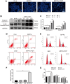
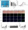
MPPα-PDT induces HOS cells cycle arrest and apoptosis. (A) HOS cells nuclei were examined for apoptotic morphological changes under a fluorescence microscope (magnification: ×200) after MPPα-PDT treatment for 12 h. (B, C) Cells were harvested after MPPα-PDT treatment for 3, 6, and 12 h and western blot used to evaluate cleaved caspase-3 and cleaved PARP levels. (DF) Apoptosis was determined by flow cytometry. (EG) Cell cycle distribution was analyzed using flow cytometry. *P < 0.05 vs. control group. All data represent the mean ± SD of 3 independent experiments.
Figure 2
Figure 2
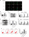
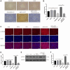
ER stress and PERK-eIF2α-ATF4 signaling pathway activation are induced by MPPα-PDT in HOS cells. (A) ER morphology was observed by TEM (magnification, ×20000). (B) Intracellular Ca2+ concentration was quantified by flow cytometry using Fluo-4-AM probe. (C, D) MPPα-PDT treated cells were harvested after 3, 6, and 12 h and PERK, p-PERK, BIP, p-eIF2α, ATF4, and CHOP, levels determined by western blot. Data are shown as mean ± SD of 3 independent experiments. *P < 0.05 vs. control group.
Figure 3
Figure 3
Autophagy can be triggered by MPPα-PDT. (A) Transmission electron microscopy (magnification, ×20000) was used to observe autophagosomes upon autophagy induction by MPPα-PDT after 12 h. (BD) Cells were harvested 3, 6, and 12 h after MPPα-PDT treatment and protein ATG5, LC3-II/LC3-I, and p62 were determined by western blot. Data are shown as mean ± SD of 3 independent experiments. *P < 0.05 vs. control group.
Figure 4
Figure 4
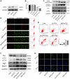
Inhibiting PERK may suppress autophagy induced by MPPα-PDT, and enhance the anti-tumor ability of MPPα-PDT in HOS cells. HOS cells in MPPα-PDT+GSK2656157 group were pretreated with 5 mM GSK2656157 for 1 h before MPPα-PDT treatment. HOS cells in MPPα-PDT + Bafilomycin A1 group were pretreated with 100 nM bafilomycin A1 for 2 h before MPPα-PDT treatment. (A, B) Immunofluorescence analysis of p-PERK levels (magnification: ×400). (CG) After indicated treatments, cells were harvested and PERK, p-PERK, ATF4, LC3-II/LC3-I, p62, cleaved caspase-3, and cleaved PARP levels determined by western blot. (H, I) Apoptotic rate was examined by flow cytometry. Data are shown as mean ± SD of 3 independent experiments. *P < 0.05.
Figure 5
Figure 5
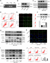
Targeting ATF4 increases the sensitivity of PDT by inhibiting autophagy. HOS cells in MPPα-PDT+GSK2656157 group were pretreated with 5 mM GSK2656157 for 1 h before MPPα-PDT treatment. HOS cells were transfected with siRNAs and then treated with MPPα-PDT. (A) Protein ATF4 or mRNA ATF4 was detected by western blot and represents the transfection efficiency. (B) Cell viabilities were detected by CCK-8 after treated with SiRNA-ATF4 (Si-ATF4), SiRNA-negative control (Si-NC), MPPα-PDT and group MPPα-PDT combined with Si-ATF4. (C) Following indicated treatments, cells were harvested and ATF4, p21, PARP and cleaved caspase-3 levels determined by western blot. (D, E) Apoptotic rate or JC-1 stain was examined by flow cytometry or fluorescence microscope (×200). (F) Following indicated treatments, cells were harvested and LC3, P62, Atg5 and Beclin1 levels were determined by western blot. (G) Adenovirus-GFP-LC3 was transfected into the HOS cells after treated, the LC3 fluorescent particles were observed by laser confocal microscope (×400). *P < 0.05 vs. control group, data are shown as mean ± SD of 3 independent experiments.
Figure 6
Figure 6
The pro-survival effect of P21 is achieved by regulating autophagy after MPPα-PDT. (A) ChIP analysis was used to detect ATF4 binding to the first intron of p21. HOS cells were transfected with siRNAs and then treated with MPPα-PDT. (B) Protein P21 were detected by western blot to represent the transfection efficiency. (C) Following indicated treatments, cells were harvested and P21 and cleaved caspase-3 levels were determined by western blot. (D, E) Apoptotic rate or JC-1 stain were examined by flow cytometry or fluorescence microscope (×200). (F) Protein P21 was detected by western blot after transfected by lentivirus-overexpress P21. (G) Following indicated treatments, cells were harvested and LC3, P62, Atg5 and Beclin1 levels were determined by western blot. (H) Adenovirus-GFP-LC3 was transfected into the HOS cells after treated, the LC3 fluorescent particles were observed by laser confocal microscope (×400). (I) Protein CHOP, cleaved-caspase3 were detected by western blot. (J) Apoptotic rate was examined by flow cytometry. *P < 0.05 vs. control group, data are shown as mean ± SD of 3 independent experiments.
Figure 7
Figure 7
GSK2656157 enhances the antitumor activity of MPPα-PDT by inhibiting PERK pathway in vivo. HOS tumor-bearing mice were treated with MPPα (15 mg/kg), LED (120 J/mm2), GSK2656157 (30 mg/kg) and MPPα-PDT (LED following MPPα, as described in Materials and Methods). (A, B) After 31 days, tumors were collected. Tumor volume was determined as described in methods. (C) Tumor volume change curves. (D, E) After treatments, tumor necrosis and apoptosis were analyzed by H&E and TUNEL analysis, respectively (magnification, ×100). (F, G) After treatments, tumors apoptosis was assessed by cleaved caspase-3 western blot analysis. Data are shown as mean ± SD of 3 independent experiments. *P < 0.05.
Figure 8
Figure 8
p-PERK in vivo levels upon indicated treatments. (A, B) IHC analysis of p-PERK levels in tumor sections (magnification, ×200). (C, D). Fluorescence analysis of p-PERK levels in tumor sections (magnification, ×400). (E, F) Following indicated treatments, tumor tissues were analyzed by western blot for p-PERK levels. Data are shown as mean ± SD of 3 independent experiments. aP < 0.01 vs. control group; bP < 0.01 vs. MPPα-PDT group.
Figure 9
Figure 9
The potential mechanisms of inhibiting Perk-ATF4-P21 pathway to enhance the efficacy of MPPα-PDT. Antitumor ability of Mppα-PDT is improved by inhibiting PERK signaling, which is achieved by P21 regulating autophagy.

Similar articles

References

    1. Whelan JS, Davis LE. Osteosarcoma, Chondrosarcoma, and Chordoma. J Clin Oncol. 2018; 36:188–93. 10.1200/JCO.2017.75.1743 - DOI - PubMed
    1. Bielack SS, Kempf-Bielack B, Branscheid D, Carrle D, Friedel G, Helmke K, Kevric M, Jundt G, Kühne T, Maas R, Schwarz R, Zoubek A, Jürgens H. Second and subsequent recurrences of osteosarcoma: presentation, treatment, and outcomes of 249 consecutive cooperative osteosarcoma study group patients. J Clin Oncol. 2009; 27:557–65. 10.1200/JCO.2008.16.2305 - DOI - PubMed
    1. Smeland S, Bielack SS, Whelan J, Bernstein M, Hogendoorn P, Krailo MD, Gorlick R, Janeway KA, Ingleby FC, Anninga J, Antal I, Arndt C, Brown KLB, et al.. Survival and prognosis with osteosarcoma: outcomes in more than 2000 patients in the EURAMOS-1 (European and American Osteosarcoma Study) cohort. Eur J Cancer. 2019; 109:36–50. 10.1016/j.ejca.2018.11.027 - DOI - PMC - PubMed
    1. Pham TC, Nguyen VN, Choi Y, Lee S, Yoon J. Recent Strategies to Develop Innovative Photosensitizers for Enhanced Photodynamic Therapy. Chem Rev. 2021; 121:13454–619. 10.1021/acs.chemrev.1c00381 - DOI - PubMed
    1. Mishchenko T, Balalaeva I, Gorokhova A, Vedunova M, Krysko DV. Which cell death modality wins the contest for photodynamic therapy of cancer? Cell Death Dis. 2022; 13:455. 10.1038/s41419-022-04851-4 - DOI - PMC - PubMed

Publication types

Substances