

Functional Enrichment Analysis of Tumor Microenvironment-Driven Molecular Alterations That Facilitate Epithelial-to-Mesenchymal Transition and Distant Metastasis
- PMID: 38318286
- PMCID: PMC10840405
- DOI: 10.1177/11779322241227722
Functional Enrichment Analysis of Tumor Microenvironment-Driven Molecular Alterations That Facilitate Epithelial-to-Mesenchymal Transition and Distant Metastasis
Abstract
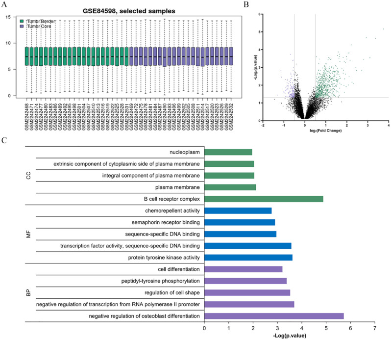
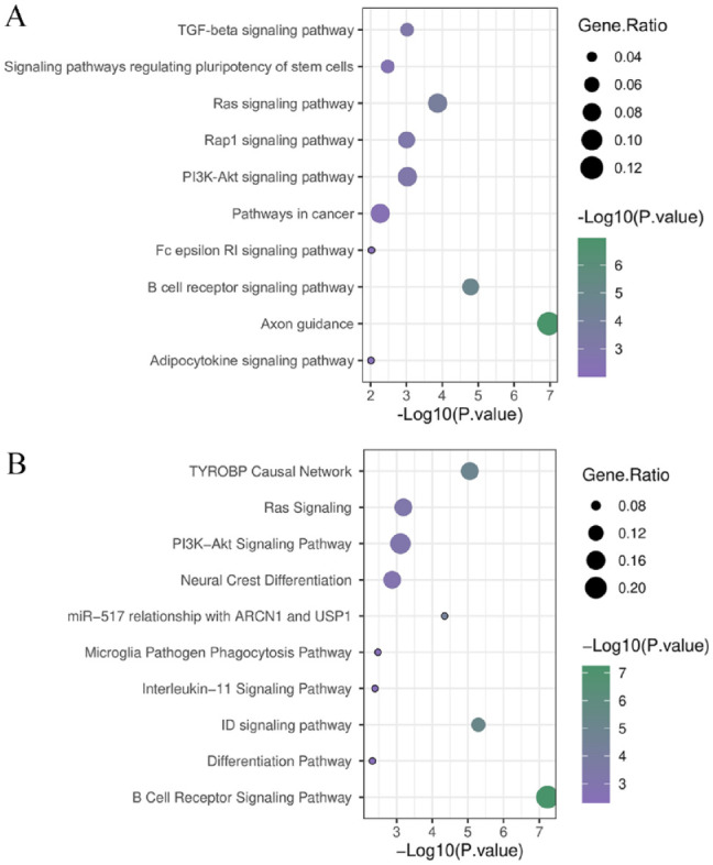
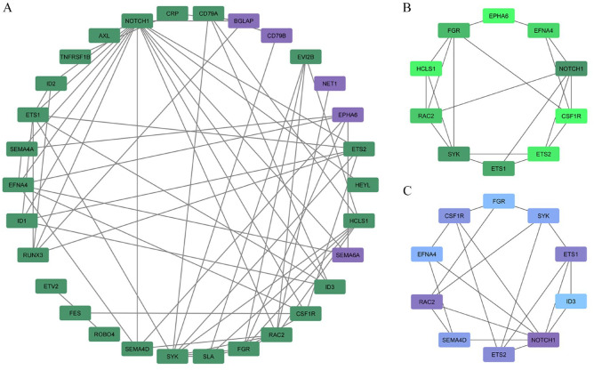
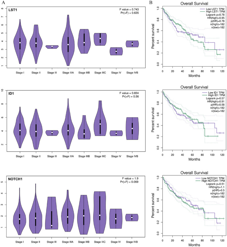
Nowadays, hepatocellular carcinoma (HCC) is the second leading cause of cancer deaths, and identifying the effective factors in causing this disease can play an important role in its prevention and treatment. Tumors provide effective agents for invasion and metastasis to other organs by establishing appropriate communication between cancer cells and the microenvironment. Epithelial-to-mesenchymal transition (EMT) can be mentioned as one of the effective phenomena in tumor invasion and metastasis. Several factors are involved in inducing this phenomenon in the tumor microenvironment, which helps the tumor survive and migrate to other places. It can be effective to identify these factors in the use of appropriate treatment strategies and greater patient survival. This study investigated the molecular differences between tumor border cells and tumor core cells or internal tumor cells in HCC for specific EMT genes. Expression of NOTCH1, ID1, and LST1 genes showed a significant increase at the HCC tumor border. Targeting these genes can be considered as a useful therapeutic strategy to prevent distant metastasis in HCC patients.
Keywords: Hepatocellular carcinoma; epithelial-to-mesenchymal transition; metastasis; tumor microenvironment.
© The Author(s) 2024.
Conflict of interest statement
The author(s) declared no potential conflicts of interest with respect to the research, authorship, and/or publication of this article.
Figures
Similar articles
-
Cyclin-Dependent Kinase 4 is expected to be a therapeutic target for hepatocellular carcinoma metastasis using integrated bioinformatic analysis.Bioengineered. 2021 Dec;12(2):11728-11739. doi: 10.1080/21655979.2021.2006942. Bioengineered. 2021. PMID: 34784846 Free PMC article.
-
Acidic Microenvironment Up-Regulates Exosomal miR-21 and miR-10b in Early-Stage Hepatocellular Carcinoma to Promote Cancer Cell Proliferation and Metastasis.Theranostics. 2019 Mar 16;9(7):1965-1979. doi: 10.7150/thno.30958. eCollection 2019. Theranostics. 2019. PMID: 31037150 Free PMC article.
-
Epithelial to mesenchymal transition is associated with shorter disease-free survival in hepatocellular carcinoma.Ann Surg Oncol. 2014 Nov;21(12):3882-90. doi: 10.1245/s10434-014-3779-2. Epub 2014 May 15. Ann Surg Oncol. 2014. PMID: 24833103
-
Epithelial-to-mesenchymal plasticity of cancer stem cells: therapeutic targets in hepatocellular carcinoma.J Hematol Oncol. 2016 Aug 30;9(1):74. doi: 10.1186/s13045-016-0307-9. J Hematol Oncol. 2016. PMID: 27578206 Free PMC article. Review.
-
Role of epithelial to mesenchymal transition in hepatocellular carcinoma.J Hepatol. 2016 Oct;65(4):798-808. doi: 10.1016/j.jhep.2016.05.007. Epub 2016 May 17. J Hepatol. 2016. PMID: 27212245 Review.
Cited by
-
Leveraging Patient-Derived Organoids for Personalized Liver Cancer Treatment.Int J Biol Sci. 2024 Oct 7;20(13):5363-5374. doi: 10.7150/ijbs.96317. eCollection 2024. Int J Biol Sci. 2024. PMID: 39430248 Free PMC article.
References
-
- Friedrich RE, Zustin J. Multiple distant metastases of hepatocellular carcinoma to the oral cavity. In Vivo. 2010;24:211-214. - PubMed
LinkOut - more resources
Full Text Sources
