. 2024 Feb 5;7(1):150.
doi: 10.1038/s42003-024-05847-w.

Arabidopsis COP1 guides stomatal response in guard cells through pH regulation

Affiliations

Arabidopsis COP1 guides stomatal response in guard cells through pH regulation

Seoyeon Cha et al. Commun Biol. .

Abstract

Plants rely on precise regulation of their stomatal pores to effectively carry out photosynthesis while managing water status. The Arabidopsis CONSTITUTIVE PHOTOMORPHOGENIC 1 (COP1), a critical light signaling repressor, is known to repress stomatal opening, but the exact cellular mechanisms remain unknown. Here, we show that COP1 regulates stomatal movement by controlling the pH levels in guard cells. cop1-4 mutants have larger stomatal apertures and disrupted pH dynamics within guard cells, characterized by increased vacuolar and cytosolic pH and reduced apoplastic pH, leading to abnormal stomatal responses. The altered pH profiles are attributed to the increased plasma membrane (PM) H+-ATPase activity of cop1-4 mutants. Moreover, cop1-4 mutants resist to growth defect caused by alkali stress posed on roots. Overall, our study highlights the crucial role of COP1 in maintaining pH homeostasis of guard cells by regulating PM H+-ATPase activity, and demonstrates how proton movement affects stomatal movement and plant growth.


Conflict of interest statement

The authors declare no competing interests.

Figures
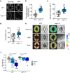
Fig. 1
Fig. 1. cop1-4 mutants exhibit altered vacuolar properties in guard cells.
a Vacuolar structures in guard cells of wild-type (WT) and cop1-4 mutants visualized with the tonoplast marker VACUOLAR H+-PYROPHOSPHATASE 1 (VHP1)-GFP after 2 h of light-induced stomatal opening, followed by treatment with or without 10 µM abscisic acid (ABA) for an hour. (Scale bar, 5 µm.) Quantitative analysis of three guard cell parameters after stomatal opening. Stomatal aperture index is calculated by dividing the stomatal width by its length (b), combined vacuolar volume (c), and vacuolar occupancy (d). Note that these were obtained from the same guard cells used in (f) for stomatal opening. ****P < 0.0001; two-sample t test; n = 30 (WT) and n = 29 (cop1-4). Boxplots show the median (center line) and the 75th and 25th percentiles (edges of the box) of the data; whiskers extend to 1.5 times the interquartile range. e Representative pseudocolor ratiometric images of vacuolar pH in guard cells of wild-type (WT) and cop1-4 mutants were obtained after 2 h of light-induced stomatal opening, followed by treatment with or without 10 µM ABA for 1 h, or 1 h of darkness. Guard cells were loaded with the pH-sensitive vacuolar loading dye 2′,7′-bis-(2-carboxyethyl)-5-(and-6)-carboxyfluorescein (BCECF-AM), and the images were generated by dividing the emission images acquired in the 488 nm channel by those obtained in the 458 nm channel. Ratiometric images (left) and bright-field (right). (Scale bar, 5 µm.) f Mean vacuolar pH values for entire vacuoles in guard cells of (e). ****P < 0.0001 and NS, not significant (P > 0.05); two-way ANOVA; Tukey’s HSD; n = 9–34.
Fig. 2
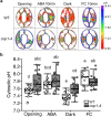
Fig. 2. Cytosolic pH of cop1-4 mutants are more alkaline than WT.
a Representative pseudocolor ratiometric images of guard cells of wild-type (WT) and cop1-4 mutants each expressing ClopHensor. Detached leaves were incubated with stomatal opening buffer for 2 h, and then treated with 10 µM abscisic acid (ABA) for 10 min or with dark for 1 h. To assess the impact of fusicoccin (FC), dark-treated leaves were incubated with 10 µM fusicoccin for 1 h. Fluorescence intensity of cytosolic pH in guard cells of WT and cop1-4 mutants was measured directly from detached leaves using confocal laser scanning microscope (Leica SP8 X). Pseudocolor ratiometric images were generated by dividing the emission images obtained at 488 nm by those acquired at 458 nm. (Scale bar, 5 µm.) b Quantification of ratiometric values for cytosolic pH in guard cells shown in (a). Different letters indicate statistically significant differences between the plants (P < 0.05); one-way ANOVA; Tukey’s HSD.
Fig. 3
Fig. 3. cop1-4 mutants exhibit constitutively acidic apoplast in leaf epidermal cells.
a, b Representative pseudocolor images of apoplastic pH in the epidermis of wild-type (WT) and cop1-4 mutants visualized using 8-hydroxypyrene-1,3,6-trisulfonic acid trisodium salt (HPTS) staining with or without abscisic acid (ABA) in the stomatal opening buffer. Staining and observation were conducted simultaneously and instantly. Ratiometric images were generated by dividing the emission images obtained in the 488 nm channel by those acquired in the 405 nm channel. Calculated values were converted into absolute pH values using the in situ HPTS calibration curve (Supplementary Fig. 3). Right panels show expansions of the left panels, respectively. (Scale bar, 20 µm.) c Mean apoplastic pH values for guard cells shown in (a, b). ****P < 0.0001 and *P < 0.05; two-way ANOVA; Tukey’s HSD; n = 17 to 27. Boxplots show the median (center line) and the 75th and 25th percentiles (edges of the box) of the data; whiskers extend to 1.5 times the interquartile range.
Fig. 4
Fig. 4. COP1 negatively regulates proton pumping activities.
a Rhizosphere acidification assays were conducted using wild-type (WT) and cop1-4 mutants. Eleven-day-old seedlings were transferred to ½ MS media containing 0.003% of pH indicator dye bromocresol purple and grown vertically. Seedlings were then subjected to dark, long-day, and continuous light conditions, respectively, as indicated, and color changes were recorded after 36 h. (Scale bar, 1 cm.) The experiment was conducted three times, and one representative experiment is shown in (a). b Plasma membrane H+-ATPase activity was measured using WT and cop1-4 mutants. Plasma membrane vesicles were isolated from the leaves of WT and cop1-4 mutant plants, and treated with reaction buffer containing pH-sensitive fluorescence probe quinacrine. H+-ATPase activation was induced by adding 3 mM ATP, and pH decrease inside the vesicles was measured using spectrofluorometer. Proton gradient inside the vesicles was collapsed by adding uncoupler nigericin. The experiment was conducted three times, and one representative experiment is shown in (b). c Mean H+-ATPase activity of WT and cop1-4 mutants of (b). H+-ATPase activity of WT was set to 100%, and the relative H+-ATPase activity of cop1-4 mutants was shown as percentage of the activity of WT. **P < 0.01; two-sample t-test; Boxplots show the median (center line) and the 75th and 25th percentiles (edges of the box) of the data; whiskers extend to 1.5 times the interquartile range.
Fig. 5
Fig. 5. Enhanced plasma membrane (PM) H+-ATPase activity of cop1-4 mutants affects plant growth and stomatal behavior.
a Detached leaves of 11-day-old wild-type (WT) and cop1-4 mutant seedlings were incubated with stomatal opening buffer for 2 h, and then treated with various conditions as Fig. 2, except for abscisic acid (ABA) treated for 1 h. Guard cells were incubated with 1 mM Na3VO4 to investigate the role of H+-ATPase on stomatal aperture. (Scale bar, 5μm.) b Quantification of stomatal apertures of guard cells from (a). Stomatal aperture index calculated by dividing the stomatal width by its length. Different letters indicate statistically significant differences between the plants (P < 0.05); one-way ANOVA; Tukey’s HSD. c 11-day-old seedlings of WT and cop1-4 mutants were transferred to ½ MS media at varying pH levels (3.6, 5.8, and 8.0) and grown vertically. Photographs and physiological assays were conducted 7 days after transferring. (Scale bar, 2 cm.) d, e Corresponding quantification of fresh weight (d) and primary root length (e) of plants from (c). *P < 0.05 and NS, not significant (P > 0.05); two-sample t test; n = 4 each.
Fig. 6
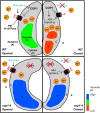
Fig. 6. A hypothetical model for the role of COP1 in regulating stomatal movement through pH regulation.
In an opened wild-type (WT) guard cell (upper left panel), proton pumping by plasma membrane (PM) H+-ATPase is downregulated by COP1. After stomata-closing signals such as ABA or dark (upper right panel), COP1 promotes ABA signaling, thereby inactivating PM H+-ATPase activity again. Thus, cytosolic acidification and apoplastic alkalinization occur, and subsequent signal transduction induces vacuolar acidification and fragmentation, and finally stomatal closure. In an opened guard cell of cop1-4 mutant (lower left panel), absence of COP1 is responsible for enhanced PM H+-ATPase activity. This elevated activity results in acidic apoplast and alkaline cytosol compared to WT. Stronger pH gradient across the PM induces signaling for stomatal opening, followed by enlarged alkaline vacuole and constitutive open stomata phenotype of cop1-4 mutants. After ABA or dark-mediated stomatal closure signals (lower right panel), only mild inhibitory effect can be posed to PM H+-ATPase due to absence of COP1, a positive regulator of ABA signaling. Therefore, cop1-4 mutant’s apoplastic pH is lower, and cytosolic pH remains higher than WT guard cells with closed stomata. Still, pH gradient across the PM is sufficient to induce stomatal opening, and the vacuole morphology and pH remain unaffected. Black arrows indicate stimulation, and blocked lines indicate inhibition. Red arrows indicate proton (H+) movement. Relative pH of cytosol and apoplast are proportional to the numbers of “H+”s inside respective regions. For graphical visibility, vacuoles at the upper side of the guard cells are omitted.

Similar articles

Cited by

References

    1. Lawson T, Matthews J. Guard cell metabolism and stomatal function. Annu. Rev. Plant Biol. 2020;71:273–302. doi: 10.1146/annurev-arplant-050718-100251. - DOI - PubMed
    1. Dubeaux G, et al. Deep dive into CO2-dependent molecular mechanisms driving stomatal responses in plants. Plant Physiol. 2021;187:2032–2042. doi: 10.1093/plphys/kiab342. - DOI - PMC - PubMed
    1. Kim TH, Böhmer M, Hu H, Nishimura N, Schroeder JI. Guard cell signal transduction network: advances in understanding abscisic acid, CO2, and Ca2+ signaling. Annu. Rev. Plant Biol. 2010;61:561–591. doi: 10.1146/annurev-arplant-042809-112226. - DOI - PMC - PubMed
    1. Schroeder JI, Raschke K, Neher E. Voltage dependence of k+ channels in guard-cell proto-plasts. Proc. Natl. Acad. Sci. 1987;84:4108–4112. doi: 10.1073/pnas.84.12.4108. - DOI - PMC - PubMed
    1. Zhang J, et al. Identification of slac1 anion channel residues required for co2/bicarbonate sensing and regulation of stomatal movements. Proc. Natl. Acad. Sci. 2018;115:11129–11137. doi: 10.1073/pnas.1807624115. - DOI - PMC - PubMed

Publication types

MeSH terms

Substances