. 2024 Jan 16:17:1296458.
doi: 10.3389/fnins.2023.1296458. eCollection 2023.

Targeted therapeutic hypothermia protects against noise induced hearing loss

Affiliations

Targeted therapeutic hypothermia protects against noise induced hearing loss

Samantha Rincon Sabatino et al. Front Neurosci. .

Abstract

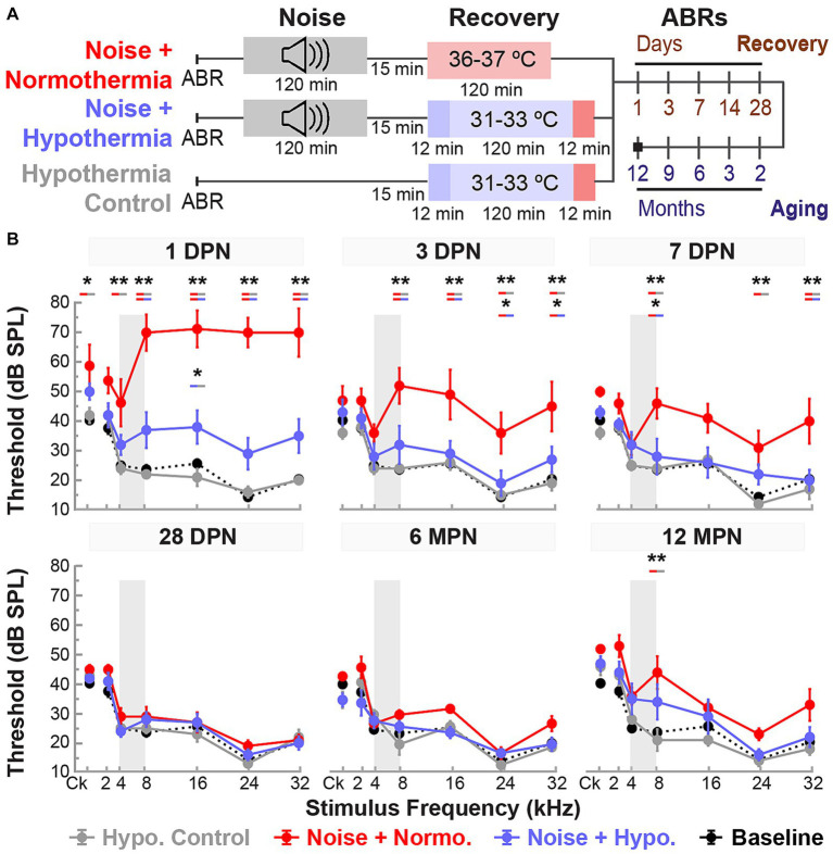
Introduction: Exposure to occupational or recreational loud noise activates multiple biological regulatory circuits and damages the cochlea, causing permanent changes in hearing sensitivity. Currently, no effective clinical therapy is available for the treatment or mitigation of noise-induced hearing loss (NIHL). Here, we describe an application of localized and non-invasive therapeutic hypothermia and targeted temperature management of the inner ear to prevent NIHL.

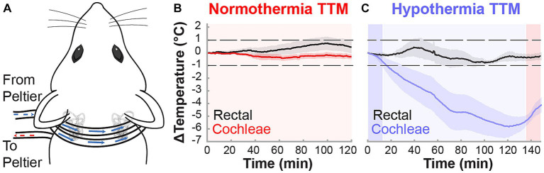
Methods: We developed a custom-designed cooling neck collar to reduce the temperature of the inner ear by 3-4°C post-injury to deliver mild therapeutic hypothermia.

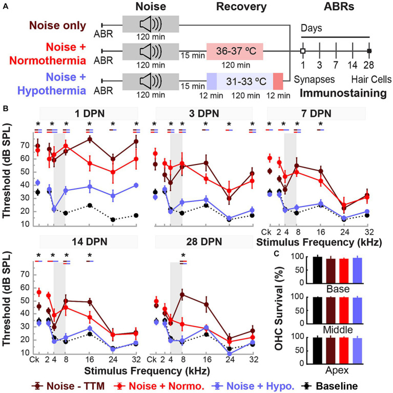
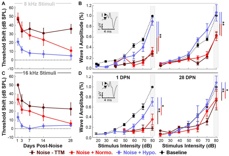
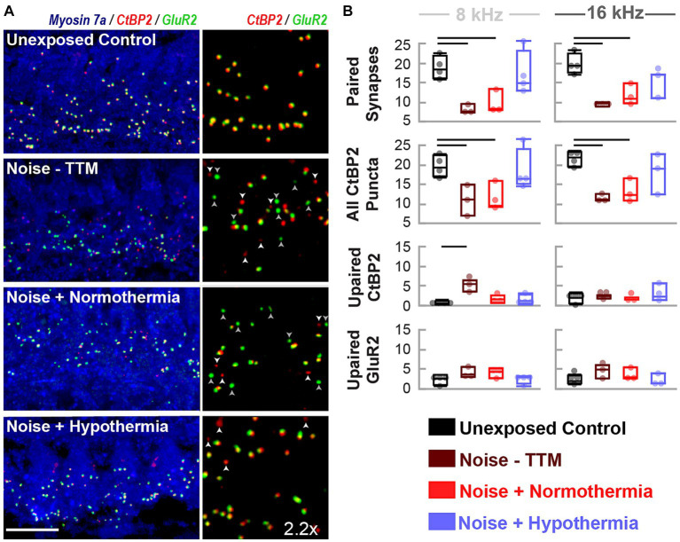
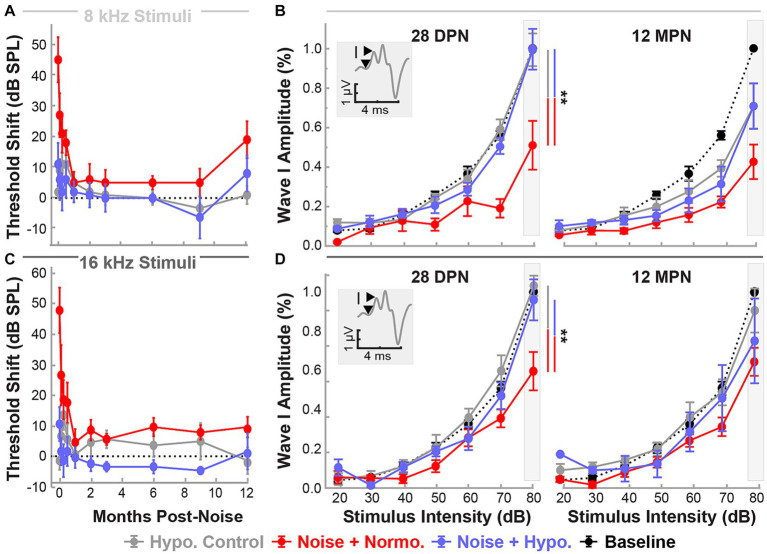
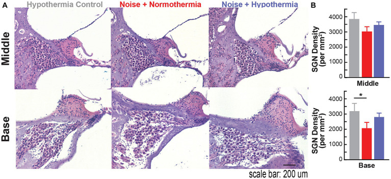
Results: This localized and non-invasive therapeutic hypothermia successfully mitigated NIHL in rats. Our results show that mild hypothermia can be applied quickly and safely to the inner ear following noise exposure. We show that localized hypothermia after NIHL preserves residual hearing and rescues noise-induced synaptopathy over a period of months.

Discussion: This study establishes a minimally-invasive therapeutic paradigm with a high potential for rapid translation to the clinic for long-term preservation of hearing health.

Keywords: hair cells; hidden hearing loss; neuroprotection; noise-induced hearing loss; synaptopathy; therapeutic hypothermia.


Conflict of interest statement

SRa and CK are inventors of the intellectual property (IP) used in this study. SRa and the University of Miami may receive royalties for the commercialization of the IP. SRa and CK are co-founders of RestorEar Devices LLC. RestorEar did not provide any financial support for the work described in this manuscript. All conflict of interests for SRa are disclosed to and managed by the University of Miami. The remaining authors declare that the research was conducted in the absence of any commercial or financial relationships that could be construed as a potential conflict of interest.

Figures

Figure 1
Figure 1
Non-invasive targeted cochlear temperature management. (A) The cooling neck collar placement was maintained above rat bullae for the duration of the TTM protocols to deliver mild therapeutic hypothermia. Validation of TTM target temperatures was performed in acute experiments (n = 3, k = 6 cochleae) measuring intracochlear and rectal temperatures (black trace) during the normothermia (B) and hypothermia (C) protocols. Temperature data are presented as mean ± SE.
Figure 2
Figure 2
Post-noise hypothermia attenuates early ABR hearing threshold shifts. (A) Experimental protocol for animals used to study of recovery of noise-induced induced injury with TTM. Animals were exposed to 2 h of continuous noise at 105 dB under isoflurane anesthesia before initiating TTM protocols of normothermia and hypothermia. For animals receiving TTM, temperature modulation commenced roughly 15 min post-noise. (B) ABR thresholds were obtained in age-matched female rats at 1, 3, 7, 14 and 28 DPN and compared to combined baseline thresholds (dashed curve). ABRs were obtained for click (Ck) and 2, 4, 8, 16, 24, and 32 kHz pure-tone stimuli. The frequency spectrum of the experimental noise is illustrated by gray vertical bars at 4–8 kHz. Results for post-noise ABR thresholds are shown for animals that recovered from noise exposure in noise only (dark red), normothermia TTM (red), and hypothermia TTM (light blue) conditions. Data presented as mean ± SE of ABR thresholds with between-group comparisons illustrated with colored bars at each stimulus frequency (*p < 0.05, **p < 0.01). (C) To assess hair cell integrity after noise exposure, FITC-Phalloidin labeled hair cell survival was quantified at 28 DPN for apical, middle, and basal cochlear regions in unexposed control (black), noise only (dark red), noise+normothermia (red), and noise+hypothermia animals (blue). Hair cell survival data for respective groups are shown as percent survival (mean ± SE).
Figure 3
Figure 3
Hypothermic preservation of ABR Wave I amplitudes post-noise. ABR threshold elevations (mean ± SE) from respective baseline measurements are highlighted for two frequencies, 8 kHz (light gray, A) and 16 kHz (dark gray, C). Thresholds shifts from 1 to 28 DPN for noise (dark red), normothermia (red), and hypothermia treated animals (blue) are shown. The dotted line visualizes the baseline. Wave I amplitudes normalized to respective baseline measures at 80 dB are presented (mean ± SE) at stimulus intensity levels from 20 to 80 for 8 (B) and 16 (D) kHz pure-tone stimuli. Normalized baseline amplitude calculations (dotted line) are shown for comparison to normalized post-noise amplitudes at 1 and 28 DPN. Figure insets portray Wave I amplitude measurement from node to peak. Between-group comparisons at suprathreshold level of 80 dB are illustrated with vertical bars (*p < 0.05, **p < 0.01).
Figure 4
Figure 4
Hypothermia applied after synaptopathic noise exposure preserves pairing of pre-and post-synaptic puncta. (A) Confocal images of paired presynaptic ribbons (anti-CtBP2, red) and post-synaptic glutamate receptors (anti-GluR2, green) in the IHC area (anti-myosin VIIa, blue) were obtained for unexposed controls, noise, normothermia TTM, and hypothermia TTM animals at 24 h post-noise. Vertical panels depict the colocalization of CtBP2 and GluR2 labeled puncta with an enlarged (2.2x) view of the IHC area. Scale bar: 10 μm (B) Visually quantified, colocalized synapses and unpaired puncta per IHC were obtained for 8 and 16 kHz areas. Comparisons to control (black) animals were made for paired and unpaired puncta in noise-exposed animals without post-noise TTM (dark red) and with post-noise normothermia (red) and hypothermia (blue) (mean ± SE, *p < 0.05, **p < 0.01).
Figure 5
Figure 5
Post-noise hypothermic induction limits temporary hearing changes associated with accelerated age-related hearing loss. (A) Experimental protocol for the study of Hypothermia TTM long-term efficacy and safety. Animals in noise groups were subjected to 2-h continuous noise exposure (4–8 kHz, 105 dB) under isoflurane anesthesia before initiating TTM protocols (normothermia or hypothermia) under anesthesia. (B) Baseline (black dotted line) and post-procedure ABR thresholds (mean ± SE) are illustrated at multiple time points to observe the progression of hearing recovery between experimental groups. Early time points at 1, 3, 7, and 28 DPN were used to observe the acute recovery response, and later time points at 6 and 12 MPN to observe aging deterioration in previously noise-exposed animals. ABR thresholds were obtained for broadband (click, Ck) stimuli and pure tones ranging from 2 to 32 kHz. Gray bands depict the narrowband 4–8 kHz frequency composition of the 2-h noise exposure. Between-group comparisons are illustrated for each tested frequency and time point (REML, *p < 0.05, **p < 0.01).
Figure 6
Figure 6
Post-noise localized hypothermic induction recovers suprathreshold ABR Wave I amplitude by 28 DPN. Recovery of ABR threshold up to 12 MPN is indicated by threshold shifts (mean ± SE), post-procedure thresholds with baseline threshold subtraction. Data are shown for frequencies 8 kHz (light gray, A) and 16 kHz (dark gray, C) for the three experimental groups hypothermia control (gray), noise+normothermia (red), and noise+hypothermia (blue). The dotted line depicts a difference from baseline threshold. Baseline-normalized Wave I amplitudes (mean ± SE) are depicted at stimulus intensities of 20 to 80 dB for respective middle frequencies (B – 8 kHz, D – 16 kHz). ABR amplitudes are normalized for each subject to its baseline amplitude at a maximal stimulus intensity of 80 dB SPL. Average Normalized baseline amplitudes for each frequency are exemplified with a black dotted line. Normalized post-procedure ABR amplitudes are compared at 28 DPN as the primary endpoint of acute noise recovery and 12 MPN as the final study endpoint. Figure insets illustrate Wave I peak and node demarcation for amplitude measurement. Between-group comparisons at suprathreshold level of 80 dB are illustrated with vertical bars (*p < 0.05, **p < 0.01).
Figure 7
Figure 7
Spiral ganglion loss associated with acute early noise-exposure. (A) Hematoxylin/eosin (H&E) stained cross-section of the middle turn in Brown Norway rats. Image is from a representative 10 μm paraffin-embedded section imaged at 20X. H&E-stained cochlear cross-section across the modiolus display spiral ganglion neurons housed in Rosenthal’s canal at different cochlear turns. Scale bar: 100 μm. (B) Quantified number of SGNs in 10 μm mid-modiolar sections at the basal (top) and middle (bottom) turns were compared across hypothermia control (gray), normothermia (red), and hypothermia (blue) female brown Norway rats at 12 months post-noise (two-way ANOVA, *p < 0.05, **p < 0.01).
Figure 8
Figure 8
Proposed neuroprotective mechanisms of mild therapeutic hypothermia intervention in NIHL. The working model highlights prominent damage pathways involved in NIHL and potential sites of MTH action that likely play a protective role in preserving sensory hair cells and synaptic elements. Studies have shown that NIHL can activate apoptosis and necroptosis cell death pathways in hair cells and lead to glutamate excitotoxicity, negatively impacting synaptic elements. Conversely, MTH has been shown to have broad neuroprotective effects against ischemic and excitotoxic damage. The protective effects of MTH treatment on several NIHL damage pathways have been highlighted with blue arrows.

Similar articles

Cited by

References

    1. Aedo C., Aguilar E. (2020). Cochlear synaptopathy: new findings in animal and human research. Rev. Neurosci. 31, 605–615. doi: 10.1515/revneuro-2020-0002, PMID: - DOI - PubMed
    1. Alva N., Carbonell T., Palomeque J. (2004). A model of deep experimental hypothermia and rewarming in rat. J. Therm. Biol. 29, 259–264. doi: 10.1016/j.jtherbio.2004.04.001 - DOI
    1. Alva N., Palomeque J., Carbonell T. (2013). Oxidative stress and antioxidant activity in hypothermia and rewarming: can RONS modulate the beneficial effects of therapeutic hypothermia? Oxidative Med. Cell. Longev. 2013, –957054. doi: 10.1155/2013/957054 - DOI - PMC - PubMed
    1. Arslan H. H., Satar B., Serdar M., Yilmaz E. (2017). Changes in proinflammatory cytokines in the cochlea in relation to hearing thresholds in noise-exposed rats. J. Int. Adv. Otol. 13, 308–312. doi: 10.5152/iao.2017.1674, PMID: - DOI - PubMed
    1. Arteaga A. A., Beatrous B., Eby T. L., Smith J., Yoder M., Meritt R., et al. . (2023). Achieving mild therapeutic hypothermia in the human cochlea. Otol. Neurotol. 44, e338–e342. doi: 10.1097/MAO.0000000000003843, PMID: - DOI - PubMed

LinkOut - more resources