

Subchondral Bone Alignment in Osteochondral Allograft Transplants for Large Oval Defects of the Medial Femoral Condyle: Comparison of Lateral versus Medial Femoral Condyle Donors
- PMID: 38282570
- PMCID: PMC11418256
- DOI: 10.1177/19476035231226218
Subchondral Bone Alignment in Osteochondral Allograft Transplants for Large Oval Defects of the Medial Femoral Condyle: Comparison of Lateral versus Medial Femoral Condyle Donors
Abstract
Objective: Supply-demand mismatch of medial femoral condyle (MFC) osteochondral allografts (OCAs) remains a rate-limiting factor in the treatment of osteochondral defects of the femoral condyle. Surface contour mapping was used to determine whether a contralateral lateral femoral condyle (LFC) versus ipsilateral MFC OCA differs in the alignment of donor:native subchondral bone for large osteochondral defects of the MFC.
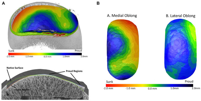
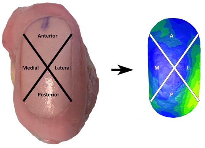
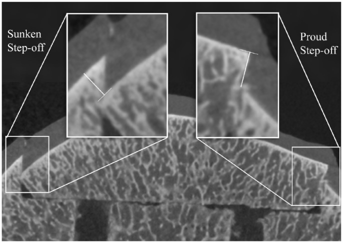
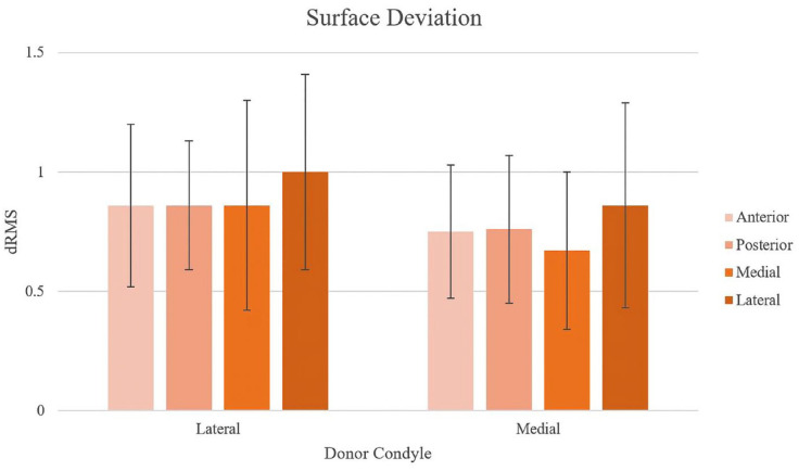
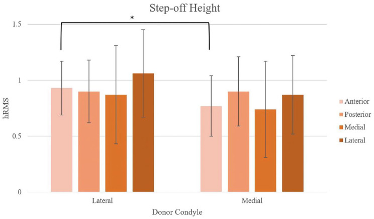
Design: Thirty fresh-frozen human femoral condyles were matched by tibial width into 10 groups of 3 condyles (MFC recipient, MFC donor, and LFC donor) each for 3 cartilage surgeons (90 condyles). The recipient MFC was imaged using nano-computed tomography scan. Donor oval grafts were harvested from each matched condyle and transplanted into a 17 mm × 36 mm defect created in the recipient condyle. Following the first transplant, the recipient condyle was imaged and superimposed on the native condyle nano-CT scan. The donor plug was removed and the process repeated for the other donor. Surface height deviation and circumferential step-off height deviation were compared between native and donor subchondral bone surfaces for each transplant.
Results: There was no statistically significant difference in mean subchondral bone surface deviation (LFC = 0.87 mm, MFC = 0.76 mm, P = 0.07) nor circumferential step-off height (LFC = 0.93 mm, MFC = 0.85 mm, P = 0.09) between the LFC and MFC plugs. There were no significant differences in outcomes between surgeons.
Conclusions: There were no significant differences in subchondral bone circumferential step-off or surface deviation between ipsilateral MFC and contralateral LFC oval-shaped OCAs for 17 mm × 36 mm defects of the MFC.
Keywords: cartilage transplant; osteochondral allograft; subchondral bone.
Conflict of interest statement
Declaration of Conflicting InterestsThe author(s) declared the following potential conflicts of interest with respect to the research, authorship, and/or publication of this article: Kelly M.R. Taylor: None declared; Conor S. Locke: None declared; Timothy S. Mologne: JRF Ortho, Arthrex; William D. Bugbee: JRF Ortho, Arthrex; John A. Grant: JRF Ortho, Arthrex, Vericel, Aesculap Biologics.
Figures
Similar articles
-
Osteochondral Allografts for Large Oval Defects of the Medial Femoral Condyle: A Comparison of Single Lateral Versus Medial Femoral Condyle Oval Grafts Versus 2 Overlapping Circular Grafts.Am J Sports Med. 2023 Feb;51(2):379-388. doi: 10.1177/03635465221139272. Epub 2022 Dec 20. Am J Sports Med. 2023. PMID: 36537663
-
Patient-Specific Distal Femoral Guides Optimize Cartilage Topography Matching in Osteochondral Allograft Transplantations.Am J Sports Med. 2024 Aug;52(10):2547-2554. doi: 10.1177/03635465241261353. Epub 2024 Aug 5. Am J Sports Med. 2024. PMID: 39101660
-
Topographic Matching of Osteochondral Allograft Transplantation Using Lateral Femoral Condyle for the Treatment of Medial Femoral Condyle Lesions: A Computer-Simulated Model Study.Arthroscopy. 2018 Nov;34(11):3033-3042. doi: 10.1016/j.arthro.2018.05.039. Arthroscopy. 2018. PMID: 30392687
-
Contralateral Lateral Femoral Condyle Allografts Provide an Acceptable Surface Match for Simulated Classic Osteochondritis Dissecans Lesions of the Medial Femoral Condyle.Orthop J Sports Med. 2020 Jan 28;8(1):2325967119898413. doi: 10.1177/2325967119898413. eCollection 2020 Jan. Orthop J Sports Med. 2020. PMID: 32064295 Free PMC article.
-
Topographic Analysis of Lateral Versus Medial Femoral Condyle Donor Sites for Oblong Medial Femoral Condyle Lesions.Arthroscopy. 2020 Nov;36(11):2900-2908. doi: 10.1016/j.arthro.2020.07.007. Epub 2020 Jul 28. Arthroscopy. 2020. PMID: 32735941
References
-
- Chu CR, Convery FR, Akeson WH, Meyers M, Amiel D. Articular cartilage transplantation: clinical results in the knee. Clin Orthop Relat Res. 1999;360:159-68. - PubMed
-
- Convery FR, Meyers MH, Akeson WH. Fresh osteochondral allografting of the femoral condyle. Clin Orthop Relat Res. 1991;273:139-45. - PubMed
-
- Cotter EJ, Hannon CP, Christian DR, Wang KC, Lansdown DA, Waterman BR, et al.. Clinical outcomes of multifocal osteochondral allograft transplantation of the knee: an analysis of overlapping grafts and multifocal lesions. Am J Sports Med. 2018;46(12):2884-93. doi:10.1177/0363546518793405. - DOI - PubMed
Publication types
MeSH terms
Grants and funding
LinkOut - more resources
Full Text Sources
