Review
. 2024 Jan 19;25(2):1251.
doi: 10.3390/ijms25021251.

Diagnostic Challenges during Inflammation and Cancer: Current Biomarkers and Future Perspectives in Navigating through the Minefield of Reactive versus Dysplastic and Cancerous Lesions in the Digestive System

Affiliations
Review

Diagnostic Challenges during Inflammation and Cancer: Current Biomarkers and Future Perspectives in Navigating through the Minefield of Reactive versus Dysplastic and Cancerous Lesions in the Digestive System

Ioannis S Pateras et al. Int J Mol Sci. .

Abstract

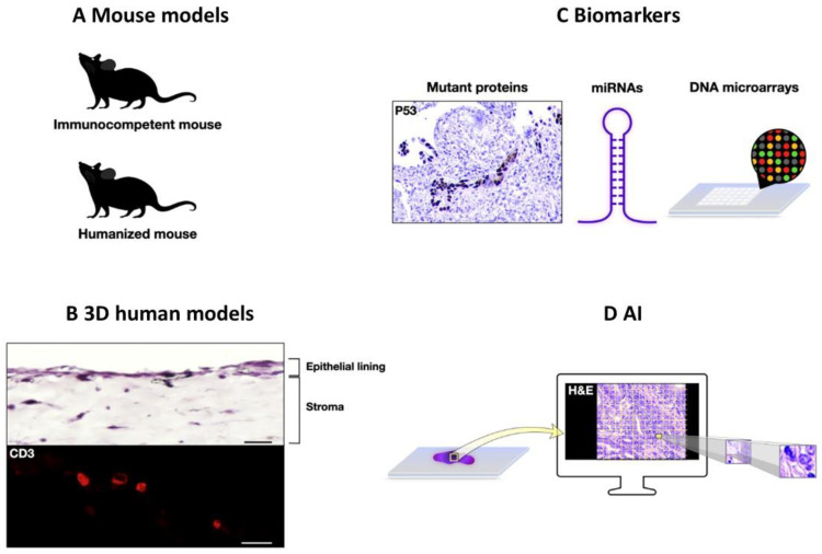
In the setting of pronounced inflammation, changes in the epithelium may overlap with neoplasia, often rendering it impossible to establish a diagnosis with certainty in daily clinical practice. Here, we discuss the underlying molecular mechanisms driving tissue response during persistent inflammatory signaling along with the potential association with cancer in the gastrointestinal tract, pancreas, extrahepatic bile ducts, and liver. We highlight the histopathological challenges encountered in the diagnosis of chronic inflammation in routine practice and pinpoint tissue-based biomarkers that could complement morphology to differentiate reactive from dysplastic or cancerous lesions. We refer to the advantages and limitations of existing biomarkers employing immunohistochemistry and point to promising new markers, including the generation of novel antibodies targeting mutant proteins, miRNAs, and array assays. Advancements in experimental models, including mouse and 3D models, have improved our understanding of tissue response. The integration of digital pathology along with artificial intelligence may also complement routine visual inspections. Navigating through tissue responses in various chronic inflammatory contexts will help us develop novel and reliable biomarkers that will improve diagnostic decisions and ultimately patient treatment.

Keywords: artificial intelligence; biomarkers; cancer; digital pathology; dysplasia; immunohistochemistry; inflammation; molecular biology; pathology; reactive atypia; tissue response.


Conflict of interest statement

The authors declare no conflicts of interest.

Figures

Figure 1
Figure 1
Tissue response upon chronic injury in the gastrointestinal tract. (A). Representative hematoxylin and eosin (H&E) staining micrograph from the oral mucosa showing pseudoepitheliomatous hyperplasia, visualized by epithelial hyperplasia along with irregular infiltrative tongue-like cords of squamous cells. The latter extend into the dermis with a pseudo-invasion pattern (arrowheads) and are accompanied by a marked inflammatory infiltrate. Periodic Acid Schiff (PAS) reaction micrograph highlights hyphae (arrows), supporting a fungal infection. Scale bar: 400 μm (H&E); 20 μm (PAS) (B). Representative hematoxylin and eosin (H&E) staining micrograph of esophageal mucosa with Barrett’s esophagus; notice the presence of intestinal metaplasia characterized by mucin-secreting goblet cells staining intensely blue with Alcian blue (AB). Scale bar: 100 μm (upper photo); 20 μm (lower photo). Downregulation of the squamous cell marker TAp63 and SRY (sex-determining region Y)-box 2 (SOX2), along with upregulation of the intestinal markers Caudal-type homeobox 2 (CDX2) and SRY (sex-determining region Y)-box 9 (SOX9), promote reprogramming of squamous cells into columnar epithelium (C). Representative hematoxylin and eosin (H&E) staining micrograph of gastric mucosa with intestinal complete (type I) (lower part) and incomplete (type II) (upper part) metaplasia; notice the presence of mucin-secreting goblet cells stained intensely blue with Alcian blue (AB). Mucin 2 (MUC2) along with CDX2 drives intestinal metaplasia phenotype. Scale bar: 100 μm (upper photo); 20 μm (lower photo). (D). Area indefinite for dysplasia in colonic biopsy in the setting of active inflammation due to inflammatory bowel disease. R-spondin-Wnt/β-catenin-LGR5 axis plays an essential role for the maintenance and expansion of intestinal stem crypt base cells; β-catenin transcriptionally induces Leucine-rich repeat-containing G protein-coupled receptor 5 (LGR5), while it represses MUC2 that is associated with loss of mucin. Arrows depict neutrophils; arrowheads demonstrate mitotic figures. Scale bar: 100 μm (upper photo); 20 μm (lower photo).
Figure 2
Figure 2
Chronic inflammation, tissue response and pancreatic ductal adenocarcinoma development. During chronic pancreatitis (CP), acinar cells may undergo acinar-to-ductal metaplasia (ADM) that is composed both of duct-like and acinar-like cells with embryonic progenitor cell properties. ADM cells stain with ductal ((Cytokeratin 19(CK19) and SRY-Box Transcription factor 9 (SOX9)), acinar (i.e., enzymes including amylase, elastase, etc.), and pancreatic progenitor ((pancreatic and duodenal homeobox 1 (Pdx1), β-catenin, and Notch)) markers. ADM is a reversible process. Upon oncogenic KRAS activation, ADM can progress towards pancreatic intraepithelial neoplastic lesion (PanIN). Macrophages have been shown to drive ADM and play a role in ADM to PanIN transition. Progression during higher-grade PanIN and pancreatic ductal adenocarcinoma (PDAC) is associated with mutations and/or allelic loss of Cyclin-Dependent kinase inhibitor A (CDKN2A), TP53, and Deleted in Pancreatic Cancer 4 (DPC4, also known as SMAD4) genes encoding the tumor suppressors P16INK4A, P14ARF, P53, and the transforming growth factor β (TGFβ) signal transducer SMAD4, respectively. Scale bar: 50 μm.
Figure 3
Figure 3
Panel of immunohistochemical markers in the differential diagnosis between reactive urothelium and urothelial carcinoma in situ. Representative hematoxylin and eosin (H&E) staining and immunohistochemistry micrographs showing CK20 expression limited to the umbrella cells, faint and patchy nuclear p53, and full-thickness CD44 immunostaining in non-neoplastic urothelium (upper photos), in contrast to full-thickness CK20, intense and diffused nuclear p53, and CD44 basal expression in urothelial carcinoma in situ (lower photos). Scale bar: 100 μm.
Figure 4
Figure 4
Future perspectives in differential diagnosis between reactive non-neoplastic and neoplastic lesions. (A) Immunocompetent mouse models including gain or loss of gene function complemented with humanized mouse models could improve our understanding of human inflammatory-associated diseases. (B) Three-dimensional human models providing a mechanistic insight into the tissue response to chronic inflammatory stimuli: representative hematoxylin and eosin (H&E) staining and immunofluorescent micrographs of a three-dimensional human colonic immunocompetent model with embedding of CD45+ cells. The localization of CD3+ lymphocytes in the organotypic 3D model was assessed by immunofluorescence, using an antibody specific for CD3 (red). Nuclei were counterstained with DAPI. Scale bar: 50 μm (upper photo); 25 μm (lower photo). (A. Bergonzini and T. Frisan, personal communication) (C) Novel biomarkers: incorporation of mutant-specific antibodies like against p53 hotspot mutants, as well as miRNAs, and DNA microarray applications could improve diagnostic accuracy. (D) Artificial intelligence (AI)-based prediction models analyzing routine histopathological H&E-stained sections.

Similar articles

Cited by

References

    1. Rocha e Silva M. A brief survey of the history of inflammation. Agents Action. 1978;8:45–49. doi: 10.1007/BF01972401. - DOI - PubMed
    1. Serhan C.N., Brain S.D., Buckley C.D., Gilroy D.W., Haslett C., O’Neill L.A.J., Perretti M., Rossi A.G., Wallace J.L. Resolution of inflammation: State of the art, definitions and terms. FASEB J. Off. Publ. Fed. Am. Soc. Exp. Biol. 2007;21:325. doi: 10.1096/fj.06-7227com. - DOI - PMC - PubMed
    1. Serhan C.N., Savill J. Resolution of inflammation: The beginning programs the end. Nat. Immunol. 2005;6:1191–1197. doi: 10.1038/ni1276. - DOI - PubMed
    1. Medzhitov R. Origin and physiological roles of inflammation. Nature. 2008;454:428–435. doi: 10.1038/nature07201. - DOI - PubMed
    1. Colotta F., Allavena P., Sica A., Garlanda C., Mantovani A. Cancer-related inflammation, the seventh hallmark of cancer: Links to genetic instability. Carcinogenesis. 2009;30:1073–1081. doi: 10.1093/carcin/bgp127. - DOI - PubMed

Grants and funding

Swedish Cancer Society grant n. 23 2814 Pj and the Kempestiftelserna grant n. 2021 JCK-3110 to TF. 9326 SARG (ELKE)/National and Kapodistrian University of Athens (NKUA) to IGP.

LinkOut - more resources