

Color Analysis of Merkel Cell Carcinoma: A Comparative Study with Cherry Angiomas, Hemangiomas, Basal Cell Carcinomas, and Squamous Cell Carcinomas
- PMID: 38275477
- PMCID: PMC10814937
- DOI: 10.3390/diagnostics14020230
Color Analysis of Merkel Cell Carcinoma: A Comparative Study with Cherry Angiomas, Hemangiomas, Basal Cell Carcinomas, and Squamous Cell Carcinomas
Abstract
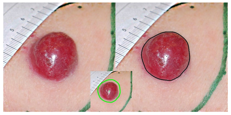
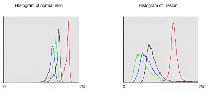
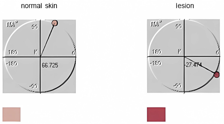
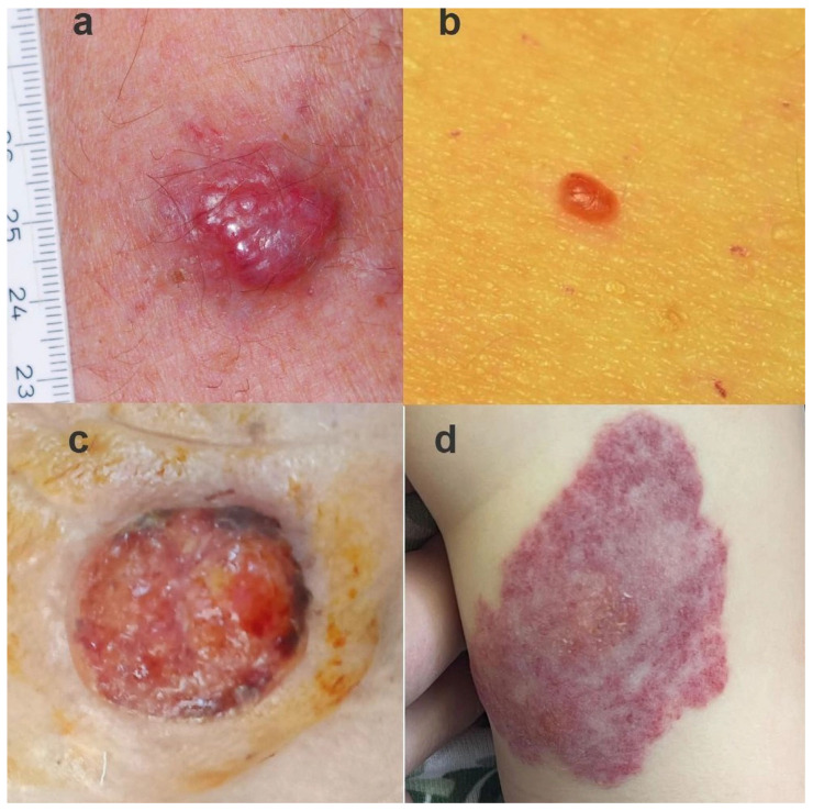
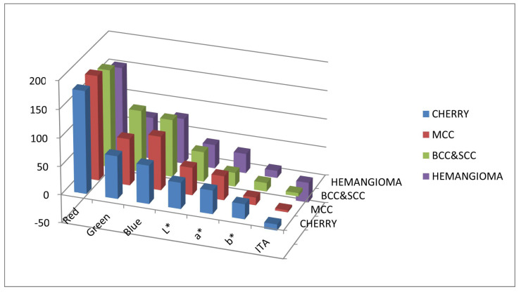
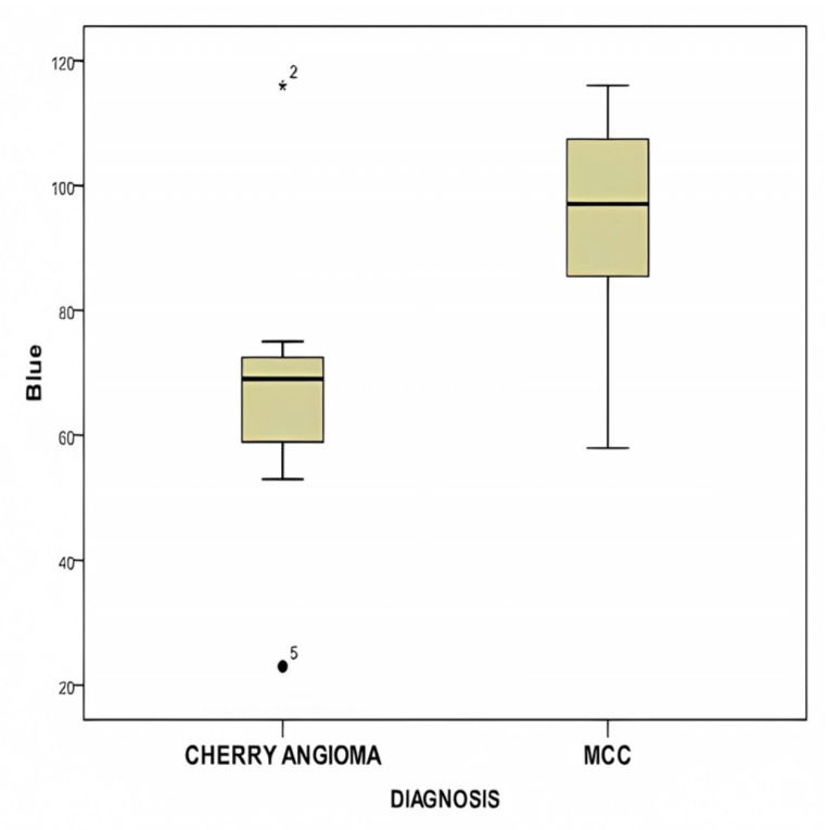
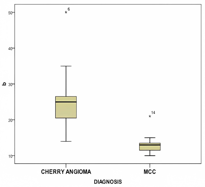
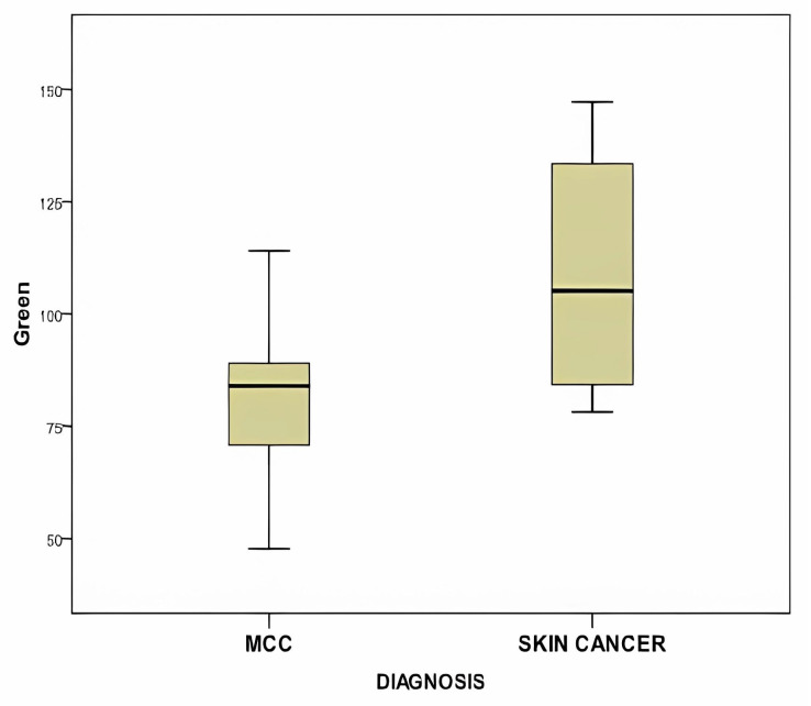
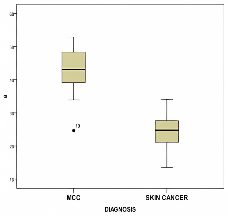
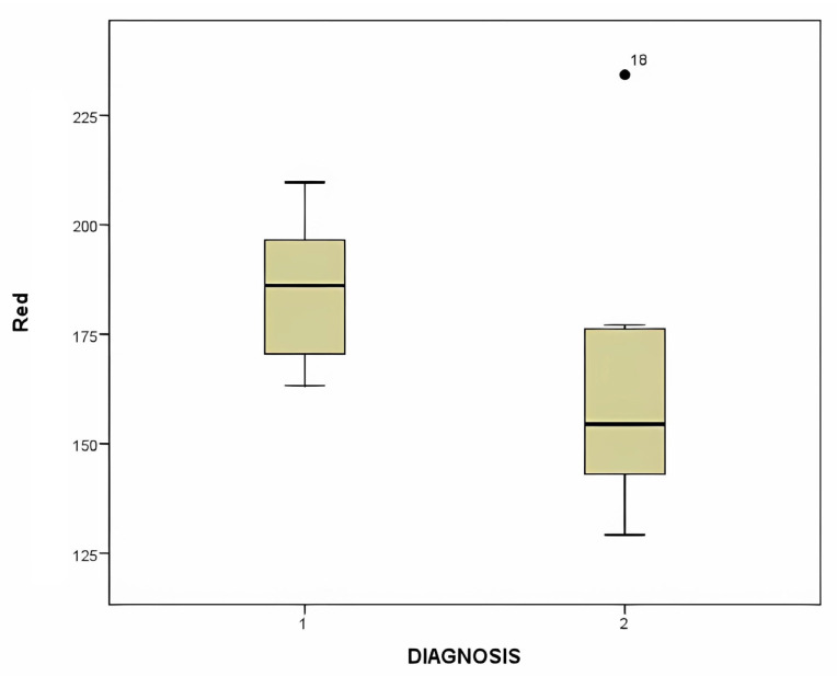
Merkel cell carcinoma (MCC) is recognized as one of the most malignant skin tumors. Its rarity might explain the limited exploration of digital color studies in this area. The objective of this study was to delineate color alterations in MCCs compared to benign lesions resembling MCC, such as cherry angiomas and hemangiomas, along with other non-melanoma skin cancer lesions like basal cell carcinoma (BCC) and squamous cell carcinoma (SCC), utilizing computer-aided digital color analysis. This was a retrospective study where clinical images of the color of the lesion and adjacent normal skin from 11 patients with primary MCC, 11 patients with cherry angiomas, 12 patients with hemangiomas, and 12 patients with BCC/SCC (totaling 46 patients) were analyzed using the RGB (red, green, and blue) and the CIE Lab color system. The Lab color system aided in estimating the Individual Typology Angle (ITA) change in the skin, and these results are documented in this study. It was demonstrated that the estimation of color components can assist in the differential diagnosis of these types of lesions because there were significant differences in color parameters between MCC and other categories of skin lesions such as hemangiomas, common skin carcinomas, and cherry hemangiomas. Significant differences in values were observed in the blue color of RGB (p = 0.003) and the b* parameter of Lab color (p < 0.0001) of MCC versus cherry angiomas. Similarly, the mean a* value of Merkel cell carcinoma (MCC) compared to basal cell carcinoma and squamous cell carcinoma showed a statistically significant difference (p < 0.0001). Larger prospective studies are warranted to further validate the clinical application of these findings.
Keywords: Merkel cell carcinoma (MCC); basal cell carcinoma; cherry angiomas; color analysis; computer-aided diagnostics; digital dermatology; image processing; neuroendocrine carcinoma; skin cancer; squamous cell carcinoma.
Conflict of interest statement
The authors declare no conflicts of interest.
Figures
Similar articles
-
Clinical and dermoscopic features of combined cutaneous squamous cell carcinoma (SCC)/neuroendocrine [Merkel cell] carcinoma (MCC).J Am Acad Dermatol. 2015 Dec;73(6):968-75. doi: 10.1016/j.jaad.2015.08.041. Epub 2015 Oct 2. J Am Acad Dermatol. 2015. PMID: 26433246 Free PMC article.
-
Lack of evidence for basal or squamous cell carcinoma infection with Merkel cell polyomavirus in immunocompetent patients with Merkel cell carcinoma.J Am Acad Dermatol. 2010 Sep;63(3):400-3. doi: 10.1016/j.jaad.2009.08.064. Epub 2010 Jun 26. J Am Acad Dermatol. 2010. PMID: 20584559 Free PMC article.
-
Quantitative analysis of Merkel cell polyomavirus (MCPyV) genome in non-melanoma skin cancer and normal tumor margins.Braz J Microbiol. 2022 Dec;53(4):1987-1994. doi: 10.1007/s42770-022-00850-x. Epub 2022 Oct 24. Braz J Microbiol. 2022. PMID: 36279096 Free PMC article.
-
Merkel cell carcinoma of the forehead area: a literature review and case report.Oral Maxillofac Surg. 2019 Sep;23(3):365-373. doi: 10.1007/s10006-019-00793-y. Epub 2019 Jul 24. Oral Maxillofac Surg. 2019. PMID: 31342210 Review.
-
Combined Merkel Cell Carcinoma and Squamous Cell Carcinoma: A Systematic Review.Cancers (Basel). 2024 Jan 18;16(2):411. doi: 10.3390/cancers16020411. Cancers (Basel). 2024. PMID: 38254900 Free PMC article. Review.
References
-
- Starrett G.J., Thakuria M., Chen T., Marcelus C., Cheng J., Nomburg J., Thorner A.R., Slevin M.K., Powers W., Burns R.T., et al. Clinical and molecular characterization of virus-positive and virus-negative Merkel cell carcinoma. Genome Med. 2020;12:30. doi: 10.1186/s13073-020-00727-4. - DOI - PMC - PubMed
-
- Paulson K.G., Park S.Y., Vandeven N.A., Lachance K., Thomas H., Chapuis A.G., Harms K.L., Thompson J.A., Bhatia S., Stang A., et al. Merkel cell carcinoma: Current US incidence and projected increases based on changing demographics. J. Am. Acad. Dermatol. 2018;78:457–463. doi: 10.1016/j.jaad.2017.10.028. - DOI - PMC - PubMed
Grants and funding
LinkOut - more resources
Full Text Sources
Research Materials
Miscellaneous
