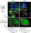
Extracellular vesicles and microvilli in the immune synapse
- PMID: 38268920
- PMCID: PMC10806406
- DOI: 10.3389/fimmu.2023.1324557
Extracellular vesicles and microvilli in the immune synapse
Abstract
T cell receptor (TCR) binding to cognate antigen on the plasma membrane of an antigen-presenting cell (APC) triggers the immune synapse (IS) formation. The IS constitutes a dedicated contact region between different cells that comprises a signaling platform where several cues evoked by TCR and accessory molecules are integrated, ultimately leading to an effective TCR signal transmission that guarantees intercellular message communication. This eventually leads to T lymphocyte activation and the efficient execution of different T lymphocyte effector tasks, including cytotoxicity and subsequent target cell death. Recent evidence demonstrates that the transmission of information between immune cells forming synapses is produced, to a significant extent, by the generation and secretion of distinct extracellular vesicles (EV) from both the effector T lymphocyte and the APC. These EV carry biologically active molecules that transfer cues among immune cells leading to a broad range of biological responses in the recipient cells. Included among these bioactive molecules are regulatory miRNAs, pro-apoptotic molecules implicated in target cell apoptosis, or molecules triggering cell activation. In this study we deal with the different EV classes detected at the IS, placing emphasis on the most recent findings on microvilli/lamellipodium-produced EV. The signals leading to polarized secretion of EV at the synaptic cleft will be discussed, showing that the IS architecture fulfills a fundamental task during this route.
Keywords: FMNL1β; T lymphocytes; actin cytoskeleton; extracellular vesicles; immune synapse; microvilli; multivesicular bodies; protein kinase C δ.
Copyright © 2024 Ruiz-Navarro, Calvo and Izquierdo.
Conflict of interest statement
The authors declare that the research was conducted in the absence of any commercial or financial relationships that could be construed as a potential conflict of interest. The author(s) declared that they were an editorial board member of Frontiers, at the time of submission. This had no impact on the peer review process and the final decision.
Figures


Similar articles
-
T Lymphocyte and CAR-T Cell-Derived Extracellular Vesicles and Their Applications in Cancer Therapy.Cells. 2022 Feb 24;11(5):790. doi: 10.3390/cells11050790. Cells. 2022. PMID: 35269412 Free PMC article. Review.
-
Inducible Polarized Secretion of Exosomes in T and B Lymphocytes.Int J Mol Sci. 2020 Apr 10;21(7):2631. doi: 10.3390/ijms21072631. Int J Mol Sci. 2020. PMID: 32290050 Free PMC article. Review.
-
Polarized release of T-cell-receptor-enriched microvesicles at the immunological synapse.Nature. 2014 Mar 6;507(7490):118-23. doi: 10.1038/nature12951. Epub 2014 Feb 2. Nature. 2014. PMID: 24487619 Free PMC article.
-
Role of Actin Cytoskeleton Reorganization in Polarized Secretory Traffic at the Immunological Synapse.Front Cell Dev Biol. 2021 Feb 4;9:629097. doi: 10.3389/fcell.2021.629097. eCollection 2021. Front Cell Dev Biol. 2021. PMID: 33614660 Free PMC article. Review.
-
Actin reorganization at the centrosomal area and the immune synapse regulates polarized secretory traffic of multivesicular bodies in T lymphocytes.J Extracell Vesicles. 2020 Jun 19;9(1):1759926. doi: 10.1080/20013078.2020.1759926. J Extracell Vesicles. 2020. PMID: 32939232 Free PMC article.
Cited by
-
Is the tumor cell side of the immunological synapse a polarized secretory domain?Front Immunol. 2024 Sep 24;15:1452810. doi: 10.3389/fimmu.2024.1452810. eCollection 2024. Front Immunol. 2024. PMID: 39380986 Free PMC article.
References
Publication types
MeSH terms
Substances
Grants and funding
LinkOut - more resources
Full Text Sources

