. 2023 Dec 18;27(1):108763.
doi: 10.1016/j.isci.2023.108763. eCollection 2024 Jan 19.

Hypoxia inducible factors inhibit respiratory syncytial virus infection by modulation of nucleolin expression

Affiliations

Hypoxia inducible factors inhibit respiratory syncytial virus infection by modulation of nucleolin expression

Xiaodong Zhuang et al. iScience. .

Abstract

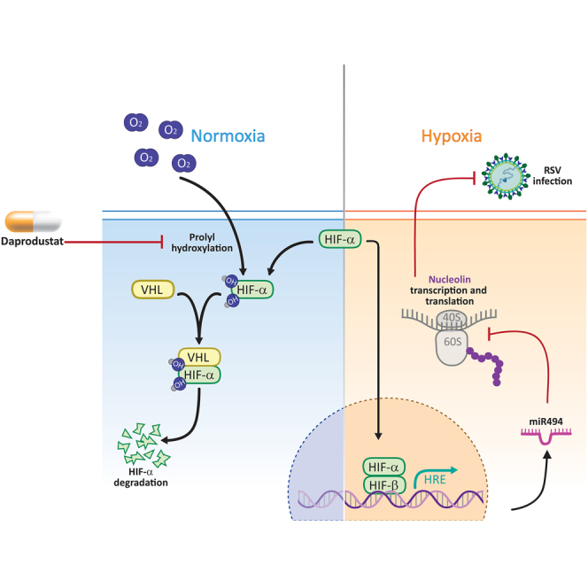
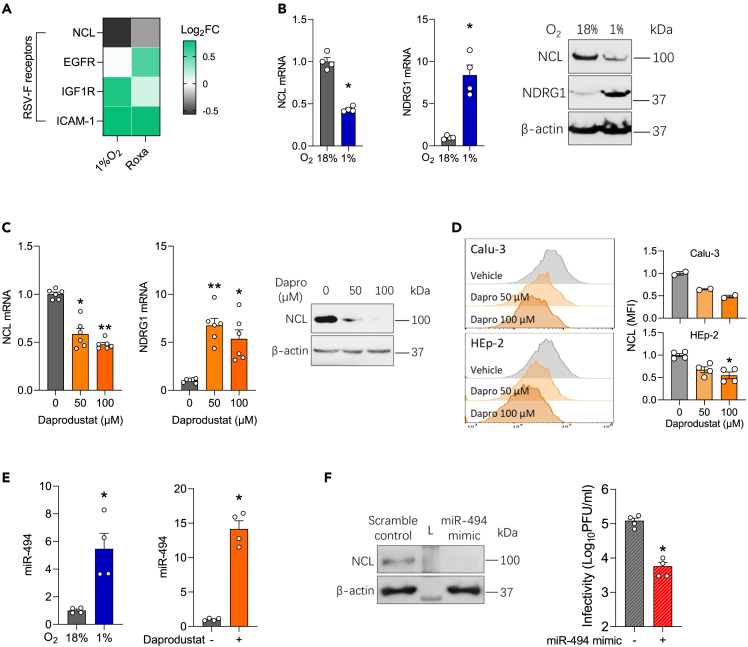
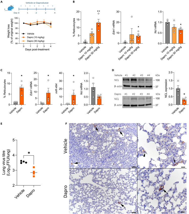
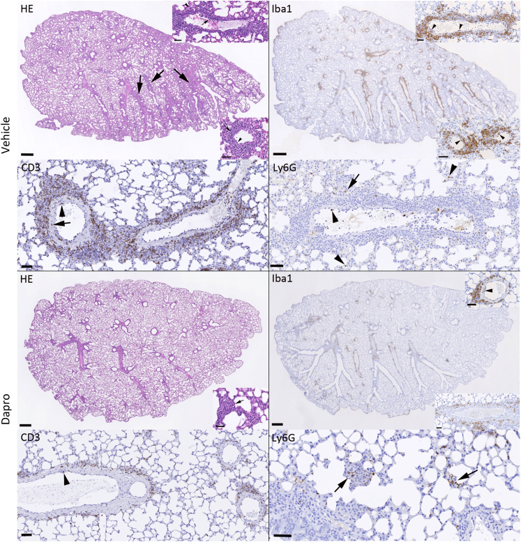
Respiratory syncytial virus (RSV) is a global healthcare problem, causing respiratory illness in young children and elderly individuals. Our knowledge of the host pathways that define susceptibility to infection and disease severity are limited. Hypoxia inducible factors (HIFs) define metabolic responses to low oxygen and regulate inflammatory responses in the lower respiratory tract. We demonstrate a role for HIFs to suppress RSV entry and RNA replication. We show that hypoxia and HIF prolyl-hydroxylase inhibitors reduce the expression of the RSV entry receptor nucleolin and inhibit viral cell-cell fusion. We identify a HIF regulated microRNA, miR-494, that regulates nucleolin expression. In RSV-infected mice, treatment with the clinically approved HIF prolyl-hydroxylase inhibitor, Daprodustat, reduced the level of infectious virus and infiltrating monocytes and neutrophils in the lung. This study highlights a role for HIF-signalling to limit multiple aspects of RSV infection and associated inflammation and informs future therapeutic approaches for this respiratory pathogen.

Keywords: Molecular biology; Omics; Transcriptomics;.


Conflict of interest statement

None of the authors have any competing interests to declare.

Figures

None
Graphical abstract
Figure 1
Figure 1
RSV infection is suppressed under hypoxic conditions and treatment with prolyl-hydroxylase inhibitors (A) Calu-3 cells were cultured at 18% (gray) or 1% (blue) oxygen (O2) for 24h prior to infecting with RSV (MOI 0.2) and the cultures maintained under normoxic or hypoxic conditions for the stated times and cells lysed for the PCR quantification of intracellular viral RNA (RSV-F), NDRG1 mRNA and infectivity at 24h or 48hpi (mean ± SEM, n = 4–8, Mann–Whitney test, Two-sided). All data are expressed relative to 18% O2 values at their respective time point. HIF-1α and β-actin protein expression were assessed by immunoblot at 48hpi. (B) Real-time monitoring of Calu-3 cells transduced with HRE-GFP were treated with PHIs (Daprodustat, Molidustat or Roxadustat at 50 μM) or infected with RSV at MOI 0.2 or 2 (mean ± SEM, n = 5), where UF = uninfected. (C) Calu-3 HRE-GFP reporter cells were treated with Daprodustat at 50 or 100 μM or infected with RSV at a range of MOIs for 48hpi and stained with anti-RSV-F-APC followed by flow cytometric analysis. (D) Calu-3 cells were pre-treated with PHIs for 24h before infecting with RSV (MOI 0.2) and intracellular viral RNA (RSV-F) and infectivity measured at 48hpi (mean ± SEM, n = 6–8, One-way ANOVA with multiple comparisons, Two-sided). HIF-1α and β-actin protein expression were assessed by immunoblot at 48hpi. (E) HEp-2 or BEAS-2B were pre-treated with Daprodustat (100 μM) for 24h before infecting with RSV (MOI 0.2) and intracellular viral RNA (RSV-F) and infectivity measured at 48 hpi (mean ± SEM, n = 4, Mann–Whitney test, Two-sided). HIF-1α and β-actin protein expression were assessed by immunoblot at 48hpi. UF = uninfected. ∗ denotes p < 0.05, ∗∗ <0.01 and ∗∗∗ <0.001.
Figure 2
Figure 2
Prolyl-hydroxylase inhibitors inhibit RSV replication (A) Calu-3 cells were pre-treated with PHIs (Daprodustat, Molidustat, Roxadustat at 50 μM) 24h prior to infection with a reporter RSV expressing GFP (MOI 0.2) and viral replication measured every 8h using an Incucyte Live-Cell Imaging System. GFP signal was normalised per image with respect to cell confluence (mean ± SEM, n = 4). (B) Effect of PHI treatment on RSV infected Calu-3 cells. Calu-3 cells were treated with Daprodustat or Molidustat (50 μM) before and after infection (pre+post) or 24hpi or 48hpi where the arrows indicate the addition of drugs and GFP expression measured using the Incucyte Live-Cell Imaging System. GFP signal and confluence were measured and the integrated intensity values are represented (mean ± SEM, n = 6). (C) Combined Daprodustat and Palivizumab treatment inhibits RSV replication. Calu-3 cells were treated with a range of Palivizumab doses in the presence or absence of Daprodustat (9.0 μM) prior to infection with RSV-GFP (MOI 0.2) and viral replication measured at 48hpi using an Incucyte Live-Cell Imaging System. GFP signal was normalized per image and cell confluence (mean ± SEM, n = 4). UF = uninfected.
Figure 3
Figure 3
Inhibition of RSV-F mediated cell-cell fusion by prolyl-hydroxylase inhibitors (A) HEK293T effector cells (expressing beta strands 1–7 of GFP), engineered to express a doxycycline-inducible RSV F protein, were cocultured with HEK293T target cells (expressing beta strands 8–11 of GFP). (B) The inhibitory activity of Daprodustat or Molidustat was measured as GFP intensity signal 48h after co-culture (mean ± SEM, n = 3). (C) HEK293T cells were treated with PHI (50 μM) in the presence of doxycycline and F expression analyzed by flow cytometry using an APC-conjugated anti-RSV-F antibody at 24hpi. (D) Calu-3 cells were infected with RSV (MOI 1) and at 48hpi RSV-F expression imaged.
Figure 4
Figure 4
Hypoxia or prolyl-hydroxylase inhibitors inhibit nucleolin via regulating miR-494 (A) Calu-3 cells were cultured at 18% or 1% O2 or treated with Roxadustat (50 μM) for 48h and cellular RNA sequenced. Differential expression analysis was performed to interrogate the expression of host entry factors that regulate RSV fusion, where the heatmap denotes Log2 Fold change (FC). (B) Calu-3 cells were cultured at 18% or 1% O2 for 48h and NCL and NDRG1 mRNA or protein expression measured by qPCR or immunoblotting, respectively (mean ± SEM, n = 4, Mann–Whitney test, Two-sided). (C) Calu-3 cells were treated with Daprodustat for 48h and NCL mRNA and protein expression determined by qPCR and immunoblotting, respectively (mean ± SEM, n = 6, One-way ANOVA with multiple comparisons, Two-sided). (D) Calu-3 or HEp-2 cells were treated with Daprodustat for 48h and total NCL protein expression determined by flow cytometry (mean ± SEM, n = 2–4, One-way ANOVA with multiple comparisons, Two-sided). (E) Calu-3 cells were cultured at 18% or 1% O2 or treated with Daprodustat (50 μM) for 48h and miR-494 expression measured by qPCR (mean ± SEM, n = 4, Mann–Whitney test, Two-sided). (F) Calu-3 cells were pre-treated with a miR-494 mimic (1 μM) for 24h before infecting with RSV (MOI 0.2) and intracellular infectivity determined at 48 hpi (mean ± SEM, n = 4, Mann–Whitney test, Two-sided). L = Molecular weight ladder. ∗ denotes p < 0.05 and ∗∗ <0.01.
Figure 5
Figure 5
Daprodustat treatment reduces RSV infection in mice (A) BALB/c mice (6–8 weeks old, female) were treated with vehicle (1% methylcellulose) or Daprodustat (10 or 30 mg/kg), twice daily via oral gavage for 4 days (B) Reticulocyte counts were measured and RNA extracted from the lung and Edn1 and Ncl mRNA levels measured by qPCR (mean ± SEM, vehicle n = 3; Daprodustat n = 4, One-way ANOVA with multiple comparisons, Two-sided). (C) BALB/c mice (6–8 weeks old, female) were treated with vehicle (1% methylcellulose) or Daprodustat (30 mg/kg), BID, via oral gavage 24h prior to intranasal infection with RSV A2 (1 × 107 PFU) and treated with drug as stated in (A) for 4 days. At the end of the experiment, reticulocyte counts were performed, and RNA extracted from the lung for the qPCR measurement of Edn1, miR-494 and Ncl mRNA levels (mean ± SEM, n = 4, Mann–Whitney test, Two-sided). (D) Lung homogenates were assessed for NCL protein expression by immunoblotting and densitometric analysis quantified. NCL in individual samples were normalised to their β-actin loading controls (mean ± SEM, n = 4, Mann–Whitney test, Two-sided). (E) RSV infectivity in lung homogenate was measured by a focus forming assay (mean ± SEM, n = 4, Mann–Whitney test, Two-sided). (F) Representative images of lungs from mice at 4 dpi. The lung of a vehicle treated mouse exhibits several small patches of alveoli with RSV antigen positive pneumocytes (left: arrows). A higher magnification (right) shows infected type I pneumocytes (arrowhead) and type II pneumocytes (arrow). The lung of a Daprodustat (30 mg/kg) treated mouse shows a single patch of viral antigen expressing pneumocytes (left: arrow). The higher magnification of the patch (right; arrow) confirms that both type I and II pneumocytes are infected. Immunohistology, hematoxylin counterstain. Bars = 50 μm (left) and 20 μm (right). ∗ denotes p < 0.05 and ∗∗ <0.01.
Figure 6
Figure 6
Daprodustat treatment reduces the RSV-associated pulmonary inflammatory responses in mice Representative images of lungs from mice at 4 days post RSV infection. As shown in the HE stained section, the lung of a vehicle treated mouse exhibits mild perivascular and peribronchial mononuclear infiltrates (arrows) associated with leukocyte rolling and emigration (bottom inset, arrowhead) and subendothelial leukocyte aggregates in particular in muscular veins (top inset, arrowhead) consistent with vasculitis. The infiltrates are dominated by monocytes/macrophages (Iba1+) which are also found rolling and attached to the endothelium (bottom inset: arrowheads) and within the subendothelial infiltrates (top inset: arrowheads). T cells (CD3+) are numerous and present rolling (arrowhead) and within the vascular wall (arrow). There are a few neutrophils (Ly6G), within the lumen of vessels and capillaries (arrowheads) and occasionally within the small parenchymal infiltrates (arrow). After Daprodustat treatment, the inflammatory response is limited to very mild perivascular mononuclear infiltrates and rare small focal parenchymal infiltrates (HE stain: inset, arrow). The infiltrate shows a similar composition as in the vehicle treated mice. It is dominated by monocytes/macrophages (Iba1+) that are occasionally found also in vascular walls (top inset: arrowhead), followed by T cells (CD3+) that are occasionally seen rolling along the vascular endothelium (arrowhead). Neutrophils (Ly6G+) are mainly seen in the rare small parenchymal infiltrates (arrows). Bars = 500 μm (whole section images) and 50 μm (all other images).

Similar articles

Cited by

References

    1. Shi T., McAllister D.A., O'Brien K.L., Simoes E.A.F., Madhi S.A., Gessner B.D., Polack F.P., Balsells E., Acacio S., Aguayo C., et al. Global, regional, and national disease burden estimates of acute lower respiratory infections due to respiratory syncytial virus in young children in 2015: a systematic review and modelling study. Lancet. 2017;390:946–958. - PMC - PubMed
    1. Fleming D.M., Taylor R.J., Lustig R.L., Schuck-Paim C., Haguinet F., Webb D.J., Logie J., Matias G., Taylor S. Modelling estimates of the burden of Respiratory Syncytial virus infection in adults and the elderly in the United Kingdom. BMC Infect. Dis. 2015;15:443. - PMC - PubMed
    1. Grayson S.A., Griffiths P.S., Perez M.K., Piedimonte G. Detection of airborne respiratory syncytial virus in a pediatric acute care clinic. Pediatr. Pulmonol. 2017;52:684–688. - PubMed
    1. Collins P.L., Graham B.S. Viral and host factors in human respiratory syncytial virus pathogenesis. J. Virol. 2008;82:2040–2055. - PMC - PubMed
    1. Homaira N., Rawlinson W., Snelling T.L., Jaffe A. Effectiveness of Palivizumab in Preventing RSV Hospitalization in High Risk Children: A Real-World Perspective. Int. J. Pediatr. 2014;2014 - PMC - PubMed

LinkOut - more resources