[Preprint]. 2024 Apr 28:2024.01.08.574656.
doi: 10.1101/2024.01.08.574656.

Lipid osmosis, membrane tension, and other mechanochemical driving forces of lipid flow

Affiliations

Lipid osmosis, membrane tension, and other mechanochemical driving forces of lipid flow

Yongli Zhang et al. bioRxiv. .

Update in

Abstract

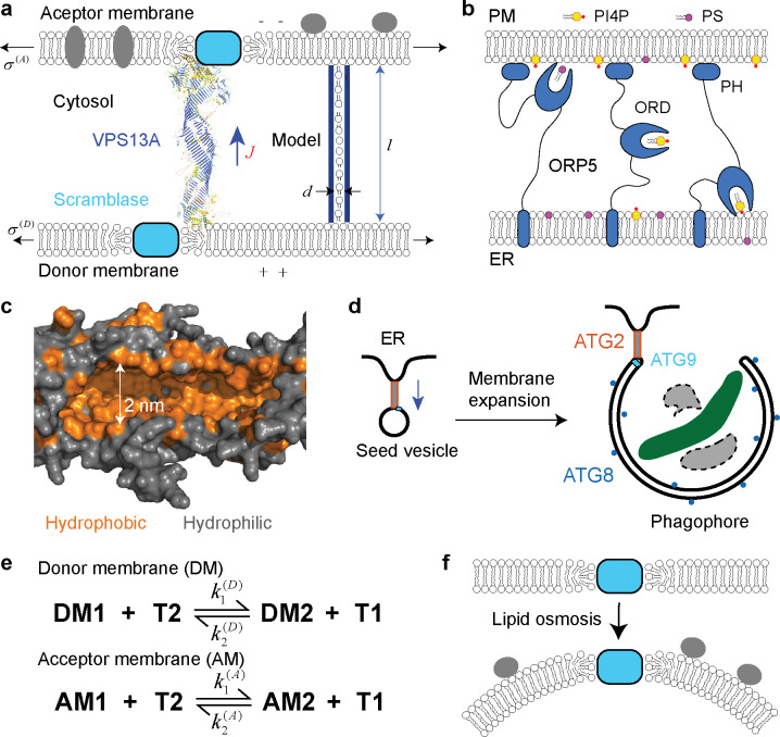
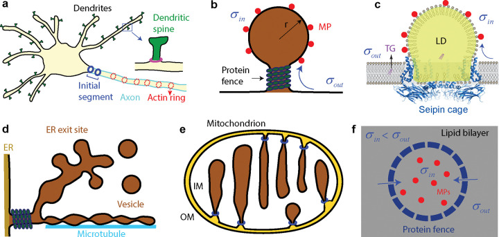
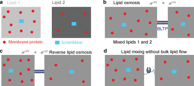
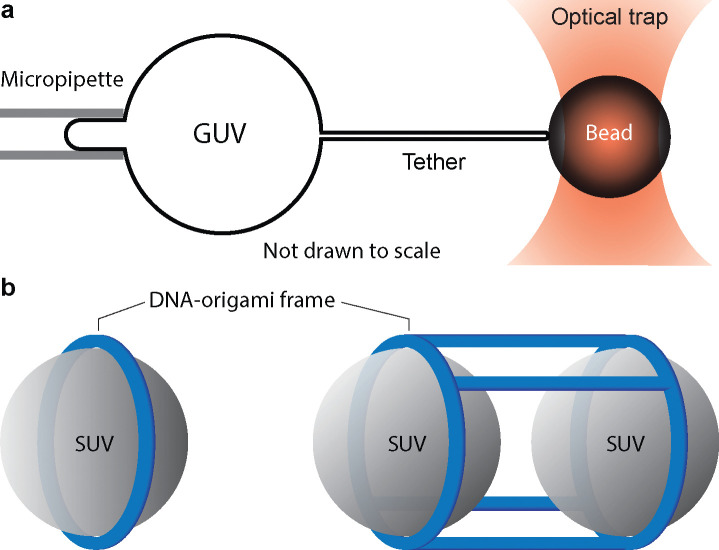
Nonvesicular lipid transport among different membranes or membrane domains plays crucial roles in lipid homeostasis and organelle biogenesis. However, the forces that drive such lipid transport are not well understood. We propose that lipids tend to flow towards the membrane area with a higher membrane protein density in a process termed lipid osmosis. This process lowers the membrane tension in the area, resulting in a membrane tension difference called osmotic membrane tension. We examine the thermodynamic basis and experimental evidence of lipid osmosis and osmotic membrane tension. We predict that lipid osmosis can drive bulk lipid flows between different membrane regions through lipid transfer proteins, scramblases, or other similar barriers that selectively pass lipids but not membrane proteins. We also speculate on the biological functions of lipid osmosis. Finally, we explore other driving forces for lipid transfer and describe potential methods and systems to further test our theory.


Conflict of interest statement

Conflict of interest The authors declare no conflict of interest.

Figures

Figure 1.
Figure 1.. Lipid transfer proteins (LTPs) and scramblases transfer lipids between different membrane leaflets.
(a) A bridge LTP (BLTP), shown here by the AlphFold structure of a representative BLTP VPS13A [56], acts as a lipid conduit to allow bulk lipid flow from one membrane to the other. The flow (with its direction indicated by the blue arrow) may be driven by the gradient of membrane tension, electric potential, or membrane protein density as indicated. The channel on the right represents an idealized BLTP used to estimate its lipid flow rate. (b) A representative shuttle LTP ORP5 transfers phospholipids PS and PI4P between the plasma membrane (PM) and the ER through two lipid-exchange processes. ORP5 contains an N-terminal pleckstrin homology (PH) domain that binds to the PM, a middle OSBP-related domain (ORP) that binds to PS or PI4P, and a C-terminal transmembrane domain in the ER. Three snapshots of the lipid transfer are shown. (c) A segment of the hydrophobic groove of the bacterial BLTP YhdP predicted by AlphaFold. (d) During macroautophagy, BLTP ATG2 and the scramblase ATG9 mediate fast lipid flow from the ER to the double-membraned phagophore for its membrane expansion, which eventually engulfs damaged organelle (green) or protein aggregations (gray) for their degradation. The enzyme-catalyzed lipidation of ATG8 proteins (blue dots) on the phagophore membrane is required for the membrane expansion. (e) The two lipid-exchange processes are characterized by two sets of rate constants, which may be equal or different, depending upon the membrane environments. DMi, AMi, and Ti with i=1 or 2 denote the i-th lipid species in the donor membrane, acceptor membrane, and shuttle LTP, respectively. (f) Lipid osmosis can occur between different leaflets of the same membrane mediated by scramblases, which may cause membrane bending.
Figure 2.
Figure 2.. Ubiquitous protein barriers separate cell membranes into domains with distinct functions and membrane protein densities and compositions.
(a) The axon and dendritic spines are separated from the neural cell body at the initial segment and dendritic spine neck, respectively, by physical barriers (blue and magenta circles) that limit protein diffusion across the membrane domains. (b) The vesicular precursor crowded with membrane proteins (red circles) is connected to the plasma membrane through a protein-coated neck. Lipid osmosis (blue arrow) may facilitate the vesicular membrane expansion. (c) Seipin forms a decameric fence-like structure in the ER membrane to mediate lipid droplet (LD) formation between the two ER leaflets. Each droplet contains a neutral lipid core (highlighted in yellow) containing triglycerides (TG) covered by a phospholipid monolayer and various membrane proteins (red dots). (d) The ER exit site is a specialized ER domain that serves as a hub for membrane trafficking. The domain is connected to the ER membrane by a neck coated by proteins including COPII and TANGO1. (e) The mitochondrial inner membrane invaginates to form cristae with protein-coated crista junctions (marked by blue circles) connected to the inner boundary membrane. (f) Lipid osmosis occurs through a protein fence that selectively passes lipids but not membrane proteins (blue dashed ring), leading to lower equilibrium tension of the membrane inside the fence with enriched proteins.
Figure 3.
Figure 3.. Areas and lipid distributions of two membranes before and after the LTP-mediated lipid transfer reaches equilibrium.
(a) The two membranes initially have identical areas but contain different lipid species (indicated by different grey scales) and densities of membrane proteins (red circles and cyan rectangles) before lipid transfer. (b) In the absence of membrane tension gradient, lipid osmosis through BLTPs causes expansion of the membrane with a higher density of membrane proteins, leading to equal densities of membrane proteins and fractions of lipids in both membranes upon equilibrium. (c) A high gradient of membrane tension counteracts lipid osmosis to expand the membrane with lower membrane protein density, leading to reverse lipid osmosis. (d) Lipid transfer mediated by shuttle LTPs causes lipid mixing without changes in membrane areas in a manner independent of the protein density gradient.
Figure 4.
Figure 4.. Methods for controlling tension, curvature, and separation of model membranes.
(a) Membrane tension of a GUV can be controlled by micropipette aspiration and measured by membrane tether pulling with optical tweezers. Different suction pressures can be applied via the micropipette to change membrane tension. The pulling force measured by the optical trap can be used to calculate the membrane tension. (b) Membrane curvature and inter-membrane distance of SUVs (grey) can be controlled by DNA-origami frames (blue). These DNA nanostructures can be modified with amphipathic molecules on the interior (not shown for clarity) and serve as templates for vesicle reconstitution. Left: a size-defined SUV formed inside a DNA ring. Right: a pair of SUVs separated by a set of rigid DNA pillars with defined lengths.

Similar articles

References

    1. Hanna M, Guillen-Samander A, De Camilli P: RBG motif bridge-like lipid transport proteins: structure, functions, and open questions. Annu. Rev. Cell. Dev. Biol. 2023, 39:409–434 - PubMed
    1. Kumar N, Leonzino M, Hancock-Cerutti W, Horenkamp FA, Li P, Lees JA, Wheeler H, Reinisch KM, De Camilli P: VPS13A and VPS13C are lipid transport proteins differentially localized at ER contact sites. J. Cell Biol. 2018, 217:3625–3639 - PMC - PubMed
    1. Reinisch KM, Prinz WA: Mechanisms of nonvesicular lipid transport. J. Cell Biol. 2021, 220:e202012058. - PMC - PubMed
    1. Wong LH, Gatta AT, Levine TP: Lipid transfer proteins: the lipid commute via shuttles, bridges and tubes. Nat. Rev. Mol. Cell Bio. 2019, 20:85–101 - PubMed
    1. Cai SJ, Wu YM, Guillen-Samander A, Hancock-Cerutti W, Liu J, De Camilli P: In situ architecture of the lipid transport protein VPS13C at ER-lysosome membrane contacts. Proc. Natl. Acad. Sci. USA 2022, 119. - PMC - PubMed

Publication types