Oregano (Origanum vulgare) Essential Oil and Its Constituents Prevent Rat Kidney Tissue Injury and Inflammation Induced by a High Dose of L-Arginine
- PMID: 38256015
- PMCID: PMC10815453
- DOI: 10.3390/ijms25020941
Oregano (Origanum vulgare) Essential Oil and Its Constituents Prevent Rat Kidney Tissue Injury and Inflammation Induced by a High Dose of L-Arginine
Abstract
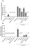
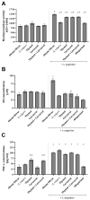
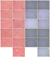
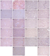
This study aimed to evaluate the protective action of oregano (Origanum vulgare) essential oil and its monoterpene constituents (thymol and carvacrol) in L-arginine-induced kidney damage by studying inflammatory and tissue damage parameters. The determination of biochemical markers that reflect kidney function, i.e., serum levels of urea and creatinine, tissue levels of neutrophil-gelatinase-associated lipocalin (NGAL), and kidney injury molecule-1 (KIM-1), as well as a panel of oxidative-stress-related and inflammatory biomarkers, was performed. Furthermore, histopathological and immunohistochemical analyses of kidneys obtained from different experimental groups were conducted. Pre-treatment with the investigated compounds prevented an L-arginine-induced increase in serum and tissue kidney damage markers and, additionally, decreased the levels of inflammation-related parameters (TNF-α and nitric oxide concentrations and myeloperoxidase activity). Micromorphological kidney tissue changes correlate with the alterations observed in the biochemical parameters, as well as the expression of CD95 in tubule cells and CD68 in inflammatory infiltrate cells. The present results revealed that oregano essential oil, thymol, and carvacrol exert nephroprotective activity, which could be, to a great extent, associated with their anti-inflammatory, antiradical scavenging, and antiapoptotic action and, above all, due to their ability to lessen the disturbances arising from acute pancreatic damage. Further in-depth studies are needed in order to provide more detailed explanations of the observed activities.
Keywords: L-arginine; Origanum vulgare; inflammatory parameters; kidney; tissue damage parameters.
Conflict of interest statement
The authors declare no conflicts of interest.
Figures


Similar articles
-
Activity of Common Thyme (Thymus vulgaris L.), Greek Oregano (Origanum vulgare L. ssp. hirtum), and Common Oregano (Origanum vulgare L. ssp. vulgare) Essential Oils against Selected Phytopathogens.Molecules. 2024 Sep 29;29(19):4617. doi: 10.3390/molecules29194617. Molecules. 2024. PMID: 39407547 Free PMC article.
-
Proteomic analysis and antibacterial resistance mechanisms of Salmonella Enteritidis submitted to the inhibitory effect of Origanum vulgare essential oil, thymol and carvacrol.J Proteomics. 2020 Mar 1;214:103625. doi: 10.1016/j.jprot.2019.103625. Epub 2019 Dec 24. J Proteomics. 2020. PMID: 31881347
-
Intraspecific divergence in essential oil content, composition and genes expression patterns of monoterpene synthesis in Origanum vulgare subsp. vulgare and subsp. gracile under salinity stress.BMC Plant Biol. 2023 Aug 7;23(1):380. doi: 10.1186/s12870-023-04387-5. BMC Plant Biol. 2023. PMID: 37550621 Free PMC article.
-
A Recent Insight Regarding the Phytochemistry and Bioactivity of Origanum vulgare L. Essential Oil.Int J Mol Sci. 2020 Dec 17;21(24):9653. doi: 10.3390/ijms21249653. Int J Mol Sci. 2020. PMID: 33348921 Free PMC article. Review.
-
Can Origanum be a hope for cancer treatment? A review on the potential of Origanum species in preventing and treating cancers.Int J Environ Health Res. 2023 Sep;33(9):894-910. doi: 10.1080/09603123.2022.2064437. Epub 2022 Apr 13. Int J Environ Health Res. 2023. PMID: 35414316 Review.
Cited by
-
Evaluation of Chemical Profile and Biological Properties of Extracts of Different Origanum vulgare Cultivars Growing in Poland.Int J Mol Sci. 2024 Aug 30;25(17):9417. doi: 10.3390/ijms25179417. Int J Mol Sci. 2024. PMID: 39273364 Free PMC article.
References
MeSH terms
Substances
Grants and funding
LinkOut - more resources
Full Text Sources
Research Materials
Miscellaneous

