Review
. 2024 Jan 22;13(1):6.
doi: 10.1186/s40164-024-00474-x.

Crosstalk between colorectal CSCs and immune cells in tumorigenesis, and strategies for targeting colorectal CSCs

Affiliations
Review

Crosstalk between colorectal CSCs and immune cells in tumorigenesis, and strategies for targeting colorectal CSCs

Qi Zhao et al. Exp Hematol Oncol. .

Abstract

Cancer immunotherapy has emerged as a promising strategy in the treatment of colorectal cancer, and relapse after tumor immunotherapy has attracted increasing attention. Cancer stem cells (CSCs), a small subset of tumor cells with self-renewal and differentiation capacities, are resistant to traditional therapies such as radiotherapy and chemotherapy. Recently, CSCs have been proven to be the cells driving tumor relapse after immunotherapy. However, the mutual interactions between CSCs and cancer niche immune cells are largely uncharacterized. In this review, we focus on colorectal CSCs, CSC-immune cell interactions and CSC-based immunotherapy. Colorectal CSCs are characterized by robust expression of surface markers such as CD44, CD133 and Lgr5; hyperactivation of stemness-related signaling pathways, such as the Wnt/β-catenin, Hippo/Yap1, Jak/Stat and Notch pathways; and disordered epigenetic modifications, including DNA methylation, histone modification, chromatin remodeling, and noncoding RNA action. Moreover, colorectal CSCs express abnormal levels of immune-related genes such as MHC and immune checkpoint molecules and mutually interact with cancer niche cells in multiple tumorigenesis-related processes, including tumor initiation, maintenance, metastasis and drug resistance. To date, many therapies targeting CSCs have been evaluated, including monoclonal antibodies, antibody‒drug conjugates, bispecific antibodies, tumor vaccines adoptive cell therapy, and small molecule inhibitors. With the development of CSC-/niche-targeting technology, as well as the integration of multidisciplinary studies, novel therapies that eliminate CSCs and reverse their immunosuppressive microenvironment are expected to be developed for the treatment of solid tumors, including colorectal cancer.

Keywords: Colorectal cancer stem cells; Immune Cells; Immunotherapy; Targeting cancer stem cells; Tumor immune microenvironment.


Conflict of interest statement

All authors declare that they have no conflicts of interest affecting this work.

Figures

Fig. 1
Fig. 1
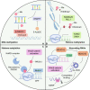
Signaling pathways in colorectal CSCs. A Wnt/β-catenin signaling pathway. In the presence of Wnt ligand proteins, the engagement of Wnt, Frizzled and LRP5/6 on the cell surface induces the activation of downstream DVL signaling, which in turn inhibits the β-catenin destruction complex (which is mainly composed of APC, Axin1/2, GSK3β and CK1α), allowing β-catenin to avoid being degraded, to accumulate in the cytoplasm and enter the nucleus. β-catenin enters the nucleus and binds with TCF/LEF to promote the transcription and expression of target genes. B Hippo/Yap signaling pathway. Upon stimulation via upstream signaling (mainly cell polarity, cell contact, and mechanical force signals), phosphorylated MST1/2 phosphorylate LATS1/2, and then, activated LATS1/2 phosphorylate YAP/TAZ to induce their degradation. In the absence of upstream signal stimulation, dephosphorylated YAP/TAZ aggregate in the nucleus and combine with the transcription factors TEAD1-4 to promote the expression of target genes. C JAK/STAT signaling pathway. When cytokines bind plasma membrane the receptors, receptors dimerize, and receptor-associated JAK kinase is activated via mutual phosphorylation. Then, the activated JAK kinase phosphorylates receptor tyrosine residues and recruits and activates SH2 domain-containing STAT, which dissociates from the receptor, forms dimers in the cytoplasm and enters the nucleus to regulate the expression of target genes. D Notch signaling pathway. Delta-like ligands (DLL1, DLL2, DLL3 and DLL4) and Jagged ligands (JAG1 and JAG2) adjacent to cells are ligands for Notch receptors (including Notch 1, Notch 2, Notch 3 and Notch 4). Through a combination of ligands and receptors, the S2 site in Notch receptors is cleaved by the ADAM10 or ADAM17 protease, resulting in the release of the extracellular portion of Notch. Then, γ-secretase cleaves the Notch receptor at the S3 site, allowing the release of the Notch intracellular domain (NICD). The NICD enters the nucleus and interacts with CSL to regulate the expression of downstream target genes
Fig. 2
Fig. 2
Epigenetic modifications of colorectal CSCs. A DNA methylation. DNA methyltransferase 1 (DNMT1) participates in the activation of the Wnt/β-catenin pathway to maintain the stemness of CRC cells. After knocking down DNMT1 or treating cells with the DMNT1 inhibitor 5-aza-2'-deoxycytidine (5-AzaDC), the transcriptional activity of β-catenin in the nucleus was significantly reduced. B Histone methylation. Repressive H3K9me2 mark wasmarks are removed by histone demethylases (KDM3A/B), which simultaneously recruit lysine methyltransferase (MLL1), which mediates for the H3K4me3 modification to and thus promotes the expression of the Wnt target genes AXIN2 and DKK1. C Histone acetylation. The Wnt/β-catenin target gene PROX1 interacts with the Notch1 promoter and recruits the nucleosome remodeling and deacetylase (NuRD) complex, which deacetylates histones and remodels chromatin to block Notch1 transcription. D Noncoding RNAs. cis-HOX (a cyclic RNA) can binds to HOXC10 mRNA in the cytoplasm to inhibit the KSRP-dependent degradation of HOXC10 mRNA. The increased HOXC10, the level of which has been increased, enters the nucleus and drives FZD3 expression to activate the Wnt/β-catenin signaling pathway
Fig. 3
Fig. 3
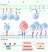
Expression profiles of immune proteins on colorectal CSCs. A The expression of MHC class I molecules on colorectal CSCs is downregulated, making them difficult to recognize and kill by CD8+ T cells. B NK cells are activated by receptors such as NKG2D and natural cytotoxicity receptors (NCRs), which recognize and kill CSCs in an MHC-independent manner. C Colorectal CSCs express high levels of the immune checkpoint PD-L1, which can inhibit the antitumor immune effect of T cells. D PD-L1 and colorectal CSC-related genes are downstream of the same transcriptional element. The ARID3B-KDM4C complex regulates chromatin state to activate downstream Notch target genes, CRC stemness genes and PD-L1 transcriptional expression. In addition, PD-L1 is the direct target gene of the Wnt/β-catenin, PI3K/Akt/mTOR, and STAT3 signaling pathways
Fig. 4
Fig. 4
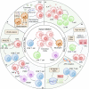
CSC-immune cell crosstalk involvement in tumor initiation. A Colorectal CSCs suppress the proliferation of T cells via high expression of IL-4. B A high-fat diet reduced the expression of MHC class II molecules in colorectal CSCs by inhibiting the PRR and IFNγ signaling pathways, thereby inhibiting the activity of CD4+ T cells and promoting CRC occurrence. C APC mutation, the major oncogenic variant in early colorectal tumorigenesis, activates the Wnt/β-catenin signaling pathway, and the β-catenin-TCF4 complex binds to the promoter of the PD-L1-encoding gene to induce PD-L1 expression, which inhibits CD8+ T cell activation. D Colorectal CSCs secrete macrophage migration inhibitory factor (MIF), which binds the CD74 receptor on tumor-associated monocytes and macrophages (TAMMs), inducing immunosuppressive signaling. In addition, TAMMs secrete prostaglandin E2 (PGE2) and promote the proliferation of colorectal CSCs via the action of the PGE2 receptor EP4. E The Wnt/β-catenin signaling pathway activates the expression of RAB27B to mediate the secretion of miR-146a exosomes, which promote the stemness of tumor cells and tumorigenicity and reduces the infiltration of CD8+ T cells. (F) Colorectal CSCs secrete exosomal RNAs to increase the number of neutrophils in the bone marrow and induce neutrophils to express IL-1β. In addition, colorectal CSCs secrete CXCL1/2 to recruit neutrophils expressing CXCR2 in bone marrow, and IL-1β secreted by neutrophils can promote tumorigenesis
Fig. 5
Fig. 5
CSC-intrinsic immune factors in CSC-immune cell crosstalk. A PD-L1 interacts with Frizzled 6 to inhibit the degradation of β-catenin via the destruction complex. Moreover, the activation of the Wnt/β-catenin pathway induces the expression of PD-L1, forming a positive feedback β-catenin/PD-L1 loop to promote the stemness and expansion of colorectal CSCs. B PD-L1 interacts with the HMGA1 to promote the proliferation of colorectal CSCs by activating the HMGA1-dependent MEK/ERK and AKT signaling pathways. C Colorectal CSCs express low levels of S100A14 (SA14) to avoid the degradation of STAT3 in CSCs, and thus PD-L1 is highly expressed as a target of STAT3. D circREEP3 recruits the chromatin-remodeling protein CHD7 to the promoter of FKBP10 to activate its transcription and promote tumor progression. In addition, circREEP3 can enhance the ubiquitination and degradation of RIG-1 mediated by RNF125, thereby inhibiting antitumor immune effects
Fig. 6
Fig. 6
Immune microenvironment regulates colorectal CSCs to drive tumor stemness. A CD4 + T cells secrete IL-22, which activates STAT3 in colorectal CSCs. The activation of STAT3 promotes the stemness of CRC and induces the expression of DOT1L, which promotes the expression of stemness-related genes by promoting H3K79me2 deposition at their promoter regions. B Treg cells act on colorectal CSCs by secreting IL-17 and promote the maintenance of CRC stemness through the AKT and MAPK signaling pathways. C CXCL1 secreted by tumor-associated dendritic cells (TADCs) promotes the expression of CD44 and CD133 and maintains the stemness of CRC. D Granulocyte myeloid-derived suppressor cells (G-MDSCs) secrete S100A9-containing exosomes that promote the stemness of colorectal CSCs by inducing STAT3 phosphorylation and NF-κB activation. E IL-1β secreted by tumor-associated macrophages (TAMs) inhibits the phosphorylation activation of GSK3β by activating the NF-κB and AKT signaling pathways, thereby abrogating the β-catenin destruction complex and activating the Wnt/β-catenin signaling pathway in colorectal CSCs
Fig. 7
Fig. 7
Colorectal CSCs remodel the immune microenvironment. A DCLK1 expressed by colorectal CSCs activates the ERK signaling pathway and promotes the expression of CXCL1/CXCL2 to recruit myeloid-derived suppressor cells (MDSCs), thereby inhibiting the activity of CD4+ T cells and CD8+ T cells. B Six1 expressed by colorectal CSCs promotes the secretion of CSF-1, CCL2/5 and VEGF and recruits TAMs to form an immunosuppressive microenvironment. IL-4 secreted by colorectal CSCs is an important factor for maintaining TAM tumor-promoting activity

Similar articles

Cited by

References

    1. Biller LH, Schrag D. Diagnosis and treatment of metastatic colorectal cancer: a review. JAMA. 2021;325(7):669–685. doi: 10.1001/jama.2021.0106. - DOI - PubMed
    1. Rawla P, Sunkara T, Barsouk A. Epidemiology of colorectal cancer: incidence, mortality, survival, and risk factors. Prz Gastroenterol. 2019;14(2):89–103. - PMC - PubMed
    1. González-Silva L, Quevedo L, Varela I. Tumor functional heterogeneity unraveled by scRNA-seq technologies. Trends Cancer. 2020;6(1):13–19. doi: 10.1016/j.trecan.2019.11.010. - DOI - PubMed
    1. Nassar D, Blanpain C. Cancer stem cells: basic concepts and therapeutic implications. Annu Rev Pathol. 2016;11:47–76. doi: 10.1146/annurev-pathol-012615-044438. - DOI - PubMed
    1. Batlle E, Clevers H. Cancer stem cells revisited. Nat Med. 2017;23(10):1124–1134. doi: 10.1038/nm.4409. - DOI - PubMed