. 2024 Jan 22;15(1):80.
doi: 10.1038/s41419-024-06423-0.

Unexpected inhibition of the lipid kinase PIKfyve reveals an epistatic role for p38 MAPKs in endolysosomal fission and volume control

Affiliations

Unexpected inhibition of the lipid kinase PIKfyve reveals an epistatic role for p38 MAPKs in endolysosomal fission and volume control

Daric J Wible et al. Cell Death Dis. .

Abstract

p38 mitogen-activated protein kinases (MAPKs) participate in autophagic signaling; and previous reports suggest that pyridinyl imidazole p38 MAPK inhibitors, including SB203580 and SB202190, induce cell death in some cancer cell-types through unrestrained autophagy. Subsequent studies, however, have suggested that the associated cytoplasmic vacuolation resulted from off-target inhibition of an unidentified enzyme. Herein, we report that SB203580-induced vacuolation is rapid, reversible, and relies on the class III phosphatidylinositol 3-kinase (PIK3C3) complex and the production of phosphatidylinositol 3-phosphate [PI(3)P] but not on autophagy per se. Rather, vacuolation resulted from the accumulation of Rab7 on late endosome and lysosome (LEL) membranes, combined with an osmotic imbalance that triggered severe swelling in these organelles. Inhibition of PIKfyve, the lipid kinase that converts PI(3)P to PI(3,5)P2 on LEL membranes, produced a similar phenotype in cells; therefore, we performed in vitro kinase assays and discovered that both SB203580 and SB202190 directly inhibited recombinant PIKfyve. Cancer cells treated with either drug likewise displayed significant reductions in the endogenous levels of PI(3,5)P2. Despite these results, SB203580-induced vacuolation was not entirely due to off-target inhibition of PIKfyve, as a drug-resistant p38α mutant suppressed vacuolation; and combined genetic deletion of both p38α and p38β dramatically sensitized cells to established PIKfyve inhibitors, including YM201636 and apilimod. The rate of vacuole dissolution (i.e., LEL fission), following the removal of apilimod, was also significantly reduced in cells treated with BIRB-796, a structurally unrelated p38 MAPK inhibitor. Thus, our studies indicate that pyridinyl imidazole p38 MAPK inhibitors induce cytoplasmic vacuolation through the combined inhibition of both PIKfyve and p38 MAPKs, and more generally, that p38 MAPKs act epistatically to PIKfyve, most likely to promote LEL fission.


Conflict of interest statement

The authors declare no competing interests.

Figures
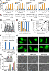
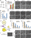
Fig. 1
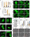
Fig. 1. Pyridinyl imidazole p38 MAPK inhibitors induce cytoplasmic vacuolation.
A, B Various cancer cell lines were treated with DMSO (control) or SB203580 (50 µM) for 2–24 h and assessed for vacuolation and cell death by phase-contrast microscopy and flow cytometry (annexin V-propidium iodide staining), respectively (†, not determined; see also Supplementary Fig. S1A). At 24 h, <3% of DMSO-treated cells were vacuolated and cell death values (%) were: DU145 (9 ± 0.4), A549 (12.8 ± 0.8), HCT116 (15.0 ± 1.6), HT-29 (10.2 ± 1.2). The number of vacuoles per cell and the average vacuole size (µm2) was determined using ImageJ analysis of phase-contrast images. C DU145 cells were stained with CFDA-SE, exposed to DMSO (shaded peaks) or SB203580 (empty peaks), and monitored for changes in cell proliferation, as described in the methods. D DU145 cells were treated with SB203580 (50 µM) for 24 h and then washed and replaced with fresh media ± SB203580 for 4–24 h. At each time point following the washout, cells were examined for vacuolation by phase-contrast microscopy (see also Supplementary Fig. S1C). E DU145 prostate cancer cells were treated with increasing concentrations of the pharmacological p38 MAPK inhibitor, SB203580 (0–100 µM), for 24 h and examined for signs of vacuolation by phase-contrast microscopy (200×). Inset: SB203580 (50 µM) inhibited p38-dependent sequential phosphorylation of MK2 and HSP27. Concentration-dependent inhibition of HSP27 phosphorylation by SB203580 (0–100 µM) was also determined by western blotting (see Supplementary Fig. S1D, E) with individual bands scanned, quantified with ImageJ software, and plotted as the percent of p-HSP27 inhibited. F, G DU145 cells were transiently transfected with expression plasmids encoding EGFP, constitutively active (D176A/F327S) p38α (EGFP-p38α-CA), or p38α-CA containing an additional mutation to the gatekeeper residue (T106M) that renders p38α resistant to SB203580 [EGFP-p38α-CA (T106M)]. EGFP-positive cells were then evaluated by fluorescence microscopy for the number of vacuoles present per cell. H, I p38α and p38β were deleted from DU145 cells using CRISPR-Cas9; and three p38 DKO clones were exposed to SB203580 (50 µM) or SB202190 (50 µM) and evaluated for vacuolation by phase-contrast microscopy using ImageJ analysis software.
Fig. 2
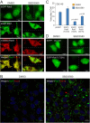
Fig. 2. SB203580 does not stimulate autophagy but does induce vacuolation in a PIK3C3-dependent manner and incorporates pre-existing autophagosome membranes.
A DU145, HEK293, A549, and MEFs were exposed to SB203580 (50 µM) ± the class III PI3 kinase inhibitor, 3-MA (5 mM), and examined for vacuolation. For each cell-type, asterisks indicate that 3-MA was significantly different from the SB treatment alone (one-way ANOVA with Student–Neumann–Keuls post hoc analysis; p < 0.05). B DU145 cells were stably transfected with scrambled control or beclin 1 shRNA plasmids, treated with DMSO (control) or SB203580 (50 µM), and examined for vacuolation and cell death. Inset: Beclin 1 expression levels were determined by immunoblotting. C DU145 cells were transiently transfected with the PI(3)P probe, PX-EGFP, or the binding mutant R57Q, and subsequently treated with (i) DMSO or (ii–iv) SB203580 ± 3-MA for 24 h. Similarly, (v) control and (vi) Beclin 1-depleted cells were also transfected with PX-EGFP and treated with SB203580 (50 µM) for 24 h and examined by fluorescence microscopy. D Various cancer cell lines and MEFs were transiently transfected with EGFP-LC3 and treated with SB203580 (50 µM) for 24 h. Some but not all LC3-labeled vacuoles are indicated with arrows. EH DU145 were reconstituted with ATG5 to restore basal autophagy; exposed to SB203580 (50 µM) or SB202190 (50 µM) for 24 h; and evaluated by phase-contrast microscopy for vacuolation.
Fig. 3
Fig. 3. SB203580-induced vacuolation results from Rab7-dependent fusion of LEs and lysosomes.
A DU145 cells were co-transfected with various combinations of EGFP or mCherry-labeled Rab5, Rab7, and/or Rab9. The cells were then treated with DMSO (control) or SB203580 (50 µM) for 24 h and examined for colocalization with the vacuoles and/or one another. B DU145 cells, treated for 24 h with DMSO (control) or SB203580 (50 µM), were evaluated by immunofluorescence microscopy using antibodies to endogenous Rab7 (green) and Rab9 (red) (see also Supplementary Fig. S3B). C, D DU145 cells were similarly transfected with either EGFP (empty vector) or dominant-negative Rab7(T22N). Cells were then exposed to DMSO or SB203580 (50 µM), and the number of vacuoles was counted in at least 50 cells by fluorescence microscopy. Each experiment was performed in triplicate, and each data point represents mean ± SEM.
Fig. 4
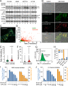
Fig. 4. SB203580 induces LEL swelling and defects in cathepsin processing and protein degradation.
A DU145, A549, HCT116, and HT-29 cells were exposed to SB203580 (50 µM) or SB202190 (50 µM) and western blotted for LC3B, p62, CTSD, or β-tubulin. Band intensities were determined by ImageJ; and the LC3B-II/β-tubulin, p62/β-tubulin, and pre-pro/mature CTSD ratios are listed below the indicated immunoblots. B DU145 cells were exposed to DMSO or SB203580 (50 µM) for 24 h, labeled with LysoTrackerTM Green and Red, and evaluated by confocal microscopy or flow cytometry. CF DU145 cells were exposed to DMSO or SB203580 (50 µM) for 24 h, loaded with LysoSensorTM Yellow/Blue DND-160 (2 µM), and evaluated 10 min later by confocal microscopy using ratiometric imaging (Ex = 360 nm; Em = 404–456 nm and 510–641 nm). LysoSensorTM-labeled vesicles/vacuoles were evaluated for volume and acidity (yellow/blue ratio), and plotted alone or against one another. GJ DU145 cells were treated with SB203580 (50 µM) for 24 h, in the presence or absence of a V-ATPase inhibitor (Bafilomycin A1, 125 nM), a Na+-H+ exchanger inhibitor (ethylisopropyl amiloride, i.e., EIPA, 5–100 nM), or a chloride channel inhibitor (5-nitro-2-(3-phenylpropyl-amino)benzoic acid, i.e., NPPB, 25–200 µM). The cells were then assayed for acidic compartments by flow cytometry (LysoTrackerTM Green) and evaluated for vacuole formation by phase-contrast microscopy.
Fig. 5
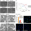
Fig. 5. Pyridinyl imidazole p38 MAPK inhibitors are direct inhibitors of the lipid kinase PIKfyve.
A DU145 cells were treated with DMSO, SB203580 (50 µM), SB202190 (50 µM), or YM201636 (1 µM) for 24 h and then evaluated for vacuolation by phase-contrast microscopy. B HCT116 cells were treated with SB203580 (50 µM) or YM201636 (1 µM) for 24 h and analyzed by transmission electron microscopy (14,000×, bars = 500 nm). C Recombinant PIKfyve was incubated with PI(3)P, phosphatidylserine, and ATP, in the presence of increasing concentrations of SB203580, SB202190, or YM201636, and assayed for ATP hydrolysis using ADP Glo luminescence reagent, as described in the methods. D DU145 cells were treated with SB202190 (50 µM), SB203580 (50 µM), or YM201636 (1 µM) for 24 h and evaluated for PI(3,5)P2 levels by immunofluorescence microscopy, as described in the methods.
Fig. 6
Fig. 6. Loss or inhibition of p38 MAPKs sensitizes cells to vacuolation induced by PIKfyve inhibitors.
A Model to explain how SB203580 and SB202190 induce vacuolation through combined inhibition of p38 MAPKs and PIKfyve. B, C p38 DKO clones were exposed to YM201636 (500 nM) or apilimod (20 nM) for 24 h and evaluated for vacuolation by phase-contrast microscopy using ImageJ analysis software. D, E p38 DKO cells were stably reconstituted with a vector control, or wild-type (WT), constitutively active (CA), or kinase dead (KD) versions of p38α, exposed to apilimod (20 nM), and evaluated for vacuolation by phase-contrast microscopy using ImageJ analysis software. F Model for epistatic relationship between PIKfyve and p38 MAPKs. GI Wild-type DU145 cells were treated with apilimod (50 nM) for 24 h, after which apilimod was removed and BIRB-796 (50 µM) was added to selectively inhibit p38 MAPKs during the “washout” phase. Vacuolation was assessed by phase-contrast microscopy over time (0–12 h) using ImageJ software to determine both the number of vacuoles per cell and percent cell area vacuolated. Inhibition of p38 MAPKs delays the resolution of vacuoles, implicating a role for p38 MAPKs in promoting LEL fission.

Update of

Similar articles

Cited by

References

    1. Cavalli V, Vilbois F, Corti M, Marcote MJ, Tamura K, Karin M, et al. The stress-induced MAP kinase p38 regulates endocytic trafficking via the GDI:Rab5 complex. Mol Cell. 2001;7:421–32. doi: 10.1016/S1097-2765(01)00189-7. - DOI - PubMed
    1. Mace G, Miaczynska M, Zerial M, Nebreda AR. Phosphorylation of EEA1 by p38 MAP kinase regulates mu opioid receptor endocytosis. EMBO J. 2005;24:3235–46. doi: 10.1038/sj.emboj.7600799. - DOI - PMC - PubMed
    1. Vergarajauregui S, San Miguel A, Puertollano R. Activation of p38 mitogen-activated protein kinase promotes epidermal growth factor receptor internalization. Traffic. 2006;7:686–98. doi: 10.1111/j.1600-0854.2006.00420.x. - DOI - PMC - PubMed
    1. Zwang Y, Yarden Y. p38 MAP kinase mediates stress-induced internalization of EGFR: implications for cancer chemotherapy. EMBO J. 2006;25:4195–206. doi: 10.1038/sj.emboj.7601297. - DOI - PMC - PubMed
    1. De Tito S, Hervas JH, van Vliet AR, Tooze SA. The Golgi as an assembly line to the autophagosome. Trends Biochem Sci. 2020;45:484–96. doi: 10.1016/j.tibs.2020.03.010. - DOI - PubMed

Publication types