. 2024 Jan 22;20(1):e1011968.
doi: 10.1371/journal.ppat.1011968. eCollection 2024 Jan.

Epitranscriptional m6A modification of rRNA negatively impacts translation and host colonization in Staphylococcus aureus

Affiliations

Epitranscriptional m6A modification of rRNA negatively impacts translation and host colonization in Staphylococcus aureus

Kathryn E Shields et al. PLoS Pathog. .

Abstract

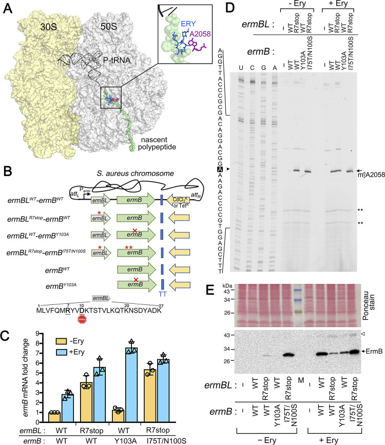
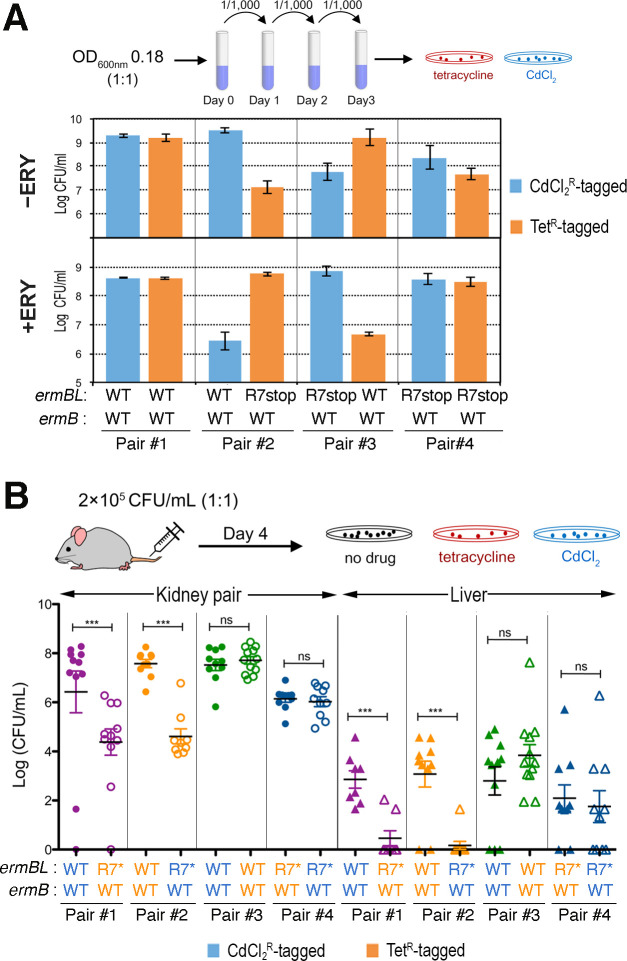
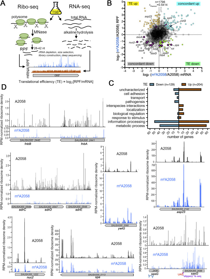
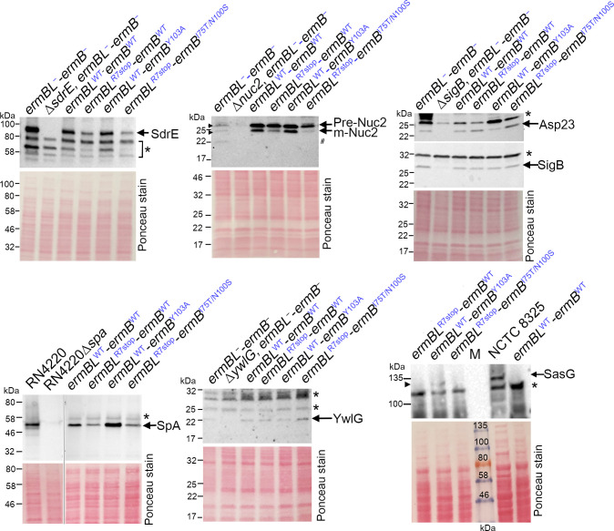
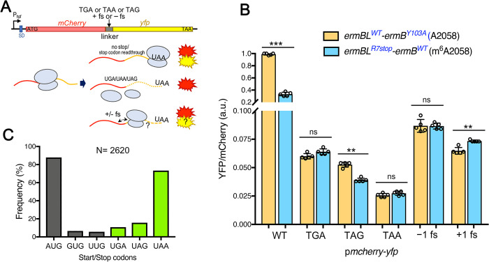
Macrolides, lincosamides, and streptogramin B (MLS) are structurally distinct molecules that are among the safest antibiotics for prophylactic use and for the treatment of bacterial infections. The family of erythromycin resistance methyltransferases (Erm) invariantly install either one or two methyl groups onto the N6,6-adenosine of 2058 nucleotide (m6A2058) of the bacterial 23S rRNA, leading to bacterial cross-resistance to all MLS antibiotics. Despite extensive structural studies on the mechanism of Erm-mediated MLS resistance, how the m6A epitranscriptomic mark affects ribosome function and bacterial physiology is not well understood. Here, we show that Staphylococcus aureus cells harboring m6A2058 ribosomes are outcompeted by cells carrying unmodified ribosomes during infections and are severely impaired in colonization in the absence of an unmodified counterpart. The competitive advantage of m6A2058 ribosomes is manifested only upon antibiotic challenge. Using ribosome profiling (Ribo-Seq) and a dual-fluorescence reporter to measure ribosome occupancy and translational fidelity, we found that specific genes involved in host interactions, metabolism, and information processing are disproportionally deregulated in mRNA translation. This dysregulation is linked to a substantial reduction in translational capacity and fidelity in m6A2058 ribosomes. These findings point to a general "inefficient translation" mechanism of trade-offs associated with multidrug-resistant ribosomes.


Conflict of interest statement

The authors have declared that no competing interests exist.

Figures

Fig 1
Fig 1. Modulation of inducible and constitutive MLS resistance by the upstream regulatory sequence in S. aureus.
(A) A simplified structure of the bacterial 70S ribosome (PDB 3J5L [117]), with a modeled nascent peptide (green) showing the interaction between the A2058 nucleotide (magenta) and erythromycin (ERY, blue) inside the ribosome exit tunnel. P-site tRNA is shown to localize to the peptidyl transferase center (PTC). (B) Reconstruction of a single copy of ermBL-ermB or ermB alone on a neutral chromosomal att site in MLS-sensitive S. aureus USA300 JE2. The integrated ermBL-ermB or ermB is under the control of its native promoter and is marked with either a cadmium chloride resistance (CdCl2R) or a tetracycline resistance (TetR) cassette. Mutant alleles, including the premature ochre codon in position Arg-7 of ermBLR7stop, catalytically inactive ermBY103A, and hyperactive ermBI75T/N100S are indicated by an asterisk or a crossmark. The ErmBL sequence containing the ribosome stalling motif VDK is shown. ERY-bound ribosomes arrest at D10 in the P-site of the PTC (stop sign)[40]. TT, transcriptional terminator. (C) RT-qPCR confirming that ermB expression is inducible by sublethal doses of ERY (1 μg/mL, 60 min) in the context of both translationally functional and defective ermBL. The relative expression of ermB was normalized by ermBLWT-ermBWT using polC as the internal reference gene. Error bars indicate ±SD. (D) Primer extension showing the magnitude of A2058 methylation in the 23S rRNA. The reverse transcriptase halts one nucleotide before the methylated site and produces a truncated cDNA that is manifested by a strong signal at A2058 analyzed on a denaturing polyacrylamide gel. ** serves as an internal reference for equal rRNA template. Each reaction contains 250 ng of rRNA input. (E) Western blots showing the steady-state protein levels of ErmB in the presence and absence of ERY. Samples were collected in parallel with the RNA extraction in Panels C-D. An open arrow marks the non-specific antibody cross-reaction. M, protein marker. A minimum of 3 independent biological replicates were performed.
Fig 2
Fig 2. In vitro and in vivo competition assays of S. aureus strains bearing ERY-inducible ermBLWT-ermBWT and constitutively expressed ermBLR7stop-ermBWT.
Each strain was tagged with either CdCl2 or a tetracycline selective marker. (A) (Top) The ermBLR7stop-ermBWT strains were >100-fold less competitive than the ermBLWT-ermBWT strains in tryptic soy broth without erythromycin (ERY, a macrolide). (Bottom) In the presence of 20 μg/mL ERY, the ermBLR7stop-ermBWT strain outgrows the ermBLWT-ermBWT strain. (B) The ermBL(R7Stop)-ermB strains were severely attenuated in a murine sepsis model (N = 12 mice/group). Bacterial burden was measured in livers and kidneys 4 days post inoculation. Each data point is the mean value ±SEM. Statistical significance was determined by 1-way ANOVA with Tukey’s test (***, p<0.005; ns, not significant). An asterisk in R7 indicates ermBLR7stop and the CdCl2-tag and a Tet-tag are color-coded in blue and orange, respectively.
Fig 3
Fig 3. Effects of m6A2058 modification on the global translatome.
(A) Experimental workflow of Ribo-seq and total mRNA-seq. Translational efficiency (TE) was calculated as the relative number of ribosome footprints to mRNA-seq reads in log2 ratios. A higher TE value represents a greater potential of mRNA for translation. (B) The log2 fold change in Ribo-seq or mRNA levels are plotted for all genes expressed under the tested conditions. The mRNA levels were calculated in reads per kilobase per million mapped reads (RPKM). Representative virulence-associated genes are marked. (C) Comparison of TEs between the m6A2058 strain and unmodified strain reveals the inventory of differentially translated mRNAs. GO analysis showed enrichment in the biological processes of gene expression, metabolic processes, and pathogenesis. (D) Representative ribosome density plots showing changes in ribosome occupancy along the gene body. The normalized reads per million mapped reads (RPM, y-axis) correspond to the average ribosome density across the most abundantly translated ORFs (>50 reads). Unexpected ribosome occupancy is detected in the 3’-end of sasG mRNA, suggesting low levels of mRNA translation downstream of the UAG codon. The potential frameshift-prone “slippery” sequence, the premature stop codon, and the epitope of anti-SasG are indicated.
Fig 4
Fig 4. Comparison of steady-state protein levels between the m6A2058 ribosome-bearing strain and its unmodified counterpart by Western blotting.
Cell lysates were prepared from strains grown under the conditions shown in Fig 3. Ponceau S staining of the membranes prior to immunoblotting served as the loading control. S. aureus NCTC 8325 encodes a full-length SasG and served as a control. The specificity of antibodies was validated by a gene knockout or a deletion mutant of a positive regulator, i.e., SigB. Both precursor (pre-Nuc2) and secreted mature forms (m-Nuc2) of Nuc2 were detected in the total cell lysates. Possible Nuc2 protein degradation (#) is observed in the ermBL-ermBstrain, resulting in weaker pre-Nuc2 and m-Nuc2 signals. Each lane corresponds to 0.1–0.2 A280 units of total proteins. Proteins were resolved on a 4–20% TGX SDS-PAGE gel and probed with the indicated antibodies. An asterisk indicates nonspecific cross-reaction of the antibodies.
Fig 5
Fig 5. Measurement of translational output and accuracy in m6A2058 ribosomes using a dual-fluorescence reporter.
(A) Construction of an S. aureus-specific mCherry-yfp reporter under the control of a constitutive hpf promoter that was adapted from a previously described E. coli system [81]. Nonsense codons and frameshift insertions were introduced between the mCherry and yfp to evaluate stop codon readthrough and ribosomal frameshifting (fs). (B) The m6A2058 ribosome-bearing strain exhibits poorer translational capacity and UAG bypassing but slightly higher +1 fs. Error bars represent ±SD (n = 5). P values were determined using unpaired t tests. ***p< 0.005, **p< 0.01; ns, not significant. (C) Genomic surveillance of start and stop codon usage in S. aureus USA300. AUG and UAA are the most frequently used start and stop codons, respectively.

Similar articles

Cited by

References

    1. Motorin Y, Helm M. RNA nucleotide methylation: 2021 update. Wiley Interdiscip Rev RNA. 2022;13(1):e1691. Epub 20211215. doi: 10.1002/wrna.1691 . - DOI - PubMed
    1. Boulias K, Greer EL. Biological roles of adenine methylation in RNA. Nat Rev Genet. 2023;24(3):143–60. Epub 20221019. doi: 10.1038/s41576-022-00534-0 ; PubMed Central PMCID: PMC9974562. - DOI - PMC - PubMed
    1. Deng X, Chen K, Luo GZ, Weng X, Ji Q, Zhou T, et al.. Widespread occurrence of N6-methyladenosine in bacterial mRNA. Nucleic Acids Res. 2015;43(13):6557–67. Epub 2015/06/13. doi: 10.1093/nar/gkv596 ; PubMed Central PMCID: PMC4513869. - DOI - PMC - PubMed
    1. Ma H, Wang X, Cai J, Dai Q, Natchiar SK, Lv R, et al.. N(6-)Methyladenosine methyltransferase ZCCHC4 mediates ribosomal RNA methylation. Nat Chem Biol. 2019;15(1):88–94. doi: 10.1038/s41589-018-0184-3 . - DOI - PMC - PubMed
    1. Liberman N O ’Brown ZK, Earl AS, Boulias K, Gerashchenko MV, Wang SY, et al.. N6-adenosine methylation of ribosomal RNA affects lipid oxidation and stress resistance. Sci Adv. 2020;6(17):eaaz4370. doi: 10.1126/sciadv.aaz4370 . - DOI - PMC - PubMed

MeSH terms