. 2024 Jan 20;9(1):17.
doi: 10.1038/s41541-024-00806-2.

Three immunizations with Novavax's protein vaccines increase antibody breadth and provide durable protection from SARS-CoV-2

Affiliations

Three immunizations with Novavax's protein vaccines increase antibody breadth and provide durable protection from SARS-CoV-2

Klara Lenart et al. NPJ Vaccines. .

Abstract

The immune responses to Novavax's licensed NVX-CoV2373 nanoparticle Spike protein vaccine against SARS-CoV-2 remain incompletely understood. Here, we show in rhesus macaques that immunization with Matrix-MTM adjuvanted vaccines predominantly elicits immune events in local tissues with little spillover to the periphery. A third dose of an updated vaccine based on the Gamma (P.1) variant 7 months after two immunizations with licensed NVX-CoV2373 resulted in significant enhancement of anti-spike antibody titers and antibody breadth including neutralization of forward drift Omicron variants. The third immunization expanded the Spike-specific memory B cell pool, induced significant somatic hypermutation, and increased serum antibody avidity, indicating considerable affinity maturation. Seven months after immunization, vaccinated animals controlled infection by either WA-1 or P.1 strain, mediated by rapid anamnestic antibody and T cell responses in the lungs. In conclusion, a third immunization with an adjuvanted, low-dose recombinant protein vaccine significantly improved the quality of B cell responses, enhanced antibody breadth, and provided durable protection against SARS-CoV-2 challenge.


Conflict of interest statement

M.G.-X., N.P., G.G., and G.S. are employees of Novavax Inc.

Figures
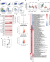
Fig. 1
Fig. 1. Limited systemic innate activation after Matrix-MTM immunization.
A Gating strategy for immunophenotyping of PBMC samples. B Fold change in frequency of different lymphocytes subsets 1 and 14 days after first immunization compared to day 0 (n = 12). C CCR7 expression on different lymphocytes subsets on days 0, 1, and 14 after first immunization (n = 6–12). D Serum cytokine levels on days 0, 1 and 14 after first immunization (n = 6). E Differentially expressed genes (DEGs) in blood, at the site of injection and in the draining lymph node 24 h after first immunization with NVX-CoV2373 (50 μg Matrix-MTM, blood) or Matrix-MTM alone (75 μg, site of injection and draining lymph node) (Blood: n = 12, Muscle and dLN: n = 3-4). F Gene set enrichment analysis of DEGs using blood transcription modules in different tissues. G Detection of soluble Spike protein in circulation at days 0 and 1 after first immunization (n = 6). LLOQ = lower limit of quantification. Data is presented as geometric mean ± geometric SD (B) or mean ± SEM (C). The dotted line represents fold change of 1 (i.e., no change from baseline) (B) or lower limit of quantification (G). Statistical analysis was performed using Wilcoxon test (G).
Fig. 2
Fig. 2. Increased breadth after the heterologous boost immunization.
A Study design and sampling schedule. Animals were immunized with NVX-CoV2373 at weeks 0 and 4 and boosted with NVX-CoV2443 at week 35. WA-1 Spike structure was obtained from PBD ID J771, with mutations in P.1 Spike compared to WA-1 Spike labeled in red. B IgG plasma binding titers to WA-1 Spike and RBD (n = 6). C Serum neutralization of WA-1/2020 SARS-CoV-2 virus (n = 6). Gray shaded area represents the NT50 of WHO international standard NIBSC 20/136 (WHO IS). D Serum neutralization using WA-1 Spike pseudotyped VSV virus particles (n = 6). E Frequency of WA-1 Spike-specific bone marrow plasma cells (BMPC) (n = 6). Representative ELISpot wells are shown on the left. All data is background subtracted based on OVA wells. F IgG binding antibodies in BAL (n = 4–6. Samples with total IgG endpoint titer <1000 were excluded from analysis). PP = pre-pandemic BAL samples. G, H Cross-reactive serum antibodies to WA-1 and P.1 Spike protein (n = 6). IgG binding titers to WA-1 Spike with and without depletion by P.1 Spike (G) and proportion of depleted antibodies (H). Serum neutralization of pseudotyped VSV viruses carrying variant Spike proteins (P.1, Omicron BA.2 and BA.5) at different timepoints (I) and the ratios between neutralization of the variants compared to WA-1 (J) (n = 6). NN = not neutralizing. Data is presented as geometric mean ± geometric SD (BF, I, J) or mean ± SEM (H). Statistical analysis was performed using Wilcoxon test (H) and repeated measures two-way ANOVA with the Geisser-Greenhouse correction and Tukey’s multiple comparisons test (I). Dose 1, dose 2, and dose 3 refer to 2 weeks after each immunization. Dotted line represents lower level of detection (B, DG, I) or the ratio of 1 (J). LLOD lower level of detection, ULOD upper level of detection.
Fig. 3
Fig. 3. Shift in RBD immunodominance with each immunization.
A Gating strategy for identification of Spike- and RBD-specific MBCs. Lineage channel contains CD3, CD11c, CD14, CD16, and CD123. Frequency of Spike- (B) and RBD-specific MBCs (C) (n = 6). D Increase in frequency of Spike- and RBD-specific MBCs 2 weeks after boost 1 and 2 compared to the frequency at the time of boost (n = 6). E The correlation between pre-existing antibody titers and the increase in MBC frequency after boost immunization (n = 6). F Proportion of RBD-binding MBCs out of all S-binding MBCs (n = 6). G Plasmablast responses in blood (n = 6). Representative ELISpot wells are shown on the left. All data is background subtracted based on OVA wells. H Ratio between Spike and RBD-specific plasmablasts after each boost (n = 6). Data is presented as mean ± SEM (B, C, F, G) or geometric mean ± geometric SD (D, H). Statistical analysis was performed using Spearman correlation (E) or Wilcoxon test (H). Boost 1 and boost 2 refer to 2 weeks after boost 1 and 2, respectively. Dotted line corresponds to fold change = 1, i.e., no change (D, H) or the lower limit of detection (E). Arrows indicate immunizations.
Fig. 4
Fig. 4. Expansion of non-RBD-specific B cell responses following boost immunizations.
WA-1 Spike-binding IgG titers in plasma before and after depletion of RBD-binding antibodies (A), and proportion of RBD-binding antibodies at different timepoints (B) (n = 6). C Correlation between proportion of RBD-specific IgG antibodies and RBD-specific MBCs in the blood (n = 6). D Neutralization potency index, defined as the ratio between WA-1 neutralizing and RBD-binding antibody titers 2 weeks after each immunization (n = 6). E Avidity index of RBD-binding IgG antibodies at different timepoints (n = 6), determined by chaotropic wash ELISA with 2 M NaSCN as the chaotropic reagent. F Schematic of RBD with representative antibodies for four defined binding classes (PDB IDs 7K90 (RBD + C144 mAb), 7BZ5 (B38 mAb), 6WPS (S309 mAb), 6W41 (CR3022 mAb)). Receptor binding site (RBS) is labeled in red. G Relative serum reactivity to each of the four defined RBD-binding antibody classes (site I-site IV, n = 6), determined through ELISA competition assay using biotinylated monoclonal antibodies listed in (F). Total RBD-binding IgG and neutralization titers are shown on the right. Dose 2 and 3 represent timepoints 4 weeks after each immunization. H Frequency of Spike- and RBD-specific MBCs 2 weeks after each immunization (n = 6). Binding to different Spike subdomains (RBD, NTD, or S2) at selected timepoints, measured by multiplexed Luminex bead-based assay (n = 6) (I) and the fold changes in the binding titers from timepoint to timepoint (Dose 1 = week 2/week 0, Dose 2 = week 6/week 2, Dose 3 = week 37/week 6) (J). Data is presented as mean ± SEM (B, D, E) or geometric mean ± geometric SD (H, I). Statistical analysis was performed using Kruskal-Wallis test with Dunn’s post hoc correction (B), Spearman correlation (C) or Friedman’s test with Dunn’s post hoc correction (D, E, H, I). Baseline refers to week 0, dose 1, dose 2 and dose 3 refer to 2 weeks after each immunization in all panels except in (G). Dotted line corresponds to lower level of detection (A), fold change = 1 (D, H). or the baseline signal at week 0 (I).
Fig. 5
Fig. 5. Maturation of the B cell response.
A Somatic hypermutation in S-specific MBCs 2 weeks after boost 1 and 2. The number below represents the number of sequences included in the analysis at each timepoint. B Somatic hypermutation in Spike+ RBD+ and Spike+ RBD- MBCs 2 weeks after boost 1 and 2. The number below represents the number of sequences included in the analysis at each timepoint. C Avidity index of Spike-binding IgG plasma antibodies at different timepoints (n = 6), determined by chaotropic wash ELISA with 2 M NaSCN as the chaotropic reagent. D Correlation between average mutational load in circulating Spike-specific memory B cells and polyclonal serum antibody avidity (n = 6). Dashed line in the violin plots represents the median and dotted lines the quartiles (A, B). Data points are presented as mean ± SEM (C). Statistical analysis was performed using Kruskal-Wallis test (A), Kruskal-Wallis test with Dunn’s post hoc correction (B) or Spearman correlation (D).
Fig. 6
Fig. 6. Durable protection induced by Novavax’s COVID-19 vaccine.
A Study design and sampling schedule of the SARS-CoV-2 challenge experiment. Viral loads in BAL assessed by subgenomic (sg)E (B) and sgN gene-targeted RT-qPCR (C) (n = 6-9 per group). D Peak sgE and sgN loads in BAL during the 14 day follow-up after challenge (n = 6–9 per group). E Kaplan-Meier survival analysis, used to evaluate the time required for the animals in different groups to fully supress viral replication (sgN copies/mL < lower level of detection) (n = 6–9 per group). Data is presented as mean (min-max) (B, C) or geometric mean ± geometric SD (D). Statistical analysis was performed using Kruskal-Wallis test with Dunn’s post hoc correction (D) or log-rank Mantel-Cox test with Bonferroni correction. Dotted line corresponds to lower level of detection (BD).
Fig. 7
Fig. 7. Rapid anamnestic responses in the lungs of immunized animals.
Expansion of Spike-specific Th1 memory T cells in blood (A) and BAL (B) after challenge (n = 6–9 per group). WA-1 Spike-binding IgG antibodies in blood (C) and BAL (D) after challenge (n = 6–9 per group). E WA-1 N protein-binding IgG antibodies in blood after challenge (n = 6–9 per group). F Serum neutralization of WA-1/2020 SARS-CoV-2 virus after challenge (n = 6–9 per group). Gray shaded area represents the NT50 of WHO international standard NIBSC 20/136 (WHO IS). Correlation between pre-existing neutralizing serum antibodies and peak sgN viral load in the lungs (BAL) (G) and nasal cavity (nasal swab) (H) (n = 6–9 per group). I Correlation between peak viral load (sgN) in the lungs and the serum antibody response against N protein 4 weeks after challenge (n = 6–9 per group). J Correlation between pre-existing Spike-binding IgG and the serum antibody response against N protein 4 weeks after challenge (n = 6–9 per group). Data is presented as mean (min-max) (AF). Dotted line corresponds to lower (CJ) and upper (F) level of detection. Statistical analysis was performed using Spearman correlation (GJ). LLOD lower level of detection, ULOD upper level of detection.

Similar articles

Cited by

References

    1. Bangaru S, et al. Structural analysis of full-length SARS-CoV-2 spike protein from an advanced vaccine candidate. Science. 2020;370:1089–1094. doi: 10.1126/science.abe1502. - DOI - PMC - PubMed
    1. Heath PT, et al. Safety and efficacy of NVX-CoV2373 Covid-19 vaccine. N. Engl. J. Med. 2021;385:1172–1183. doi: 10.1056/NEJMoa2107659. - DOI - PMC - PubMed
    1. Dunkle LM, et al. Efficacy and safety of NVX-CoV2373 in adults in the United States and Mexico. N. Engl. J. Med. 2021;386:531–543. doi: 10.1056/NEJMoa2116185. - DOI - PMC - PubMed
    1. Baden LR, et al. Efficacy and safety of the mRNA-1273 SARS-CoV-2 vaccine. N. Engl. J. Med. 2020;384:403–416. doi: 10.1056/NEJMoa2035389. - DOI - PMC - PubMed
    1. Polack FP, et al. Safety and efficacy of the BNT162b2 mRNA Covid-19 vaccine. N. Engl. J. Med. 2020;383:2603–2615. doi: 10.1056/NEJMoa2034577. - DOI - PMC - PubMed