. 2024 Apr 5;137(7):846-858.
doi: 10.1097/CM9.0000000000002946. Epub 2024 Jan 19.

GRK2-YAP signaling is implicated in pulmonary arterial hypertension development

Affiliations

GRK2-YAP signaling is implicated in pulmonary arterial hypertension development

Peng Ye et al. Chin Med J (Engl). .

Abstract

Background: Pulmonary arterial hypertension (PAH) is characterized by excessive proliferation of small pulmonary arterial vascular smooth muscle cells (PASMCs), endothelial dysfunction, and extracellular matrix remodeling. G protein-coupled receptor kinase 2 (GRK2) plays an important role in the maintenance of vascular tone and blood flow. However, the role of GRK2 in the pathogenesis of PAH is unknown.

Methods: GRK2 levels were detected in lung tissues from healthy people and PAH patients. C57BL/6 mice, vascular smooth muscle cell-specific Grk2 -knockout mice ( Grk2ΔSM22 ), and littermate controls ( Grk2flox/flox ) were grouped into control and hypoxia mice ( n = 8). Pulmonary hypertension (PH) was induced by exposure to chronic hypoxia (10%) combined with injection of the SU5416 (cHx/SU). The expression levels of GRK2 and Yes-associated protein (YAP) in pulmonary arteries and PASMCs were detected by Western blotting and immunofluorescence staining. The mRNA expression levels of Grk2 and Yes-associated protein ( YAP ) in PASMCs were quantified with real-time polymerase chain reaction (RT-PCR). Wound-healing assay, 3-(4,5)-dimethylthiahiazo (-z-y1)-3,5-di-phenytetrazoliumromide (MTT) assay, and 5-Ethynyl-2'-deoxyuridine (EdU) staining were performed to evaluate the proliferation and migration of PASMCs. Meanwhile, the interaction among proteins was detected by immunoprecipitation assays.

Results: The expression levels of GRK2 were upregulated in the pulmonary arteries of patients with PAH and the lungs of PH mice. Moreover, cHx/SU-induced PH was attenuated in Grk2ΔSM22 mice compared with littermate controls. The amelioration of PH in Grk2ΔSM22 mice was accompanied by reduced pulmonary vascular remodeling. In vitro study further confirmed that GRK2 knock-down significantly altered hypoxia-induced PASMCs proliferation and migration, whereas this effect was severely intensified by overexpression of GRK2 . We also identified that GRK2 promoted YAP expression and nuclear translocation in PASMCs, resulting in excessive PASMCs proliferation and migration. Furthermore, GRK2 is stabilized by inhibiting phosphorylating GRK2 on Tyr86 and subsequently activating ubiquitylation under hypoxic conditions.

Conclusion: Our findings suggest that GRK2 plays a critical role in the pathogenesis of PAH, via regulating YAP expression and nuclear translocation. Therefore, GRK2 serves as a novel therapeutic target for PAH treatment.


Conflict of interest statement

None.

Figures

Figure 1
Figure 1
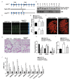
GRK2 is associated with PAH and PAH disease severity. (A) Western blotting analysis of GRK2 expression in pulmonary arterial vessels of healthy donors and patients with different systolic pulmonary arterial pressures (n = 3). (B) GRK2 (red) and α-SMA (green) immunofluorescence staining in small pulmonary arteries from humans with or without PAH. Nuclei are counterstained with DAPI (blue) (n = 8). Scale bar, 50 µm. (C) GRK2 expression in lung tissues of mice with hypoxia (cHx)/SU5416 (Su)-induced PAH compared to that in control mouse lungs (n = 8). (D) GRK2 (red) and α-SMA (green) immunofluorescence staining in small pulmonary arteries from mice with or without cHx/SU-induced PAH (n = 8). Scale bar, 100 µm. (E) The level of GRK2 protein in human PASMCs stimulated with hypoxia at various time points (n = 3). (F) GRK2 expression in sub-cultured PASMCs derived from the lungs of three normal controls (normal) and three patients with PAH (PAH = 78, 89, and 92 mmHg) (n = 3). *P <0.05 and P <0.001 vs. normal, P <0.001 vs. mild-PAH, §P <0.001 vs. Ctl, ||P <0.01 and P <0.001 vs. Ctl, **P <0.01 vs. normal. The data represent the means ± SEM. α-SMA: α-smooth muscle actin; cHx/SU: Hypoxia + SU416; Ctl: Control; DAPI: 4′,6-diamidino-2-phenylindole; GAPDH: Glyceraldehyde 3-phosphate dehydrogenase; GRK2: G protein-coupled receptor kinase 2; PAH: Pulmonary arterial hypertension; PASMCs: Pulmonary arterial smooth muscle cells; SEM: Standard error of the mean; SPAP: Systolic pulmonary artery pressure.
Figure 2
Figure 2
GRK2 down-regulation in PASMCs ameliorates PAH features in vivo. (A) Representative images of RVSP waves in control + NC shRNA (Ctl + NC sh) mice, Ctl + GRK2 shRNA (Ctl + GRK2 sh) mice, cHx/SU + NC shRNA (cHx/SU + NC sh) mice, and cHx/SU+ GRK2 shRNA (cHx/SU + GRK2 sh) mice (n = 8). (B) Assessment of RVSP in each group (n = 8). (C) RVHI calculated by the right ventricle to left ventricle + septum (RV/LV + S) weight ratios in each group (n = 8). (D) Representative images of pulmonary angiograms of cHx/SU + NC mice and cHx/SU + GRK2 shRNA mice (n = 6). (E) H&E-stained sections of small pulmonary arteries from each group (n = 8). Scale bar, 50 µm. (F) None, partial, and full muscularization of pulmonary arteries are shown as percentages (n = 8). (G) Assessment of the pulmonary arterial wall thickness as a percentage of the luminal diameter (n = 8). (H) RT-PCR assessment of the mRNA level of SM22α, SMMHC, MCP-1, MMP2, MMP9, CTGF, Collagen I, Collagen III, and Vimentin in the lungs of each group (n = 8). *P <0.001 vs. Ctl + NC sh. §P <0.05, P <0.01 and P <0.001 vs. cHx/SU + NC sh. The data represent the means ± SEM. AAV9: Adeno-associated virus 9; cHx/SU: Hypoxia/SU5416; Ctl: Control; CTGF: Connective tissue growth factor; GRK2: G protein-coupled receptor kinase 2; H&E: Hematoxylin and eosin; MCP-1: Monocyte chemoattractant protein 1; MMP2: Matrix metallopeptidase 2; MMP9: Matrix metallopeptidase 9; mRNA: Messenger Ribonucleic acid; NC: Null control; PAH: Pulmonary arterial hypertension; PASMCs: Pulmonary arterial smooth muscle cells; RT-PCR: Real-time Polymerase Chain Reaction; RVHI: Right ventricle hypertrophy index; RVSP: Right ventricular systolic pressure; SEM: Standard error of the mean; SM22α: Smooth muscle protein 22-α; SMMHC: Smooth muscle myosin heavy chain.
Figure 3
Figure 3
GRK2 up-regulation in PASMCs exacerbates PAH in vivo. (A) Representative images of RVSP waves in Ctl mice, AAV9-mediated SMC-specific GRK2 over-expression (Ctl + SMC-GRK2) mice, cHx/SU mice, and cHx/SU + SMC-GRK2 mice (n = 8). (B) Assessment of RVSP in each group (n = 8). (C) RVHI calculated in each group (n = 8). (D) Representative pulmonary angiograms of Ctl, cHx/SU, and cHx/SU+SMC-GRK2 groups (n = 5). (E) Representative H&E-stained sections of small pulmonary arteries from the lungs of four groups (n = 8). Scale bar, 50 µm. (F) None, partial, and full muscularization of pulmonary arteries are shown as percentages (n = 8). (G) Pulmonary arterial wall thickness as a percentage of the luminal diameter (n = 8). (H) mRNA level of SM22α, SMMHC, MCP-1, MMP2, MMP9, CTGF, Collagen I, Collagen III, and Vimentin in the lungs of each group (n = 8). *P <0.05, P <0.01 and §P <0.001 vs. Ctl. P <0.05, P <0.01, and ||P <0.001 vs. cHx/SU. The data represent the means ± SEM. AAV9: Adeno-associated virus 9; Ctl: Control; cHx/SU: Hypoxia/SU5416; CTGF: Connective tissue growth factor; GRK2: G protein-coupled receptor kinase 2; H&E: Hematoxylin and eosin; MCP-1: Monocyte chemoattractant protein-1; MMP2: Matrix metallopeptidase 2; MMP9: Matrix metallopeptidase 9; mRNA: Messenger Ribonucleic acid; PAH: Pulmonary arterial hypertension; PASMCs: Pulmonary arterial smooth muscle cells; RVHI: Right ventricle hypertrophy index; RVSP: Right ventricular systolic pressure; SEM: Standard error of the mean; SM22α: Smooth muscle protein 22-α; SMC: Smooth muscle cell; SMMHC: Smooth muscle myosin heavy chain.
Figure 4
Figure 4
Specific SMC-Grk2–/– mice are resistant to PAH development. (A) Schematic of the transgenic mice used to breed SMC-specific Grk2 knockout (Grk2ΔSM22) mice. (B) Genotypic identification of Grk2ΔSM22 mice. (C) Representative images of RVSP waves in Grk2f/f-Ctl mice, Grk2f/f-cHx/SU mice, and Grk2ΔSM22-cHx/SU mice (n = 10). (D) Assessment of RVSP in each group (n = 10). (E) RVHI calculated in each group (n = 10). (F) Representative pulmonary angiograms of Grk2f/f-cHx/SU mice and Grk2ΔSM22-cHx/SU mice. (G) Representative H&E-stained sections of small pulmonary arteries from the lungs of the four groups (n = 6). Scale bar, 50 µm. (H) None, partial, and full muscularization of pulmonary arteries are shown as percentages. (I) Pulmonary arterial wall thickness as a percentage of the luminal diameter (n = 10). (J) mRNA level of SM22α, SMMHC, MCP-1, MMP2, MMP9, CTGF, Collagen I, Collagen III, and Vimentin in lungs of each group (n = 10). *P <0.001 vs.Grk2f/f-Ctl. P <0.05, P <0.01, and §P <0.001 vs.Grk2f/f-cHx/SU. The data represent the mean ± SEM. bp: Base pair; cHx/SU: Hypoxia/SU5416; CRE: Cyclization recombination; Ctl: Control; CTGF: Connective tissue growth factor; Grk2: G protein-coupled receptor kinase 2; H&E: Hematoxylin and eosin; MCP-1: Monocyte chemoattractant protein-1; MMP2: Matrix metallopeptidase 2; MMP9: Matrix metallopeptidase 9; mRNA: Messenger Ribonucleic acid; PAH: Pulmonary arterial hypertension; RVHI: Right ventricle hypertrophy index; RVSP: Right ventricular systolic pressure; SEM: Standard error of the mean; SM22α: Smooth muscle protein 22-α; SMC: Smooth muscle cell; SMMHC: Smooth muscle myosin heavy chain.
Figure 5
Figure 5
GRK2 promotes hypoxia-induced PASMCs proliferation and migration. (A) Wound healing assay in Ctl + si-NC, hypoxia + si-NC, and hypoxia + si-GRK2 groups. (B, C) The proliferative activity of PASMCs assessed by MTT assay and EdU staining. Representative images of three independent experiments are shown. (D–F) Cell migration and proliferation activity assessed by wound healing assay, MTT assay, and EdU staining in PASMCs from patients with PAH transfected with or without si-GRK2 (n = 3). Representative images of three independent experiments are shown. (G–I) Representative images of the wound healing assay, MTT assay, and EdU staining in Ctl + Ad-NC, hypoxia + Ad-NC, and hypoxia + Ad-GRK2 groups (n = 3). Representative images of three independent experiments are shown. *P <0.001 vs. Ctl + si-NC, P <0.01 vs. Hypoxia + si-NC, P <0.001 vs. PAH-PASMC, §P <0.001 vs. Ctl + Ad-NC, ||P <0.001 vs. Hypoxia + Ad-NC. The data represent the mean ± SEM. Ad: Adenovirus; Ctl: Control; DAPI: 4′,6-diamidino-2-phenylindole; EdU: 5-Ethynyl-2′-deoxyuridine; GRK2: G protein-coupled receptor kinase 2; MTT: Dimethyl thiazolyl diphenyl tetrazolium salt; NC: Normal control; PAH: Pulmonary arterial hypertension; PASMC: Pulmonary arterial smooth muscle cell; SEM: Standard error of the mean; si: Small interfering.
Figure 6
Figure 6
The GRK2-YAP signaling pathway is involved in PAH. (A) Western blotting analysis of GRK2, YAP, and p-AKT473 in pulmonary arteries derived from the lungs of human subjects with or without PAH (n = 3). (B) Western blotting analysis of YAP and p-AKT473 in PASMCs transfected with Ad-GRK2 or not and then exposed to hypoxia or normoxia (n = 3). (C) Nuclear protein was extracted and the content of YAP was detected by Western blotting (n = 3). (D) Western blotting analysis of YAP and p-AKT473 in PASMCs transfected with si-GRK2 or not and exposed normoxia or hypoxia (n = 3). (E) Nuclear protein was extracted and the content of YAP was detected by Western blotting. (F) Western blotting analysis of YAP and p-AKT473 in PASMCs transfected with si-YAP or not and then exposed to hypoxia or normoxia (n = 3). (G) mRNA level of YAP in PASMCs transfected with si-GRK2 or si-NC and exposed normoxia or hypoxia (n = 3). (H) Western blotting analysis of YAP protein under hypoxia or normoxia conditions with or without treatment of MG132 (n = 3). *P <0.01 vs. Normal, P <0.05, **P <0.01 and §P <0.001 vs. Ctl, P <0.05, P <0.01 and ||P <0.001 vs. Hypoxia, ††P <0.05 vs. Hypoxia + MG132. The data represent the mean ± SEM. Ad: Adenovirus; GAPDH: Glyceraldehyde 3-phosphate dehydrogenase; GRK2: G protein-coupled receptor kinase 2; MG132: Proteasome inhibitor; mRNA: Messenger Ribonucleic acid; PAH: Pulmonary arterial hypertension; PASMC: Pulmonary arterial smooth muscle cell; p-AKT 473: Phosphorylated AKT 473; PASMCs: Pulmonary arterial smooth muscle cells; p-LATS1: Phospho-large tumor suppressor 1; SEM: Standard error of the mean; si: Small interfering; YAP: Yes-associated protein.
Figure 7
Figure 7
Hypoxia inhibits ubiquitination degradation of GRK2 in PASMCs. (A) mRNA level of GRK2 in PASMC treated with hypoxia for the indicated time points (n = 3). (B) Western blotting analysis of GRK2 in PASMCs treated with cycloheximide at different time points and exposed to normoxia or hypoxia (n = 3). (C) PASMCs were transfected with WT-GRK2 and HA-Ub plasmids and then exposed to normoxia or hypoxia pretreated with MG132. The proteins were immunoprecipitated with the antibody GRK2 and analyzed by Western blotting (n = 3). (D) Western blotting analysis of p-GRK2 at different sites in PASMCs stimulated with hypoxia at various time points (n = 3). *P <0.05 and P <0.01 vs. Ctl. The data represent the mean ± SEM. Ctl: Control; GAPDH: Glyceraldehyde 3-phosphate dehydrogenase; GRK2: G protein-coupled receptor kinase 2; HA-Ub: HA-tagged ubiquitin; IP: Immunoprecipitation; mRNA: Messenger Ribonucleic acid; NS: no statistical significance; PASMCs: Pulmonary arterial smooth muscle cells; p-GRK2: Phospho-G protein-coupled receptor kinase 2; SEM: Standard error of the mean; WT: Wild type.

Similar articles

References

    1. Galie N Humbert M Vachiery JL Gibbs S Lang I Torbicki A, et al. . 2015 ESC/ERS Guidelines for the diagnosis and treatment of pulmonary hypertension: The Joint Task Force for the Diagnosis and Treatment of Pulmonary Hypertension of the European Society of Cardiology (ESC) and the European Respiratory Society (ERS): Endorsed by: Association for European Paediatric and Congenital Cardiology (AEPC), International Society for Heart and Lung Transplantation (ISHLT). Eur Heart J 2016;37:67–119. doi: 10.1093/eurheartj/ehv317. - PubMed
    1. Lau EMT, Giannoulatou E, Celermajer DS, Humbert M. Epidemiology and treatment of pulmonary arterial hypertension. Nat Rev Cardiol 2017;14:603–614. doi: 10.1038/nrcardio.2017.84. - PubMed
    1. Sitbon O Sattler C Bertoletti L Savale L Cottin V Jais X, et al. . Initial dual oral combination therapy in pulmonary arterial hypertension. Eur Respir J 2016;47:1727–1736. doi: 10.1183/13993003.02043-2015. - PubMed
    1. Lan NSH, Massam BD, Kulkarni SS, Lang CC. Pulmonary arterial hypertension: Pathophysiology and treatment. Diseases 2018;6:38. doi: 10.3390/diseases6020038. - PMC - PubMed
    1. Hussain MB, Marshall I. Characterization of alpha1-adrenoceptor subtypes mediating contractions to phenylephrine in rat thoracic aorta, mesenteric artery and pulmonary artery. Br J Pharmacol 1997;122:849–858. doi: 10.1038/sj.bjp.0701461. - PMC - PubMed