. 2024 Mar 7;84(5):839-853.e12.
doi: 10.1016/j.molcel.2023.12.036. Epub 2024 Jan 18.

Mechanisms of RNF168 nucleosome recognition and ubiquitylation

Affiliations

Mechanisms of RNF168 nucleosome recognition and ubiquitylation

Qi Hu et al. Mol Cell. .

Abstract

RNF168 plays a central role in the DNA damage response (DDR) by ubiquitylating histone H2A at K13 and K15. These modifications direct BRCA1-BARD1 and 53BP1 foci formation in chromatin, essential for cell-cycle-dependent DNA double-strand break (DSB) repair pathway selection. The mechanism by which RNF168 catalyzes the targeted accumulation of H2A ubiquitin conjugates to form repair foci around DSBs remains unclear. Here, using cryoelectron microscopy (cryo-EM), nuclear magnetic resonance (NMR) spectroscopy, and functional assays, we provide a molecular description of the reaction cycle and dynamics of RNF168 as it modifies the nucleosome and recognizes its ubiquitylation products. We demonstrate an interaction of a canonical ubiquitin-binding domain within full-length RNF168, which not only engages ubiquitin but also the nucleosome surface, clarifying how such site-specific ubiquitin recognition propels a signal amplification loop. Beyond offering mechanistic insights into a key DDR protein, our study aids in understanding site specificity in both generating and interpreting chromatin ubiquitylation.

Keywords: 53BP1; BRCA1-BARD1; DNA damage response; NMR spectroscopy; RNF168; UbcH5c; X-ray crystallography; chromatin; cryo-EM; nucleosome; ubiquitin ligase.


Conflict of interest statement

Declaration of interests The authors declare no competing interests.

Figures

Figure 1.
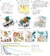
Figure 1.. Interactions of RNF168 with the ubiquitylated NCP
(A) Top: Domain structure of RNF168. Bottom: Schematics of nucleosomal H2A K13 and K15 ubiquitylation catalyzed by RNF168, illustrating signal amplification by recognition of H2AK13ub or H2AK15ub. (B and C) Fluorescence polarization binding curves of fluorescently-labeled NCP or NCP carrying H2AK13ub and H2AK15ub (NCPub) with full-length RNF168 (RNF168FL) and RNF168 MIU2-LRM (aa 430–481) (RNF168ML) (B), and with WT RNF168ML and mutants (C). Data are mean ± s.d. for each data point (n = 3 technical replicates). Kd values are indicated. ND means not determined. (D) Cryo-EM reconstruction (left and middle) and structural model (right) of the RNF168FL-NCPub complex displayed in two orientations. While the complex was assembled with RNF168FL, the only density detected was for RNF168 MIU2-LRM. (E) Close-up view of polar interactions (yellow dashes) between RNF168 LRM R477 and indicated acidic patch residues in H2A, and hydrophobic interactions between LRM Y474 and indicated residues in H2A and H2B. (F) Close-up view of RNF168 MIU2-LRM interactions with the NCP and ubiquitin linked to H2A K15, highlighting canonical interaction of ubiquitin with MIU2 and proximity of MIU2 to the NCP surface. See also Figures S1 and S2.
Figure 2.
Figure 2.. Structure and activity of RNF168R-UbcH5c with the NCP
(A) Fluorescence polarization binding curves of RNF168R, WT and mutants, with fluorescently-labeled NCP. Data are mean ± s.d. for each data point (n = 3 technical replicates). Kd values are indicated. ND means not determined. (B) Left: Quantification of the NCP or H2A-H2B (WT or mutants) ubiquitylation from n = 3 independent experiments. Bar graphs show the mean and s.d. for each data point. P values were calculated using a two-sample two-tailed Student t-test; *P < 0.05, **P < 0.01, ***P < 0.001, NS means not significant. Right: Representative SDS-PAGE gels of ubiquitylation assays of the NCP, WT H2A-H2B, H2AK13S-H2B and H2AK15S-H2B using UBA1, UbcH5c and RNF168R. (C) Cryo-EM reconstruction (left and middle) and structural model (right) of RNF168R-UbcH5c-NCP complex displayed in two orientations. Locations of H2A K13 and K15 (yellow spheres) and active site C85 of UbcH5c (red sphere) are circled in red. (D–G) Close-up views of the interactions between RNF168R and H2A-H2B (D and E), RNF168R and UbcH5c (F), and active site of UbcH5c and H2A-H2B (G). Relevant side chains, hydrogen bonds (yellow dashed lines) and Zn+2 (purple spheres) are highlighted. (H) Ubiquitylation reaction profiles of H2AK13S-H2B and H2AE61A/E64A/D91A-H2BE114A catalyzed by WT RNF168R and mutants. Reactions were probed by monitoring the NMR signal intensity changes of unreacted 15N-labeled ubiquitin. See also Figures S3–S6.
Figure 3.
Figure 3.. Cryo-EM characterization of RNF168R-UbcH5c~Ub-NCP and RNF168R-UbcH5c-UbB-NCP
(A) Cryo-EM reconstruction (left and middle) and structural model (right) of RNF168R-UbcH5c~Ub-NCP complex displayed in two orientations. Locations of H2A K13 and C15 (yellow spheres) and active site C85 of UbcH5c (red sphere) are circled in red. (B) Self-ubiquitylation of UbcH5c at indicated lysine residues detected by mass spectrometry. An abundance ratio for each site was determined by comparing the number of fragments containing the specific lysine with ubiquitylation to the total number of fragments containing the same lysine, both with and without ubiquitylation. (C) Proposed mechanism for UbcH5c self-ubiquitylation at K144 based on the crystal structure of UbcH5b~Ub self-assembly mediated by ubiquitin backside interaction (PDB 3A33). K144 in red is proximal to the UbcH5c active site. (D) Cryo-EM reconstruction (left and middle) and structural model (right) of RNF168R-UbcH5c-UbB-NCP complex displayed in two orientations. Zn+2 ions are purple spheres. See also Figures S6–S10.
Figure 4.
Figure 4.. Cryo-EM structures of post-reaction states where the NCP has been ubiquitylated by RNF168R-UbcH5c
(A) Cryo-EM reconstruction (left and middle) and structure (right) of post-reaction state 1 (class 1) of the NCPub-RNF168R-UbcH5c complex. Density is visible for RNF168R, but barely noticeable for UbcH5c. (B) Cryo-EM reconstruction (left and middle) and structure (right) of post-reaction state 2 (class 2) of the NCPub-RNF168R-UbcH5c complex. UbcH5c density matches that in the pre-reaction state in Figure 2C. See also Figure S11.
Figure 5.
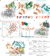
Figure 5.. Conformational dynamics of RNF168R-UbcH5c
(A) Overlay of three RNF168R-UbcH5c structures (from crystal structures of RNF168R-UbcH5c-H2A-H2B and the cryo-EM structure of RNF168R-UbcH5c-NCP), aligned with respect to RNF168R. UbcH5c samples multiple conformations, pivoting by as much as ~55° from the N-terminus of UbcH5c α-helix 1. (B) Top: Representative crystal structure (conformation 1 or open state) of RNF168R-UbcH5c and cryo-EM structure (conformation 2 or closed state) of RNF168R-UbcH5c-NCP both from A showing only the RNF168R-UbcH5c moiety. Selected residues are labeled and approximate distances between RNF168R and UbcH5c in both conformations are indicated. Bottom: RNF168R-UbcH5c NMR signals that are affected by the RNF168R and UbcH5c interaction (see spectral overlay in C (left)) define interfaces consistent with both X-ray and cryo-EM structures. (C) Left: Overlaid 1H-15N HSQC spectra of 15N-labeled RNF168R-UbcH5c, RNF168R and UbcH5c. RNF168R-UbcH5c signals that differ from corresponding signals in isolated RNF168R and UbcH5c are highlighted by arrows or boxed (and enlarged in B (bottom)). Right: Plot of the chemical shift differences between the spectra of RNF168R-UbcH5c and RNF168R (top) and between the spectra of RNF168R-UbcH5c and UbcH5c (bottom). One and two standard deviations (1σ and 2σ) above the mean chemical shift are indicated. Inset: Surface representation of RNF168R-UbcH5c. RNF168R-UbcH5c residues with ≥ 0.1 ppm chemical shift deviation from those in isolated RNF168R and UbcH5c are colored red. (D) Summary of a 1-μs molecular dynamics (MD) simulations of RNF168R-UbcH5c bound to H2A-H2B using the cryo-EM structure of RNF168R-UbcH5c-NCP as starting model. Fifty simulated RNF168R-UbcH5c models (gray) are overlaid relative to their E3 components. For comparison, RNF168R-UbcH5c conformations 1 (open state from a crystal structure) and 2 (closed state in the cryo-EM structure) are also included. (E) Determination of the ubiquitylation zone of RNF168R-UbcH5c using the MD simulations in D. Five-hundred atomic coordinates sampled from the 1-μs MD simulation were used to evaluate changes in atomic positions and predict which residues could be ubiquitylated if they were lysines. Ellipsoids generated using the “measure inertia” function of Chimera were used to approximate the spatial distributions of atoms from selected residues in the MD simulation. The ellipsoids shown are for UbcH5c active site C85 SG (blue), H2A K13 and K15 NZ (yellow), H2A non-lysine residues A12 to S19 CA (orange), and H2B K120 NZ (red). (F) H2A-H2B ubiquitylation by RNF168R, UbcH5c and UBA1 at indicated H2A lysine residues monitored using SDS-PAGE. See also Figure S12.
Figure 6.
Figure 6.. NCPub recognition and ubiquitylation by RNF168 and BRCA1-BARD1
(A) Cryo-EM structures of RNF168R-UbcH5c-NCP and BRCA1R-BARD1R-UbcH5c-NCP. The NCPs are oriented similarly to highlight the opposite positioning of UbcH5c by ~180° in the two complexes. (B and C) Close-up views of the interactions between BRCA1R and H2A-H2B acidic patch (B), and between BARD1R and H2B C-terminal α-helix (C). (D) Cryo-EM reconstructions (top left and right) and structural models (bottom left and right) illustrating ubiquitin recognition in NCPub by RNF168 and BARD1. The side chains of ubiquitin I44 and K63 are shown to highlight the radically different ubiquitin recognition modes. The side chain of D729 in BARD1 forms a polar interaction with K63 (yellow dashed line).
Figure 7.
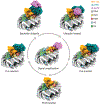
Figure 7.. Reaction cycle of RNF168
Reaction cycle of RNF168. Various modes of interactions of RNF168, RNF168R-UbcH5c, RNF168R-UbcH5c~Ub or RNF168R-UbcH5c-UbB with the NCP or NCPub are interpreted as different steps or states of the reaction cycle, leading to H2AK13ub and H2AK15ub signal amplification in chromatin.

Similar articles

Cited by

References

    1. Stewart GS, Panier S, Townsend K, Al-Hakim AK, Kolas NK, Miller ES, Nakada S, Ylanko J, Olivarius S, Mendez M, et al. (2009). The RIDDLE syndrome protein mediates a ubiquitin-dependent signaling cascade at sites of DNA damage. Cell 136, 420–434. 10.1016/j.cell.2008.12.042 - DOI - PubMed
    1. Doil C, Mailand N, Bekker-Jensen S, Menard P, Larsen DH, Pepperkok R, Ellenberg J, Panier S, Durocher D, Bartek J, et al. (2009). RNF168 binds and amplifies ubiquitin conjugates on damaged chromosomes to allow accumulation of repair proteins. Cell 136, 435–446. 10.1016/j.cell.2008.12.041 - DOI - PubMed
    1. Pinato S, Scandiuzzi C, Arnaudo N, Citterio E, Gaudino G, and Penengo L (2009). RNF168, a new RING finger, MIU-containing protein that modifies chromatin by ubiquitination of histones H2A and H2AX. BMC Mol Biol 10, 55. 10.1186/1471-2199-10-55 - DOI - PMC - PubMed
    1. Mattiroli F, Vissers JH, van Dijk WJ, Ikpa P, Citterio E, Vermeulen W, Marteijn JA, and Sixma TK (2012). RNF168 ubiquitinates K13–15 on H2A/H2AX to drive DNA damage signaling. Cell 150, 1182–1195. 10.1016/j.cell.2012.08.005 - DOI - PubMed
    1. Gatti M, Pinato S, Maspero E, Soffientini P, Polo S, and Penengo L (2012). A novel ubiquitin mark at the N-terminal tail of histone H2As targeted by RNF168 ubiquitin ligase. Cell Cycle 11, 2538–2544. 10.4161/cc.20919 - DOI - PMC - PubMed