. 2024 Jan 23;121(4):e2313677121.
doi: 10.1073/pnas.2313677121. Epub 2024 Jan 19.

Structure of saguaro cactus virus 3' translational enhancer mimics 5' cap for eIF4E binding

Affiliations

Structure of saguaro cactus virus 3' translational enhancer mimics 5' cap for eIF4E binding

Manju Ojha et al. Proc Natl Acad Sci U S A. .

Abstract

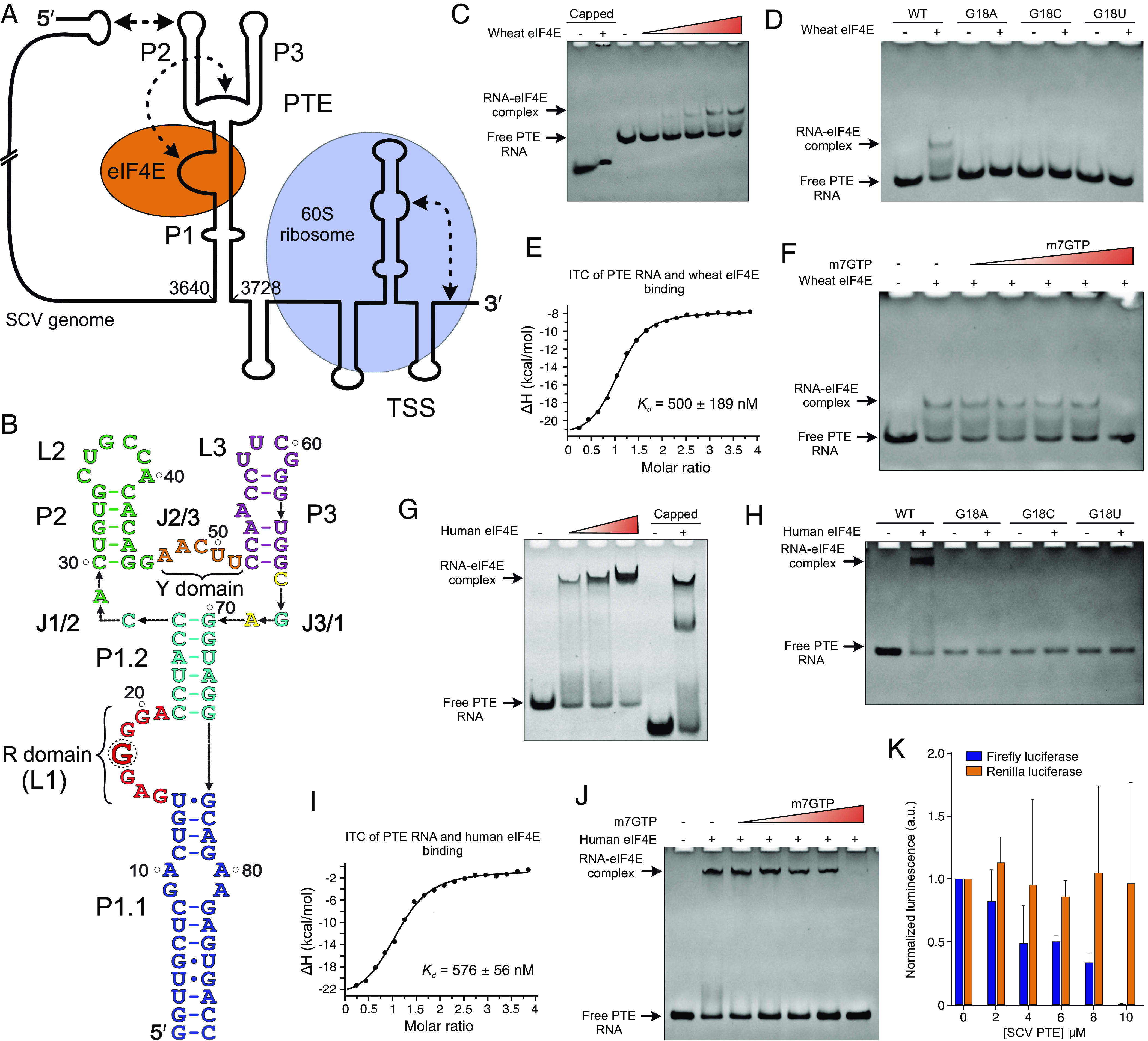
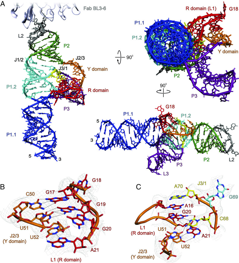
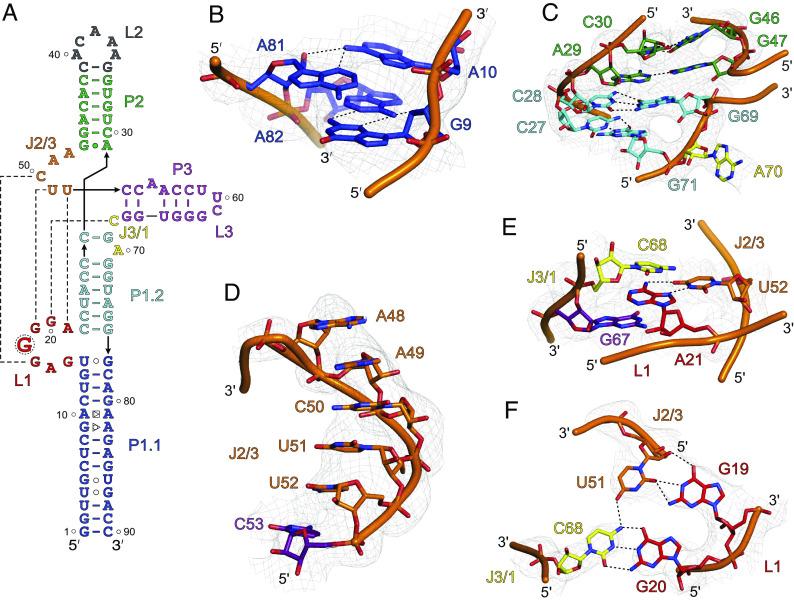
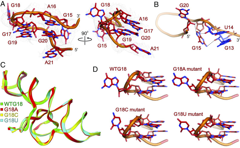
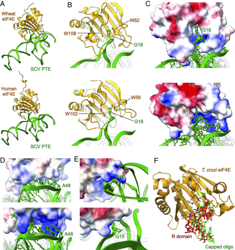
The genomes of several plant viruses contain RNA structures at their 3' ends called cap-independent translation enhancers (CITEs) that bind the host protein factors such as mRNA 5' cap-binding protein eIF4E for promoting cap-independent genome translation. However, the structural basis of such 5' cap-binding protein recognition by the uncapped RNA remains largely unknown. Here, we have determined the crystal structure of a 3' CITE, panicum mosaic virus-like translation enhancer (PTE) from the saguaro cactus virus (SCV), using a Fab crystallization chaperone. The PTE RNA folds into a three-way junction architecture with a pseudoknot between the purine-rich R domain and pyrimidine-rich Y domain, which organizes the overall structure to protrude out a specific guanine nucleotide, G18, from the R domain that comprises a major interaction site for the eIF4E binding. The superimposable crystal structures of the wild-type, G18A, G18C, and G18U mutants suggest that the PTE scaffold is preorganized with the flipped-out G18 ready to dock into the eIF4E 5' cap-binding pocket. The binding studies with wheat and human eIF4Es using gel electrophoresis and isothermal titration calorimetry, and molecular docking computation for the PTE-eIF4E complex demonstrated that the PTE structure essentially mimics the mRNA 5' cap for eIF4E binding. Such 5' cap mimicry by the uncapped and structured viral RNA highlights how viruses can exploit RNA structures to mimic the host protein-binding partners and bypass the canonical mechanisms for their genome translation, providing opportunities for a better understanding of virus-host interactions and non-canonical translation mechanisms found in many pathogenic RNA viruses.

Keywords: 3′ cap-independent translation enhancers; RNA crystallography; RNA–eIF4E interactions; crystallization chaperones; viral RNAs.


Conflict of interest statement

Competing interests statement:The authors declare no competing interest.

Figures

Fig. 1.
Fig. 1.
SCV genome organization and the PTE–eIF4E interactions. (A) Schematic representation of the SCV (+)-sense RNA genome with the predicted RNA structures at the 5′ and 3′ ends. Proposed binding sites for the 60S ribosome (TSS) and eIF4E (PTE) are indicated. The dotted arrows depict long-range RNA–RNA interactions. (B) The 89-nt SCV PTE construct (wild-type) was used in this study. Key features of the PTE elements are labeled, and a dotted circle indicates the SHAPE-hypermodifiable G18 within the R domain. The native polyacrylamide gel electrophoresis (nPAGE) assays showing the binding of the (C) wild-type and (D) G18 mutant PTEs with wheat eIF4E. (E) The ITC measurements for the PTE binding with wheat eIF4E. (F) The nPAGE of wheat eIF4E-PTE interactions in the presence of the m7GTP. Similar experiments were performed with human eIF4E. The nPAGE showing the binding of the (G) wild-type and (H) G18 mutant PTEs with human eIF4E. (I) The ITC measurements for the PTE binding with human eIF4E and (J) the nPAGE of the eIF4E-PTE interactions in the presence of m7GTP. (K) The expression levels (average ± SD, n = 6) of firefly (cap-dependent) and renilla (HCV IRES-dependent) luciferases in the presence of the PTE. The ability of the SCV PTE RNA to bind wheat and human eIF4Es effectively, the obvious displacement of the PTE RNA from the eIF4E-RNA complex by the m7GTP, and inhibition of the eIF4E-requiring cap-dependent translation in vitro strongly suggest similar binding interactions of the PTE and mRNA 5′ cap.
Fig. 2.
Fig. 2.
The crystal structure of SCV PTE. (A) The overall structure of the SCV PTE RNA in complex with a crystallization chaperone Fab BL3-6 solved at 3.13 Å resolution. For clarity, Fab is obscured in the rotated views of the PTE RNA (see SI Appendix, Fig. S12 for the complete structure of the Fab-RNA complex). (B) Specific interactions between the R (L1 nucleotides) and Y domain (J2/3 nucleotides). (C) The J3/1 nucleotides interact with the R and Y domains through hydrogen bonding and base-stacking. Gray mesh in (B) and (C) represents the composite simulated anneal-omit 2|Fo|-|Fc| electron density map at contour level 1σ and carve radius 2.5 Å. The dotted black lines depict the heteroatoms within the hydrogen bonding distances (≤3.0 Å). Figure panels and labels are colored analogously for facile comparison.
Fig. 3.
Fig. 3.
The unique features of SCV PTE crystal structure. (A) The crystal-derived secondary structure of the PTE RNA. The dotted lines represent tertiary interactions between the nucleotides (B) The non-canonical G9•A82 (sugar-edge-Hoogsteen) and A10•A81 (Hoogsteen-sugar-edge) pairs within the P1.1 helix. (C) The structure and helical stacking interactions within the J1/2. (D) Helical base-stacking of the J2/3 nucleotides that interact with L1 nucleotides to form a pseudoknot structure. (E) Additional interactions between the J2/3. The U52 base pairs with the A21, which sandwiches between the cross-strand stacking of the G67 and C68. (F) The J3/1 C68 nucleotide forms a canonical base pair with G20 and contacts U51 through a hydrogen bond that creates a planer tetrad structure involving G19 and G20, U51, and C68. Gray mesh represents the composite simulated anneal-omit 2|Fo|-|Fc| electron density map at contour level 1σ and carve radius 2.5 Å, and dotted black lines depict the heteroatoms within hydrogen bonding distances (≤3.0 Å).
Fig. 4.
Fig. 4.
The structural features of the R domain and G18 mutant SCV PTE. (A) The overall structure of the SCV PTE’s R domain showing the flipped-out G18 (major binding site for the 5′ cap-binding protein, eIF4E). (B) Specific interactions of the G13, U14, G15, and G20 nucleotides within the R domain. (C) Superposition of the G18A (red), G18C (yellow), and G18U (cyan) mutant PTE crystal structures with the wild-type PTE. (D) Comparisons of the R domain structures for wild-type and G18 mutants showing that SCV PTE adopts a preorganized structure with flipped-out G18 that constitutes a ready-made platform for the eIF4E binding. Gray mesh represents the composite simulated anneal-omit 2|Fo|-|Fc| electron density map at contour level 1σ and carve radius 2.5 Å, and dotted black lines depict the heteroatoms within hydrogen bonding distances (≤3.0 Å).
Fig. 5.
Fig. 5.
Molecular docking studies of SCV PTE with eIF4E. (A) The overall model with wheat eIF4E (Top) and human eIF4E (Bottom). (B) The docking models for both wheat (Top) and human (Bottom) eIF4E showed that the R domain G18 occupies the same binding pocket as the 5′ cap and emulates similar interactions with the protein. (C) The electrostatic model of eIF4E shows that the PTE R domain fits neatly within the binding pocket of wheat (Top) and human (Bottom) eIF4E. The human eIF4E binding pocket (Bottom) appears more open compared to that of wheat, with three additional positively charged lysine residues around the binding pocket. (D) The Y domain of the SCV PTE also fits in a groove formed by the eIF4E loops β1-β2, α2-β3, and β5-β6. Compared to the wheat eIF4E (Top), the homologous channel of the human eIF4E (Bottom) is more open and more positively charged. (E) The α6-β8 loop near the wheat eIF4E C-terminal interacts with the G15 R domain (Top). However, five residues of the loop are omitted as indeterminate in the published structure of human eIF4E (Bottom, PDB: 1IPC) that might contribute to this binding interaction. (F) The alignment of the PTE G18 with the 5′ cap (m7GTP) of an oligonucleotide (green) within the cocrystal structure with T. cruzi eIF4E (PDB: 6O7Y) resulted in PTE’s R domain (red) exiting the binding pocket in the same orientation as the oligonucleotide. The key nucleotides and residues involved in the PTE–eIF4E interactions are labeled.

Similar articles

Cited by

References

    1. Richter J. D., Sonenberg N., Regulation of cap-dependent translation by eIF4E inhibitory proteins. Nature 433, 477–480 (2005). - PubMed
    1. Jia Y., Polunovsky V., Bitterman P. B., Wagner C. R., Cap-dependent translation initiation factor eIF4E: An emerging anticancer drug target. Med. Res. Rev. 32, 786–814 (2012). - PMC - PubMed
    1. Aitken C. E., Lorsch J. R., A mechanistic overview of translation initiation in eukaryotes. Nat. Struct. Mol. Biol. 19, 568–576 (2012). - PubMed
    1. Browning K. S., Bailey-Serres J., Mechanism of cytoplasmic mRNA translation. Arabidopsis Book 13, e0176 (2015). - PMC - PubMed
    1. Kneller E. L., Rakotondrafara A. M., Miller W. A., Cap-independent translation of plant viral RNAs. Virus Res. 119, 63–75 (2006). - PMC - PubMed

LinkOut - more resources