Review
. 2024 Jan;14(1):67-86.
doi: 10.1016/j.apsb.2023.07.020. Epub 2023 Jul 21.

Allosteric modulation of G protein-coupled receptors as a novel therapeutic strategy in neuropathic pain

Affiliations
Review

Allosteric modulation of G protein-coupled receptors as a novel therapeutic strategy in neuropathic pain

Chunhao Zhu et al. Acta Pharm Sin B. 2024 Jan.

Abstract

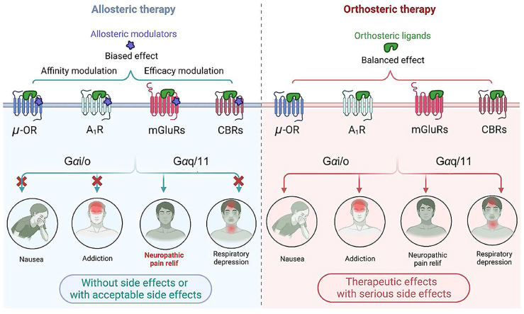
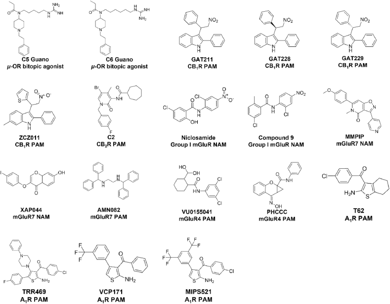
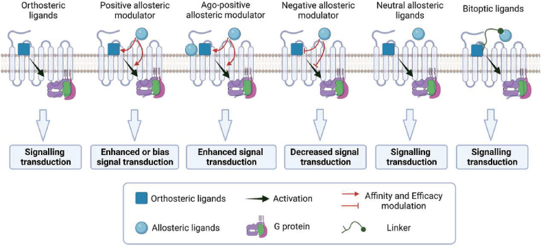
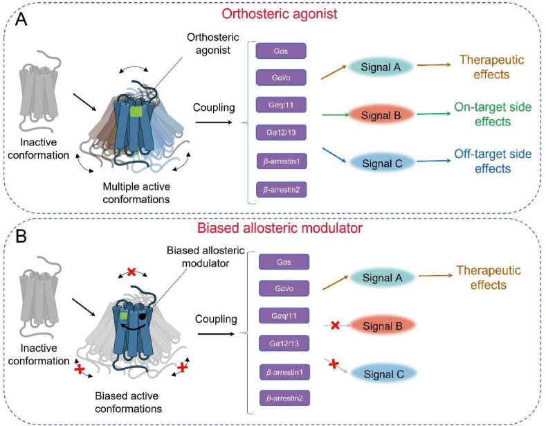
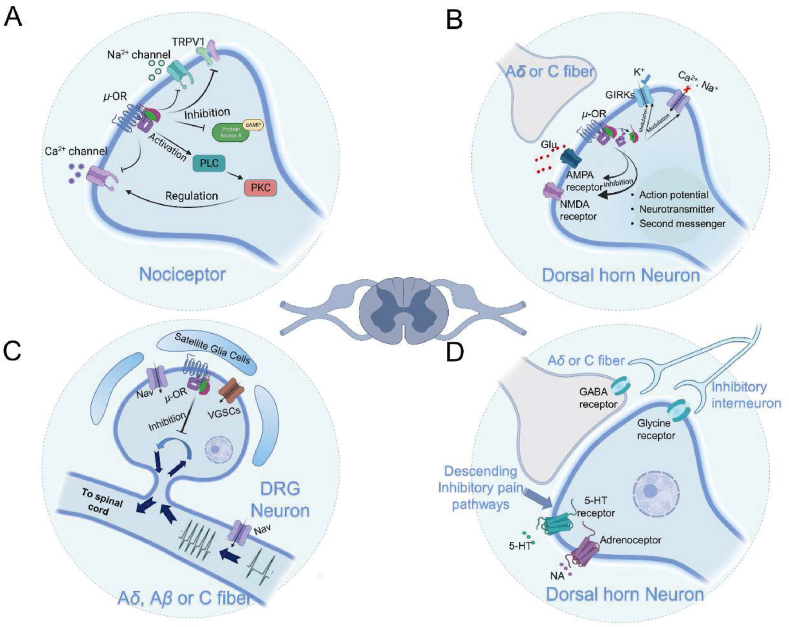
Neuropathic pain is a debilitating pathological condition that presents significant therapeutic challenges in clinical practice. Unfortunately, current pharmacological treatments for neuropathic pain lack clinical efficacy and often lead to harmful adverse reactions. As G protein-coupled receptors (GPCRs) are widely distributed throughout the body, including the pain transmission pathway and descending inhibition pathway, the development of novel neuropathic pain treatments based on GPCRs allosteric modulation theory is gaining momentum. Extensive research has shown that allosteric modulators targeting GPCRs on the pain pathway can effectively alleviate symptoms of neuropathic pain while reducing or eliminating adverse effects. This review aims to provide a comprehensive summary of the progress made in GPCRs allosteric modulators in the treatment of neuropathic pain, and discuss the potential benefits and adverse factors of this treatment. We will also concentrate on the development of biased agonists of GPCRs, and based on important examples of biased agonist development in recent years, we will describe universal strategies for designing structure-based biased agonists. It is foreseeable that, with the continuous improvement of GPCRs allosteric modulation and biased agonist theory, effective GPCRs allosteric drugs will eventually be available for the treatment of neuropathic pain with acceptable safety.


Conflict of interest statement

The authors declare no conflicts of interest.

Figures

Image 1
Graphical abstract
Figure 1
Figure 1
Chemical structures of allosteric ligands of GPCRs discussed in the main text.
Figure 2
Figure 2
Modulation of GPCRs by orthosteric and biased allosteric modulators. (A) The binding of orthosteric ligands to GPCRs stabilizes the receptor conformation in multiple active states that activate different downstream effector proteins, including heterotrimers of Gαβγ (e.g., Gαs, Gαi/o, Gαq/11, Gα12/13) and β-arrestin. These effector proteins induce downstream signaling pathways (e.g., cAMP accumulation, Ca2+ localization, and activation of Erk) that regulate various physiological activities of the cell. (B) In contrast, biased conformation modulators stabilize only one active conformation of the GPCR, producing a more limited physiological effect than orthosteric ligands. While most biased conformation modulators require an orthosteric ligand to regulate the active conformation of the receptor, there are also biased conformation modulators that induce receptor activation alone.
Figure 3
Figure 3
Five types of allosteric modulators modulate the function of GPCRs. Both PAMs and NAMs can modulate the affinity of the orthosteric binding pocket for an orthosteric ligand or affect the intrinsic efficacy of an orthosteric agonist to engage downstream signalling mechanisms. Ago-PAMs possess the properties of both orthosteric and allosteric ligands. NALs bind to allosteric sites and have no influence on orthosteric ligand responses; nonetheless, they compete with PAMs and NAMs for allosteric site binding and inhibit their effects. Bitopic ligands are a class of compounds that exhibit high selectivity and affinity for specific target receptors. They are formed by connecting orthosteric and allosteric ligands via specific linker molecules. The use of such linkers results in a distinct and selective binding mode of the ligands to the receptor.
Figure 4
Figure 4
Mechanisms of analgesic action of μ-opioid receptor in the peripheral nervous system (PNS) and central nervous system (CNS). (A) In nociceptive receptors, the activation of μ-opioid receptor (μ-OR) reduces the release of nociceptive substances and decreases Ca2+ production following nerve injury by interacting with TRPV1, H1R, and NK1R. (B) In dorsal root ganglion neurons, μ-OR inhibits the transmission of nociceptive information to the central nervous system by blocking Nav and VGSCs. (C) In spinal dorsal horn neurons, μ-OR induces cell membrane hyperpolarization by inhibiting Nav and VGSCs-mediated Ca2+ influx and activating GIRKs-mediated K+ influx. Moreover, μ-OR modulates ionotropic glutamate receptors, resulting in central analgesic effects. (D) The downstream pain inhibitory pathway activated by μ-OR inhibits the upstream transmission of nociceptive information by modulating 5-HT and norepinephrine receptors, as well as glycine receptors, on spinal dorsal horn neurons.
Figure 5
Figure 5
Mechanisms of μ-opioid receptor-mediated addiction and analgesic tolerance. (A) The neural circuit mechanisms underlying opioid-induced addiction mainly involve the inhibition of GABAergic neurons within the ventral tegmental area (VTA) through sustained activation of the μ-opioid receptor (μ-OR) in the rostromedial tegmental nucleus (RMTg), as well as the induction of the medial prefrontal cortex (mPFC) and sustained dopamine release in the nucleus accumbens (NAc). (B) The activation process of μ-OR primarily involves the conversion of GDP to GTP on Gα proteins, leading to the dissociation of activated Gα proteins from Gβγ and the conversion of intracellular ATP to cAMP, and the internalization process of μ-OR occurs through the recruitment of β-arrestin under the action of GRK phosphorylation, resulting in the depression of the plasma membrane at the receptor site to form an endosome. A portion of the receptor located on the endosome is subsequently re-inserted into the plasma membrane.
Figure 6
Figure 6
Synaptic and immune suppressive actions mediated by cannabinoid receptors in neurons and glial cells. The activation of CB1R reduces Ca2+ release from presynaptic neurons by inhibiting VGSCs at the presynaptic membrane, resulting in decreased Ca2+ influx in postsynaptic neurons. CB1R activation localized on astrocytes induces Ca2+ release by the Gαq pathway. The activation of CB2R localized on microglia reduces cytokine release, indicating its role in immune suppression.
Figure 7
Figure 7
The bitopic ligand, C5 guano and C6 guano, occupies both the orthosteric binding site and the conserved Na + binding site of the μ-opioid receptor (PDB ID: 7U2L, 7U2K). (A) Schematic diagram depicting the C5 guano‒μ-OR-Gαβγ protein complex. (B) and (C) depict the binding modes of the bitopic ligand C5 guano and μ-OR, respectively. (D) Schematic diagram depicting the C6 guano‒μ-OR-Gαβγ protein complex. (E) and (F) depict the binding modes of the bitopic ligand C6 guano and μ-OR, respectively. Hydrogen bond interactions are shown in blue dashed lines, while cation‒π interactions are depicted in black dashed lines.
Figure 8
Figure 8
Cryo-EM structure of the human A1R-Gαi2 protein complex bound to adenosine (Endogenous agonist) and MIPS521 (PAM) (PDB ID: 7LD3). The black squares showed the orthosteric binding site of adenosine in the central of TMD (left). Adenosine is shown as blue sticks. Residues interacting with adenosine are shown as purple sticks. The black squares showed the allosteric binding site of MIPS521 in the intersection of the TMD and lipid interface (right).

Cited by

References

    1. IASP-Pain.org [homepage on the Internet]. Washington: International Association for th-e Study of Pain; [updated 2021; cited 2022 June 9]. Available from: http://www.iasp-pain.org/Education/Content.aspx? ItemNumber=1698&navItem....
    1. Attal N., Lanteri-Minet M., Laurent B., Fermanian J., Bouhassira D. The specific disease burden of neuropathic pain: results of a French nationwide survey. Pain. 2011;152:2836–2843. - PubMed
    1. Doth A.H., Hansson P.T., Jensen M.P., Taylor R.S. The burden of neuropathic pain: a systematic review and meta-analysis of health utilities. Pain. 2010;149:338–344. - PubMed
    1. van Hecke O., Austin S.K., Khan R.A., Smith B.H., Torrance N. Neuropathic pain in the general population: a systematic review of epidemiological studies. Pain. 2014;155:654–662. - PubMed
    1. Zhu C., Liu N., Tian M., Ma L., Yang J., Lan X., et al. Effects of alkaloids on peripheral neuropathic pain: a review. Chin Med. 2020;15:106. - PMC - PubMed