. 2024 Apr 12;52(6):3346-3357.
doi: 10.1093/nar/gkae005.

NAC and Zuotin/Hsp70 chaperone systems coexist at the ribosome tunnel exit in vivo

Affiliations

NAC and Zuotin/Hsp70 chaperone systems coexist at the ribosome tunnel exit in vivo

Thomas Ziegelhoffer et al. Nucleic Acids Res. .

Abstract

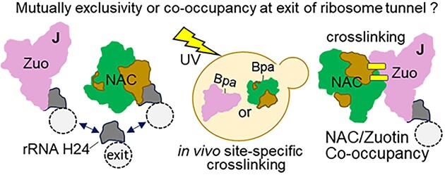
The area surrounding the tunnel exit of the 60S ribosomal subunit is a hub for proteins involved in maturation and folding of emerging nascent polypeptide chains. How different factors vie for positioning at the tunnel exit in the complex cellular environment is not well understood. We used in vivo site-specific cross-linking to approach this question, focusing on two abundant factors-the nascent chain-associated complex (NAC) and the Hsp70 chaperone system that includes the J-domain protein co-chaperone Zuotin. We found that NAC and Zuotin can cross-link to each other at the ribosome, even when translation initiation is inhibited. Positions yielding NAC-Zuotin cross-links indicate that when both are present the central globular domain of NAC is modestly shifted from the mutually exclusive position observed in cryogenic electron microscopy analysis. Cross-linking results also suggest that, even in NAC's presence, Hsp70 can situate in a manner conducive for productive nascent chain interaction-with the peptide binding site at the tunnel exit and the J-domain of Zuotin appropriately positioned to drive stabilization of nascent chain binding. Overall, our results are consistent with the idea that, in vivo, the NAC and Hsp70 systems can productively position on the ribosome simultaneously.


Figures

Graphical Abstract
Graphical Abstract
Figure 1.
Figure 1.
Hsp70 Ssb1 cross-links to NAC in vivo. (A and C) Cells expressing Ssb1 variants with Bpa incorporated at indicated positions were exposed to UV light (+) or left unexposed (−). Cross-linking was analyzed by immunoblotting after SDS–PAGE using antibodies specific for Ssb, NACα, NACβ or uL29, as indicated (anti). Ssb1Bpa cross-link products to NAC, Ssz1 and uL29 are indicated with arrowheads, and non-cross-linked proteins and migration of molecular weight markers (kDa) by dashes. WT, wild-type gene. (A) The two Ssb1 Bpa variants subjected to mass spectrometric analysis (see Supplementary Table S3) were compared using immunoblot analysis. Δ, deletion of EGD1 gene encoding NACβ. 7.5-12% gradient gel was used. (B) Schematic of the ATP-dependent Hsp70 peptide binding cycle. Nucleotide binding domain (NBD); substrate binding domain (SBD). ATP hydrolysis results in dissociation of both SBD subdomains from the NBD, allowing the αSBD lid subdomain to close over the transiently bound substrate. Dotted circle indicates segment of αSBD shown at bottom—AlphaFold model (AF-P11484-F1) of Ssb1(ATP). Positions having Bpa incorporated in A and B shown as spheres with the cross-link partner in parentheses. (C) Immunoblot analysis of Bpa variants in WT ZUO1 and zuo1H128Q (H>Q) backgrounds.
Figure 2.
Figure 2.
Zuo1Bpa cross-links to NAC in vivo. (A) Zuo1 and its association with the ribosome. (Top left) Line diagram of Zuo1 domains, highlighting the more N-terminal regions that are positioned at the 60S subunit near the tunnel exit. J-domain (JD); Zuotin Homology Domain (ZHD), which associates directly with the 60S. Residue position above and interaction partner below diagram. ZHD structure (residues 166–285) shown as cartoon with the three helices indicated; two arginines required for H24 binding shown in stick representation. (Top right) Residues 75–285 of AlphaFold modeled structure of Zuo1 (AF-P32527-F1) in surface representation; colors as in the left panel, with critical HPD motif of JD needed for stimulation of Hsp70 ATPase activity and arginine residues (R247 and R251) known to be critical for ribosome association via interaction with H24 in stick representation. (Bottom) Schematic of the ribosome without (left) or with (right) Zuo1 bound. Select ribosomal proteins near tunnel exit (dotted circle) shown in dark gray; position of the tip of rRNA H24 indicated. Zuo1 bound to ribosome color coded as in line diagram, with ZHD positioned at H24 and eL31. (B and C) Cells expressing Zuo1 variants with Bpa incorporated at indicated positions were exposed to UV light (+) or left unexposed (−). Cross-linking was analyzed by immunoblotting after SDS–PAGE using antibodies specific for (anti) Zuo1, NACα or NACβ, as indicated. Zuo1Bpa–NAC cross-link products are indicated with arrowheads, and non-cross-linked proteins and migration of molecular weight markers (kDa) by dashes. (B) Identification of Zuo1Bpa variants that cross-link to NAC. Absence (Δ) or presence (WT) of gene encoding NACβ. (C) Inhibition of translation initiation: no treatment (−); auxin analog NAA to reduce eIF3B levels (eIF down arrow); glucose starvation (GS). (D) Cartoon representation of Zuo1 residues 75–285. Positions that cross-link to NAC when Bpa is incorporated are represented as spheres. HPD of JD and arginine residues known to be critical for ribosome association via interaction with H24 shown in stick representation.
Figure 3.
Figure 3.
NAC globular domain cross-links to Zuo1 in vivo. (A) Model of NAC heterodimer (DOI: 10.5452/ma-bak-cepc-0495). Bracket designates the globular domain into which Bpa was incorporated, with the beginning and end of domain indicated by arrows and residue for each subunit indicated; expanded view of globular domain with β-strands designated (S1, S2, etc.) below. (B and C) Cells expressing NACα and NACβ variants with Bpa incorporated at indicated positions were exposed to UV light (+) or left unexposed (−). Cross-linking was analyzed by immunoblotting after SDS–PAGE using antibodies specific for (anti) NACα, NACβ or Zuo1. NACBpa–Zuo1 cross-link products indicated with arrowheads, and non-cross-linked proteins and migration of molecular weight markers (kDa) by dashes; (B) absence (Δ) or presence (WT) of gene encoding Zuo1; (C) NAC globular domain with positions of Bpa incorporation giving cross-links to Zuo1 represented as spheres. (D) Inhibition of translation initiation: no treatment (−); NAA to reduce eIF3b (eIF down arrow); glucose starvation (GS). (E) Positioning of NAC and Zuo1. Based on the Zuo1 Bpa positions that cross-linked to both NACα and NACβ (Figure 2) and NACα and NACβ Bpa positions that cross-linked to Zuo1 as in panel (D), as well as the established interaction of NACβ N-terminus with the ribosome; NAC was positioned relative to Zuo1 and then refined using PyRosetta (40). Positive Bpa positions shown as spheres. Only Zuo1 positions that cross-link to both NACα and NACβ are shown. (F and G) Comparison of models of NAC positioning in the presence and absence of Zuo1 with the N-terminus of NACβ included. For clarity, only the ZHD of Zuo1 is shown. (F) Expanded view of positioning in panel (E), rotated 90 degrees. (G) Position of NAC at the ribosome in the absence of Zuo1. Mammalian cryo-EM structure of RNC–NAC complex (10) (PDBID: 7QWR) was superimposed on the ribosome–Zuo1 model to illustrate incompatibility of the coexistence of Zuo1 and NAC in this orientation. Upper right: box indicates zoomed in segment showing region of clash between Zuo1 and NAC. Transparency of Zuo1 increased for clarity with NAC residues in conflict indicated in black.
Figure 4.
Figure 4.
Both αSBD and βSBD of Ssb1 cross-link to NAC subunits. (A) Model of Ssb1(ATP) at the ribosome based on its interaction with the J-domain of Zuo1. Ssb1 in cartoon representation. Positions in αSBD that cross-link to NAC as in Figure 1 shown as spheres. Ribosome and Zuo1 (residues 75–285) shown in surface representation. Ribosome: ribosomal proteins discussed in manuscript in are labeled, while rRNA and other ribosomal proteins are light and dark gray, respectively. The Ssb1–Zuo1 complex was predicted using ColabFold (v1.5.2) implementation (43) of AlphaFold-Multimer-v3 (44) and positioned on the ribosome by fitting the Zuo1 and ribosome structure (PDBID: 6TNU) to cryo-EM map of RNC complex in complex with Zuo1 and Ssz1 (EMDB: EMD-32977). (B and D) Cells expressing Ssb1 variants with Bpa incorporated at indicated positions were exposed to UV light (+) or left unexposed (−). Cross-linking was analyzed by immunoblotting after SDS–PAGE using antibodies specific for (anti) Ssb, NACα or NACβ. Ssb1Bpa–NAC cross-link products are indicated with arrowheads, and non-cross-linked proteins and migration of molecular weight markers (kDa) by dashes. (B) Δ, deletion of gene encoding NACβ; H>Q, H128Q substitution in Zuo1 J-domain. (C) Left: Ssb1 SBD with positions in βSBD that cross-link to NAC indicated by spheres; right: further enlarged βSBD with positions labeled. (D) Ssb1N467Bpa cross-linking after inhibition of translation initiation: no treatment (−); NAA treatment to reduce eIF3b (eIF down arrow); glucose starvation (GS).
Figure 5.
Figure 5.
Juxtaposition of NAC and Zuo1/Ssb at the ribosome. (A and C) Cells expressing NACα, NACβ or Ssb1 variants with Bpa incorporated at indicated positions were exposed to UV light (+) or left unexposed (−). Cross-linking was analyzed by immunoblotting after SDS–PAGE using antibodies specific for Ssb, NACα or NACβ, as indicated (anti). Cross-link products are indicated with arrowheads, and migration of molecular weight markers (kDa) by dashes. (A) Bpa incorporated into NACα or NACβ. Δ, SSB1 SSB2 deletion; H>Q, H128Q substitution in Zuo1 J-domain; WT, wild-type SSB1, SSB2 and ZUO1. Asterisk (*) indicates cross-link of NACβBpa to an unidentified protein that increases when Ssb is absent. (B) NAC globular domain with NACα or NACβ positions forming cross-links represented as spheres: to Zuo1 (as shown in Figure 3); to Ssb (as shown in A and Supplementary Figure S6). (C) (Top) Side by side comparison of migration of NACαBpa and Ssb1Bpa cross-link products. Two prestained markers were used as discussed in the ‘Materials and methods’ section. (Bottom) NAC globular domain as in panel (B), with putative identification of NACα cross-links to Hsp70 αSBD and βSBD subdomains deduced from migration of cross-link products indicated by brackets. (D) Model of co-occupancy of Zuo1/NAC/Ssb on the 60S ribosomal subunit. (i) Joint occupancy of Zuo1 and NAC. Though capable of binding independently, Zuo1/Ssz1 (RAC) and NAC can occupy the ribosome at the same time. Note: Only globular domain of NAC is depicted. Brackets indicate the flexibility of the N-terminal segment of Zuo1, which binds tightly to the atypical Hsp70 Ssz1, tethering it to the ribosome. (ii) Positioning of Ssb(ATP). Ssb(ATP) interacts with Ssz1 and with Zuo1’s J-domain. Note: Loops of SBDβ near tunnel exit are in close proximity to NAC. (iii) After binding of nascent chain in the peptide binding cleft and hydrolysis of ATP stimulated by J-domain, the SBD of Ssb(ADP) binds directly to the ribosome interacting with uL24, uL29 and rRNA. Note: This positioning does not preclude binding of Zuo1 and/or NAC.

Similar articles

Cited by

References

    1. Balchin D., Hayer-Hartl M., Hartl F.U.. In vivo aspects of protein folding and quality control. Science. 2016; 353:aac4354. - PubMed
    1. Kramer G., Shiber A., Bukau B.. Mechanisms of cotranslational maturation of newly synthesized proteins. Annu. Rev. Biochem. 2019; 88:337–364. - PubMed
    1. Deuerling E., Gamerdinger M., Kreft S.G.. Chaperone interactions at the ribosome. Cold Spring Harb. Perspect. Biol. 2019; 11:a033977. - PMC - PubMed
    1. Zhang Y., Sinning I., Rospert S.. Two chaperones locked in an embrace: structure and function of the ribosome-associated complex RAC. Nat. Struct. Mol. Biol. 2017; 24:611–619. - PubMed
    1. Wiedmann B., Sakai H., Davis T.A., Wiedmann M.. A protein complex required for signal-sequence-specific sorting and translocation. Nature. 1994; 370:434–440. - PubMed