. 2024 Jan;48(1):40-51.
doi: 10.1016/j.jgr.2023.06.008. Epub 2023 Jul 4.

Ginsenoside 20(S)-Rg3 reduces KIF20A expression and promotes CDC25A proteasomal degradation in epithelial ovarian cancer

Affiliations

Ginsenoside 20(S)-Rg3 reduces KIF20A expression and promotes CDC25A proteasomal degradation in epithelial ovarian cancer

Rong Zhang et al. J Ginseng Res. 2024 Jan.

Abstract

Background: Ginsenoside 20(S)-Rg3 shows promising tumor-suppressive effects in ovarian cancer via inhibiting NF-κB signaling. This study aimed to explore the downstream tumor suppressive mechanisms of ginsenoside Rg3 via this signaling pathway.

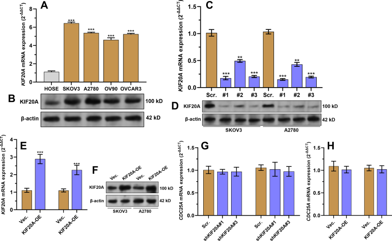
Materials and methods: A systematical screening was applied to examine the expression profile of 41 kinesin family member genes in ovarian cancer. The regulatory effect of ginsenoside Rg3 on KIF20A expression was studied. In addition, we explored interacting proteins of KIF20A and their molecular regulations in ovarian cancer. RNA-seq data from The Cancer Genome Atlas (TCGA) was used for bioinformatic analysis. Epithelial ovarian cancer cell lines SKOV3 and A2780 were used as in vitro and in vivo cell models. Commercial human ovarian cancer tissue arrays were used for immunohistochemistry staining.

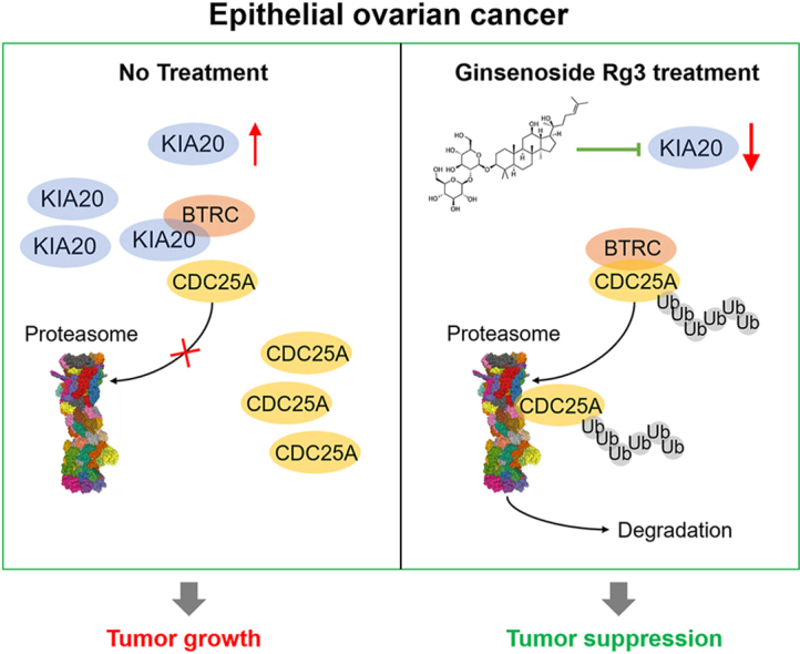
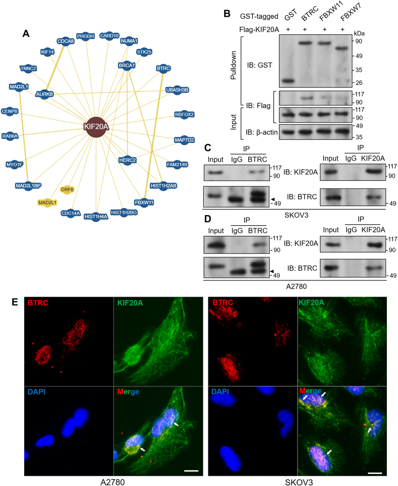
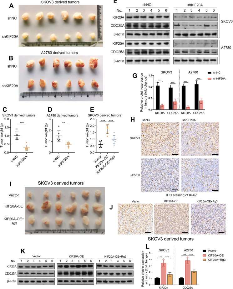
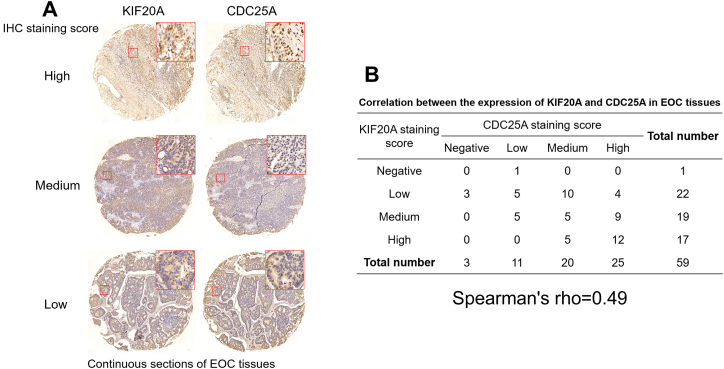
Results: KIF20A is a biomarker of poor prognosis among the kinesin genes. It promotes ovarian cancer cell growth in vitro and in vivo. Ginsenoside Rg3 can suppress the transcription of KIF20A. GST pull-down and co-immunoprecipitation (IP) assays confirmed that KIF20A physically interacts with BTRC (β-TrCP1), a substrate recognition subunit for SCFβ-TrCP E3 ubiquitin ligase. In vitro ubiquitination and cycloheximide (CHX) chase assays showed that via interacting with BTRC, KIF20A reduces BTRC-mediated CDC25A poly-ubiquitination and enhances its stability. Ginsenoside Rg3 treatment partly abrogates KIF20A overexpression-induced CDC25A upregulation.

Conclusion: This study revealed a novel anti-tumor mechanism of ginsenoside Rg3. It can inhibit KIF20A transcription and promote CDC25A proteasomal degradation in epithelial ovarian cancer.

Keywords: CDC25A; KIF20A; ginsenoside Rg3; ovarian cancer; ubiquitination.


Conflict of interest statement

The authors declare that they have no known competing financial interests or personal relationships that could have appeared to influence the work reported in this paper.

Figures

Image 1
Graphical abstract
Fig. 1
Fig. 1
Systematic screening identifies a link between KIF20A upregulation and unfavorable prognosis of ovarian cancer, A-B. K-M plotter was generated to compare the difference in PFS (C) and OS (D) between patients with high and low KIF20A expression with survival data from the Kaplan-Meier plotter. C. Representative images of KIF20A expression in commercial ovarian cancer tissue arrays. Enlarged tissue areas were provided to show the subcellular distribution of KIF20A.
Fig. 2
Fig. 2
Knockdown of KIF20A impairs ovarian cancer cell proliferation, A-D. CCK-8 assays showing the effects of KIF20A knockdown (A-B) or overexpression (C-D) on the viability of SKOV3 (A and C) and A2780 (B and D) cells. E-I. Colony formation (E-G) and flow cytometric analysis (H–I) were performed to show the effect of KIF20A knockdown or overexpression on colony formation and cell-cycle distribution of SKOV3 and A2780 cells. Data are expressed as mean ± SD. ∗, comparison between scramble (scr.) control and shKIF20A. # comparison between vector (vec.) and KIF20A-OE. ## and∗∗p < 0.01, ### and ∗∗∗p < 0.001.
Fig. 3
Fig. 3
Ginsenoside Rg3 suppresses KIF20A expression in ovarian cancer cells, A. A plot chart showing the expressional correlation between KIF20A and RELA in primary tumor cases in TCGA-OV. B–C. QRT-PCR (B) and western blotting assay (C) were performed to detect the expression of KIF20A in SKOV3 and A2780 cells 48 h after ginsenoside Rg3 treatment. D-H. QRT-PCR (D-E) and western blotting assay (F–H) were performed to detect the expression of RELA and KIF20A in SKOV3 and A2780 cells with RELA overexpression alone or in combination with ginsenoside Rg3 treatment (50 ng/ml for 48 h). ∗∗∗p < 0.001.
Fig. 4
Fig. 4
KIF20A directly interacts with BTRC, A. Bioinformatic prediction of KIF20A interacting proteins. Prediction was performed using BioGRID. Only the proteins with minimum evidence ≥2 were identified as the candidates. B. Binding affinity of flag-KIF20A to the predicted E3 ligases. After various GST-fused proteins were expressed, a GST pull-down assay was performed. C-D. Co-IP was performed to show the interaction between KIF20A and BTRC at the endogenous level in SKOV3 (C) and A2780 (D) cells. IP was conducted with an anti-BTRC or anti-KIF20A antibody. Mouse IgG served as a negative control. Arrowheads: IgG heavy chain bands. E. Immunofluorescent staining was performed to visualize BTRC (red) and KIF20A (green) in SKOV3 and A2780 cells. Scale bar: 10 μm.
Fig. 5
Fig. 5
Ginsenoside Rg3 promotes CDC25A proteasomal degradation via suppressing KIF20A expression, A. SKOV3 cells were subjected to lentiviral mediated KIF20A knockdown for 48 h, with or without the presence of MG132 (10 μM for 2 h before harvest). B–C. Western blot of in vitro ubiquitination assay. SKOV3 cells were infected with indicating selective combination of pLenti-puro-HA-Ubiquitin, myc-BTRC and/or flag-tagged KIF20A (B) or were infected with indicating selective combination of pLenti-puro-HA-Ubiquitin and shBTRC or shKIF20A (C). 48 h later, cells were lysed and the cell lysates were subjected to IP with an anti-CDC25A antibody. D-G. SKOV3 cells were infected for KIF20A overexpression (D-E) or knockdown (F-G). Then, the cells were incubated in the presence of 20 μM CHX for the indicated times (n = 3). Proteins were then analyzed by western blotting with the indicated antibodies. Relative levels of CDC25A (E and G) were determined from western blots using the ImageJ program based on the respective blots. H–I. Representative images (top panels) and quantitation (bottom panels) of western blotting analysis of KIF20A and CDC25A expression in SKOV3 and A2780 cells were subjected to ginsenoside Rg3 treatment for 48 h, with (I) or without (H) the presence of MG132 (10 μM for 2 h before harvest). J. SKOV3 and A2780 cells were subjected to lentiviral-mediated KIF20A knockdown. 48 h later, cells in indicated groups were treated with ginsenoside Rg3 for another 48 h. Then, cell samples were collected for western blotting analysis of KIF20A and CDC25A expression. Data are expressed as mean ± SD. Compared with the control group: ∗p < 0.05, ∗∗p < 0.01, ∗∗∗p < 0.001.
Fig. 6
Fig. 6
The in vivo regulatory effect of KIF20A and its correlation with CDC25A in human epithelial ovarian cancer tissues, A-D. Xenograft tumors (A-D) and their weight (C-D), derived from SKOV3 (A) and A2780 (B) cells with or without KIF20A knockdown (n = 6). F-G. Representative images (F) and quantitation (G) of KIF20A and CDC25A proteins in tumor tissues in panel A, detected by western blotting. β-actin was used as a loading control. H. Representative images of immunohistochemical staining for Ki-67 in xenograft tumor tissues in panel A. I and E. Xenograft tumors (I) and their weight (E), derived from SKOV3 cells with KIF20A overexpression alone or in combination with ginsenoside Rg3 treatment (n = 6). J. Representative images of immunohistochemical staining for Ki-67 in xenograft tumor tissues in panel I. K-L. Representative images (K) and quantitation (L) of KIF20A and CDC25A proteins in tumor tissues in panel I, detected by western blotting. β-actin was used as a loading control. Data are expressed as mean ± SD. Compared with the control group: ∗∗p < 0.01, ∗∗∗p < 0.001. Panels H and J, scale bar: 100 μm.
Fig. 7
Fig. 7
The correlation between KIF20A and CDC25A expression in human epithelial ovarian cancer tissues, A. Representative IHC staining images of high, medium, and low KIF20A (left panel) and CDC25A (right panel) expression in two continuous ovarian cancer tissue microarrays (A). B. Correlation between KIF20A and CDC25A staining scores in 59 ovarian cancer tissues. Spearman's correlation analysis was conducted.
Figure S1
Figure S1
Systematic screening identifies KIF20A upregulation in ovarian cancer, A. A heatmap was generated to show the expression profile of 41 KIF family genes in ovarian cancer tissue (N = 419) in TCGA-OV and normal ovary and fallopian tubes (N = 93) in GTEx. B. A heatmap was generated to show the expression profile of 14 significantly dysregulated KIF family genes by using the following criteria: SEOC vs. normal (log2 fold change ≥2) and adj. p < 0.05.
Figure S2
Figure S2
KIF20A expression in ovarian cancer tissues in the HPA, IHC staining images (top panels) and quantitation (bottom panels) of KIF20A expression in 12 ovarian cancer cases in the HPA. Enlarged tissue areas were provided in the right panel. Images were obtained from: v21, proteinatlas.org, https://www.proteinatlas.org/ENSG00000112984-KIF20A/pathology/ovarian+cancer#ihc.
Figure S3
Figure S3
Relevant qRT-PCR and western blotting data, A-B. QRT-PCR (A) and western blotting assay (B) were performed to detect the expression of KIF20A in HOSE cells and multiple ovarian cancer cell lines (SKOV3, A2780, OV90 and OVCAR3). C–F. QRT-PCR (C and E) and western blotting assay (D and F) were performed to detect the expression of KIF20A in SKOV3 and A2780 cells 48 h after lentiviral shKIF20A or flag-KIF20A (KIF20A-OE) infection. G-H. QRT-PCR was performed to detect the expression of CDC25A in SKOV3 and A2780 cells 48 h after lentiviral shKIF20A (G) or flag-KIF20A (KIF20A-OE) (H) infection. ∗∗p < 0.01, ∗∗∗p < 0.001.

Similar articles

Cited by

References

    1. Shen L., Xia M., Zhang Y., Luo H., Dong D., Sun L. Mitochondrial integration and ovarian cancer chemotherapy resistance. Exp Cell Res. 2021;401(2) doi: 10.1016/j.yexcr.2021.112549. Epub 2021/03/01. PubMed PMID: 33640393. - DOI - PubMed
    1. Coughlan A.Y., Testa G. Exploiting epigenetic dependencies in ovarian cancer therapy. Int J Cancer. 2021;149(10):1732–1743. doi: 10.1002/ijc.33727. Epub 2021/07/03. PubMed PMID: 34213777. - DOI - PMC - PubMed
    1. Crean-Tate K.K., Braley C., Dey G., Esakov E., Saygin C., Trestan A., et al. Pretreatment with LCK inhibitors chemosensitizes cisplatin-resistant endometrioid ovarian tumors. J Ovarian Res. 2021;14(1):55. doi: 10.1186/s13048-021-00797-x. Epub 2021/04/24. PubMed PMID: 33888137; PubMed Central PMCID: PMCPMC8063392. - DOI - PMC - PubMed
    1. Franzese E., Diana A., Centonze S., Pignata S., De Vita F., Ciardiello F., et al. PARP Inhibitors in First-Line Therapy of Ovarian Cancer: Are There Any Doubts? Front Oncol. 2020;10:782. doi: 10.3389/fonc.2020.00782. Epub 2020/07/01. PubMed PMID: 32596142; PubMed Central PMCID: PMCPMC7303974. - DOI - PMC - PubMed
    1. Bu H., Chen J., Li Q., Hou J., Wei Y., Yang X., et al. BRCA mutation frequency and clinical features of ovarian cancer patients: A report from a Chinese study group. J Obstet Gynaecol Res. 2019;45(11):2267–2274. doi: 10.1111/jog.14090. Epub 2019/08/15. PubMed PMID: 31411802. - DOI - PubMed