. 2024 Feb 9;52(3):1471-1482.
doi: 10.1093/nar/gkad1231.

Structural basis for transcription activation by the nitrate-responsive regulator NarL

Affiliations

Structural basis for transcription activation by the nitrate-responsive regulator NarL

Dmytro Kompaniiets et al. Nucleic Acids Res. .

Abstract

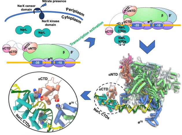
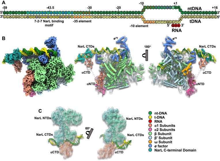
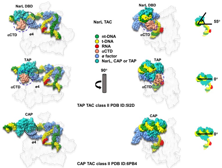
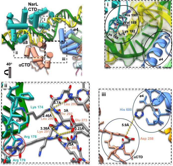
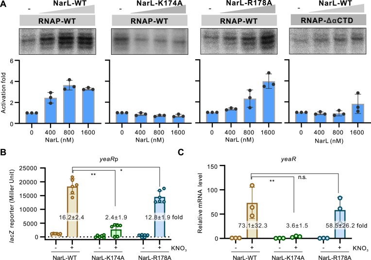
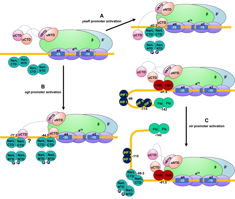
Transcription activation is a crucial step of regulation during transcription initiation and a classic check point in response to different stimuli and stress factors. The Escherichia coli NarL is a nitrate-responsive global transcription factor that controls the expression of nearly 100 genes. However, the molecular mechanism of NarL-mediated transcription activation is not well defined. Here we present a cryo-EM structure of NarL-dependent transcription activation complex (TAC) assembled on the yeaR promoter at 3.2 Å resolution. Our structure shows that the NarL dimer binds at the -43.5 site of the promoter DNA with its C-terminal domain (CTD) not only binding to the DNA but also making interactions with RNA polymerase subunit alpha CTD (αCTD). The key role of these NarL-mediated interactions in transcription activation was further confirmed by in vivo and in vitro transcription assays. Additionally, the NarL dimer binds DNA in a different plane from that observed in the structure of class II TACs. Unlike the canonical class II activation mechanism, NarL does not interact with σ4, while RNAP αCTD is bound to DNA on the opposite side of NarL. Our findings provide a structural basis for detailed mechanistic understanding of NarL-dependent transcription activation on yeaR promoter and reveal a potentially novel mechanism of transcription activation.


Figures

Graphical Abstract
Graphical Abstract
Figure 1.
Figure 1.
The cryo-EM structures of E. coli NarL-TAC. (A) Schematic representation of yeaR promoter scaffold: forest green, non-template DNA (ntDNA); yellow, template DNA (tDNA); red, de novo RNA transcript. Transcription start site (TSS), discriminator region, −35 and −10 promoter elements, and NarL binding site are highlighted in white, magenta, red and blue, respectively. (B) Overview of cryo-EM reconstruction map and model of the E. coli NarL-TAC at overall resolution 3.2 Å. Individual subunits are labeled with different colors. (C) The docked NarL/DNA binding site with the locally refined map at resolution 4.4 Å. NarL is colored in light sea green. NarL-TAC, NarL-dependent transcription activation complex; αNTD, amino-terminal domain of the alpha subunit; αCTD, carboxyl-terminal domain of the alpha subunit.
Figure 2.
Figure 2.
Comparison of E. coli NarL-TAC and E. coli class II CAP-TAC. Two complexes (surface representations) are compared in the relationship of transcription factor binding position and interaction with σ70 domain 4 (σ4). Interaction regions are encircled with black dash. When setting CAP binding position as a starting point, NarL binds DNA within a different plane being rotated ∼55° anticlockwise. CAP interacts with σ4 while NarL does not, although it is spatially close to σ4. The subunit color codes are the same as shown in Figure 1B except for CAP dimer in light sea green and others are colored in gray and transparent.
Figure 3.
Figure 3.
NarL CTD–αCTD–DNA interactions within the E. coli NarL-TAC. Cartoon view of the NarL CTD binding region with specific residues depicted as sticks. The color scheme is the same as Figure 1. (i) Zoom view of NarL/DNA interface. NarL/DNA and σ4/DNA regions are encircled. A typical HTH domain of NarL where Lys188, Val189, and Lys192 of α9 contact the base of the major groove and His190 interacts with a DNA backbone. NarL does not interact with σ4 and affects σ4/DNA interactions. (ii) Zoom view of NarL/αCTD interface. NarL Lys174 forms hydrogen bonds with αCTD Leu290, Tre292 and Leu295, stabilizing the loop involving αCTD/DNA interactions. NarL Arg178 forms hydrogen bonds with αCTD Glu273, Lys291 and Glu288. NarL Arg179 is highlighted with a red circle. (iii) Zoom view of αCTD-σ4 interface. The closest side chain distance is around 5.9 Å, suggesting no direct interaction between them.
Figure 4.
Figure 4.
Roles of key residues of NarL involved in interacting with RNAP in transcription activation. (A) Roles of NarL–RNAP interaction in NarL-activated transcription. In vitro transcription using purified E. coli RNAP with wild-type (WT) or mutated NarL protein on yeaR promoter was applied. The Lys174 residue was mutated to alanine and named as K174A. The Arg178 residue was mutated to alanine and named as R178A. The αCTD deleted RNAP (RNAP–ΔαCTD) was also purified and applied in this assay. RNA products were quantified from three experiments and are shown as mean ± SD in the bottom panel. (B) Activities of yeaRp in E. coli strain expressing WT or mutated NarL protein. The yeaRp was fused with lacZ reporter gene and the expression of lacZ was measured by β-galactosidase test. Bacteria were grown to late logarithmic growth phase at 37°C with or without 1% KNO3. Individual values of biological replicates (n = 6) are shown as dots, and the mean ± SD values are displayed as error bars. ** P < 0.01.* P < 0.05. (C) Transcriptional level of yeaR gene in E. coli strain expressing wild type or mutated NarL protein under the same conditions as in panel B. The transcriptional levels were analyzed by RT-qPCR assay. The mRNA levels of 16S rRNA in each strain was used for normalization, and the relative level of yeaR mRNA in E. coli WT strain without KNO3 induction was normalized to 1. In panels B and C, the activation fold by WT or mutated NarL to yeaRp activity in the presence of 1% KNO3 compared with WT strain without KNO3 was indicated in each column.
Figure 5.
Figure 5.
Proposed models of NarL-dependent transcription activation. (A) Activation mode on yeaR promoter. yeaR promoter is a NarL-dependent promoter, on which transcription is activated by an ‘unusual’ class II mechanism. NarL is phosphorylated by sensor kinase NarX with major repositioning of its NTD, binds DNA with help in recruiting RNAP, and thereby facilitates transcription initiation. (B) Activation mode on ogt promoter. NarL adopts a cooperative mode. NarL is phosphorylated by sensor kinase NarX and binds at positions −44.5 and −77.5 to activate transcription on the ogt promoter. (C) Activation mode on nir promoter. NarL utilizes a cooperative anti-repression mechanism. NarL is phosphorylated by sensor kinase NarX to bind promoter DNA at position −69.5, interferes with the binding site of IHF I, and removes Fis repression, allowing FNR to activate transcription.

Similar articles

Cited by

References

    1. Browning D.F., Butala M., Busby S.J.W.. Bacterial transcription factors: regulation by pick “N” mix. J. Mol. Biol. 2019; 431:4067–4077. - PubMed
    1. Browning D.F., Busby S.J.W.. Local and global regulation of transcription initiation in bacteria. Nat. Rev. Microbiol. 2016; 14:638–650. - PubMed
    1. Libis V., Delépine B., Faulon J.L.. Sensing new chemicals with bacterial transcription factors. Curr. Opin. Microbiol. 2016; 33:105–112. - PubMed
    1. Philips S.J., Canalizo-Hernandez M., Yildirim I., Schatz G.C., Mondragón A., O’Halloran T.V.. TRANSCRIPTION. Allosteric transcriptional regulation via changes in the overall topology of the core promoter. Science. 2015; 349:877–881. - PMC - PubMed
    1. Boyaci H., Chen J., Jansen R., Darst S.A., Campbell E.A.. Structures of an RNA polymerase promoter melting intermediate elucidate DNA unwinding. Nature. 2019; 565:382–385. - PMC - PubMed

MeSH terms

LinkOut - more resources