. 2024 Jan 9;14(3):2016-2026.
doi: 10.1039/d3ra05046g. eCollection 2024 Jan 3.

Effect of injectable calcium alginate-amelogenin hydrogel on macrophage polarization and promotion of jawbone osteogenesis

Affiliations

Effect of injectable calcium alginate-amelogenin hydrogel on macrophage polarization and promotion of jawbone osteogenesis

Tingting Zhao et al. RSC Adv. .

Abstract

Due to persistent inflammation and limited osteogenesis, jawbone defects present a considerable challenge in regenerative medicine. Amelogenin, a major protein constituent of the developing enamel matrix, demonstrates promising capabilities in inducing regeneration of periodontal supporting tissues and exerting immunomodulatory effects. These properties render it a potential therapeutic agent for enhancing jawbone osteogenesis. Nevertheless, its clinical application is hindered by the limitations of monotherapy and its rapid release characteristics, which compromise its efficacy and delivery efficiency. In this context, calcium alginate hydrogel, recognized for its superior physicochemical properties and biocompatibility, emerges as a candidate for developing a synergistic bioengineered drug delivery system. This study describes the synthesis of an injectable calcium amelogenin/calcium alginate hydrogel using calcium alginate loaded with amelogenin. We comprehensively investigated its physical properties, its role in modulating the immunological environment conducive to bone healing, and its osteogenic efficacy in areas of jawbone defects. Our experimental findings indicate that this synthesized composite hydrogel possesses desirable mechanical properties such as injectability, biocompatibility, and biodegradability. Furthermore, it facilitates jawbone formation by regulating the bone-healing microenvironment and directly inducing osteogenesis. This research provides novel insights into the development of bone-tissue regeneration materials, potentially advancing their clinical application.


Conflict of interest statement

There are no conflicts to declare.

Figures
Scheme 1
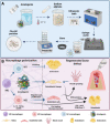
Scheme 1. Schematic illustration for the preparation and application of particles. (A) Chemical structure of the CA + AM particles. (B) Schematic illustration depicting the mechanism of particles used in extraction wounds to promote bone regeneration. Created at https://BioRender.com.
Fig. 1
Fig. 1. Physical characterization of calcium alginate–amelogenin hydrogel (CA + AM) (a) visual observation of calcium alginate (CA) hydrogel. (b) Electron microscopic image of CA hydrogel and CA + AM. (c) Particle size comparison. (d) Swelling ratio. (e) Degradation ratio. (f and g) Compressive strength. Data represent mean ± standard deviation; ns: no significant difference, *p < 0.05, **p < 0.01, ***p < 0.001, and ****p < 0.0001.
Fig. 2
Fig. 2. Cell toxicity and proliferative activity at different concentrations of the hydrogel. (a) Bone marrow-derived macrophages (BMDMs) and (b) bone marrow mesenchymal stem cells (BMSCs) viability in the CA + AM group was performed and analyzed by CCK-8 assay. (c) Calcein/propidium iodide fluorescent staining of BMSCs proliferation. Green staining indicates living cells. (d) BMDMs and (e) BMSC viability in the CA group were performed and analyzed by CCK-8 assay. Data represent the mean ± standard deviation. ns: no significant difference, *p < 0.05, **p < 0.01, ***p < 0.001, and ****p < 0.0001.
Fig. 3
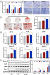
Fig. 3. Effect of CA + AM on macrophage morphology and expression of inflammatory and osteogenic genes and proteins. (a) CA + AM promotes the polarization of macrophages to M2. (b) Alkaline phosphatase staining. (c) Alizarin red staining and quantitative analysis. (d) Expression of osteogenesis-associated genes. (e) Quantitative analysis and expression of osteogenesis-related proteins. Data represent mean ± standard deviation. ns: no significant difference, *p < 0.05, **p < 0.01, ***p < 0.001, and ****p < 0.0001.
Fig. 4
Fig. 4. Animal model construction. Dental extraction of the lower central incisor in Sprague Dawley rats to evaluate the effects of different treatments on the (a) extraction socket healing, (b) bleeding time, and (c) amount of blood loss. *p < 0.05, **p < 0.01, ***p < 0.001, and ****p < 0.0001.
Fig. 5
Fig. 5. Degree of resorption of the alveolar ridge after tooth extraction trauma. (a and b) Absorption of the width and height of the sagittal alveolar ridge. (c and d) Osteogenesis in the socket 28 days after tooth extraction. *p < 0.05, **p < 0.01, ***p < 0.001, and ****p < 0.0001.
Fig. 6
Fig. 6. New bone formation in the extraction sockets of Sprague-Dawley rats. (a) New bone formation; (b) bone mineral density (BMD); (c) bone volume/total volume (BV/TV); (d) trabecular thickness (Tb.Th), and (e) trabecular separation (Tb.Sp) in the tooth-extracted socket at the indicated time points. Error bars represent the mean ± standard deviation. *p < 0.05, **p < 0.01, ***p < 0.001, and ****p < 0.0001.
Fig. 7
Fig. 7. Histological staining in the extraction sockets of Sprague-Dawley rats. (a) H&E staining sections on days 7 and 28 after tooth extraction. Black, green, and red arrows indicate new bone, fibrous tissue, and blood clots, respectively. (b) Masson's staining sections on days 7 and 28. Blue and red colours indicate fibrous tissue and mature bone tissue, respectively.
Fig. 8
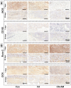
Fig. 8. IHC analysis in the extraction sockets of Sprague-Dawley rats. (a) The expression of markers (iNOS and CD163) of BMDM polarization on days 7 and 28. (b) The expression of osteogenic markers (runx-2 and OCN) after 7 and 28 days.

Similar articles

Cited by

References

    1. Wang X. Li C. He T. Q. Zheng W. H. Liu W. Zhang Y. Y. Chen X. L. Zhou Y. Q. Shui C. Y. Ning Y. D. Cai Y. C. Jiang J. Sun R. H. Wang W. Zhonghua Er Bi Yan Hou Tou Jing Wai Ke Za Zhi. 2021;56:89–92. - PubMed
    1. Guo J. Yao H. Li X. Chang L. Wang Z. Zhu W. Su Y. Qin L. Xu J. Bioact. Mater. 2023;21:175–193. - PMC - PubMed
    1. Filippi M. Born G. Chaaban M. Scherberich A. Front. Bioeng. Biotechnol. 2020;8:474. - PMC - PubMed
    1. Yao H. Guo J. Zhu W. Su Y. Tong W. Zheng L. Chang L. Wang X. Lai Y. Qin L. Xu J. Pharmaceutics. 2022:14. - PMC - PubMed
    1. Feng Q. Xu J. Zhang K. Yao H. Zheng N. Zheng L. ACS Cent. Sci. 2019;5:440–450. - PMC - PubMed