. 2024 Jan 9;20(1):18.
doi: 10.1186/s12917-023-03813-y.

Neurons cytoskeletal architecture remodeling during the replication cycle of mouse coronavirus MHV-JHM: a morphological in vitro study

Affiliations

Neurons cytoskeletal architecture remodeling during the replication cycle of mouse coronavirus MHV-JHM: a morphological in vitro study

Michalina Bartak et al. BMC Vet Res. .

Abstract

Nowadays, the population is still struggling with a post-COVID19 syndrome known as long COVID, including a broad spectrum of neurological problems. There is an urgent need for a better understanding and exploration of the mechanisms of coronavirus neurotropism. For this purpose, the neurotropic strain of mouse hepatitis virus (MHV-JHM) originating from the beta-coronavirus genus, the same as severe acute respiratory syndrome coronavirus 2 (SARS-CoV-2), has been used. The role of the cytoskeleton during virus replication in neurons in vitro was determined to understand the mechanisms of MHV-JHM neuroinfection. We have described for the first time the changes of actin filaments during MHV-JHM infection. We also observed productive replication of MHV-JHM in neurons during 168 h p.i. and syncytial cytopathic effect. We discovered that the MHV-JHM strain modulated neuronal cytoskeleton during infection, which were manifested by: (i) condensation of actin filaments in the cortical layer of the cytoplasm, (ii) formation of microtubule cisternae structures containing viral antigen targeting viral replication site (iii) formation of tunneling nanotubes used by MHV-JHM for intercellular transport. Additionally, we demonstrated that the use of cytoskeletal inhibitors have reduced virus replication in neurons, especially noscapine and nocodazole, the microtubule shortening factors.

Keywords: Actin filaments; MHV-JHM; Microtubules; Neurons; Neurotropism.


Conflict of interest statement

The authors declare no competing interests.

Figures
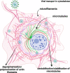
Fig. 1
Fig. 1
Schematic presentation of MHV-JHM entry and transport. Possible events occurring during virion transport in the cytoskeleton. Own work created with Biorender
Fig. 2
Fig. 2
RT-qPCR analysis of MHV-JHM viral RNA copies per µg during 168 h p.i. in murine neurons
Fig. 3
Fig. 3
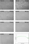
Real-time cell growth analysis of MHV-JHM infected primary murine neurons performed by using live image move analyser JuLi™Br. Cultures were observed from initial seeding for 168 h. The generated graph shows the percentage of cells' confluence level [%] during complete analysis [hours]. All images were recorded every 5 min and analysed monolayer confluence. Objective magnification × 40
Fig. 4
Fig. 4
Actin cytoskeleton network morphology of non-infected primary murine neurons. Various forms of actin fibres structures were presented and highlighted by white arrowheads: tunnelling nanotube (main image), filopodium (a) and longitudinal actin fibres (a’), stress fibres (b), peripheral stress fibres (c), lamellipodium (d), and growth cone (e). Indirect and direct immunofluorescence staining; merge images: actin filaments – red; cell nuclei – blue. Microscope magnification 60x, scale 20 μm
Fig. 5
Fig. 5
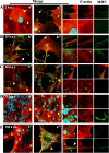
Primary culture of murine neurons infected with MHV-JHM virus. Representative confocal images of neurons obtained at 2 (A,a’,a’’), 24 (B,b’,b’’), 48 (C,c’,c’’), 72 (D,d’,d’’), and 168 h p.i (E,e’,e’’). Green arrowheads point to the presence of viral antigens in actin structures. Yellow arrowheads show areas of changes in f-actin filaments resulting from MHV-JHM infection. White arrowheads point to tunnelling nanotube (TNTs) structures. White asterisk show lamellipodia. Yellow boxes indicate the overlapping presence of actin filaments fluorescence with viral antigen and magnified area. Indirect and direct immunofluorescence staining; merge images: actin filaments - red; cell nuclei - blue; viral antigen - green. Microscope magnification 60x, scale 20 μm
Fig. 6
Fig. 6
Primary culture of murine neurons infected with MHV-JHM virus. Representative confocal images of neurons obtained at 2 h p.i. (A) and 168 h p.i (B). White asterisk represents the occurrence of a F-actin ring structures – juxtanuclear ring (A) and submembranous ring (B); green boxes show magnified areas; green arrows indicate viral antigen; double yellow arrows show juxtanuclear ring and single yellow arrow points submembrane ring. Indirect and direct immunofluorescence staining; merge images: actin filaments - red; cell nuclei - blue; viral antigen - green. Microscope magnification 60x, scale 20 μm and 10 μm
Fig. 7
Fig. 7
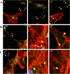
Primary culture of murine neurons infected with MHV-JHM after 168 h p.i.. Representative confocal images of specific filamentous actin structures – tunnelling nanotubes (TNTs) (a,a’,a’’,b,b’,c; white arrows); depolymerised (b’ yellow arrow) and highly polymerised rings structures (b’’ yellow arrows) spider-web-like actin structures (c’,c’’ yellow arrows). Green boxes indicate magnified area. Indirect and direct immunofluorescence staining; merge images: actin filaments - red; cell nuclei - blue; viral antigen - green. Microscope magnification 60x, scale 30 μm and 20 μm
Fig. 8
Fig. 8
Colocalization analysis of viral antigen present inside tunnelling nanotubule structures. Histograms compared correlations of TNTs with MHV-JHM antigen from the 2 h p.i. until 168 h p.i with uninfected cells (mock-infected) showing Pearson’s correlation coefficient (PCC) and Meander’s coefficients (M1 and M2) from ≥ 100 cells (data represented as mean ± SEM from TNTs structurers). The degrees of correlation were indicated as perfect for values near ± 1; strong for values between ± 0.50 and ± 1; medium for values between ± 0.30 and ± 0.49, and low for values below + 0.29. Obtained with JACoP BIOP analysis
Fig. 9
Fig. 9
Microtubule network morphology of non-infected primary murine neurons. Various forms of β-tubulin rich structures were presented and highlighted by white boxes: dendrite (a), soma (b), tubulin bundles (c), and axon growth cone (d). Indirect and direct immunofluorescence staining; merge images: β-tubulin- red; cell nuclei – blue. Microscope magnification 60x, scale 10 μm
Fig. 10
Fig. 10
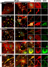
Primary culture of murine neurons infected with MHV-JHM virus. Representative confocal images of neurons obtained at 2 (A,a’,a’’), 24 (B,b’,b’’), 48 (C,c’,c’’), 72 (D,d’,d’’), and 168 (E,e’,e’’) hours post infection. Green arrowheads point the presence of viral antigens in actin structures; Yellow arrowheads show areas of changes in β-tubulin resulting from MHV-JHM infection. White arrowheads point to syncytia formation. Purple arrowheads indicate cisternae microtubule structures. Yellow boxes indicate the overlapping presence of β-tubulin fluorescence with viral antigen. Indirect and direct immunofluorescence staining; merge images: β-tubulin - red; cell nuclei - blue; viral antigen - green. Microscope magnification 60x, scale 20 μm
Fig. 11
Fig. 11
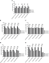
The effect of pre-treatment and post-treatment incubation with cytoskeletal inhibitors on MHV-JHM replication in primary murine neurons culture. Presented time points: 2 h (A), 24 h (B), 48 h (C), 72 h (D), 168 h (E). Viral RNA was quantified by RT real-time PCR. Data are presented as the mean ± standard deviation (SD) (n = 3). Two-way Anova tests were used to analyse statistical significance compared with the untreated control: *, significant (P ≤ 0.05); **, highly significant (P ≤ 0.01); and ***, extremely significant (P ≤ 0.001)

Similar articles

Cited by

References

    1. Zhang XY, Huang HJ, Zhuang DL, Nasser MI, Yang MH, Zhu P, et al. Biological, clinical and epidemiological features of COVID-19, SARS and MERS and AutoDock simulation of ACE2. Infect Dis Poverty. 2020;9(1):99. doi: 10.1186/s40249-020-00691-6. - DOI - PMC - PubMed
    1. Kubitschke H, Schnauss J, Nnetu KD, Warmt E, Stange R, Kaes J. Actin and microtubule networks contribute differently to cell response for small and large strains. New J Phys. 2017;19(9):093003. doi: 10.1088/1367-2630/aa7658. - DOI
    1. Walsh D, Naghavi MH. Exploitation of cytoskeletal networks during early viral infection. Trends Microbiol. 2019;27(1):39–50. doi: 10.1016/j.tim.2018.06.008. - DOI - PMC - PubMed
    1. Kłyszejko-Stefanowicz L. Cytobiochemia – biochemia niektórych struktur komórkowych. 3. Warsaw: PWN SA; 2015. pp. 58–62.
    1. Wen Z, Zhang Y, Lin Z, Shi K, Jiu Y. Cytoskeleton—a crucial key in host cell for coronavirus infection. Yao X, editor. J Mol Cell Biol. 2021;12(12):968–79. doi: 10.1093/jmcb/mjaa042. - DOI - PMC - PubMed

Substances