. 2024 Feb 20;98(2):e0174923.
doi: 10.1128/jvi.01749-23. Epub 2024 Jan 8.

N-terminal acetyltransferase 6 facilitates enterovirus 71 replication by regulating PI4KB expression and replication organelle biogenesis

Affiliations

N-terminal acetyltransferase 6 facilitates enterovirus 71 replication by regulating PI4KB expression and replication organelle biogenesis

Hang Yang et al. J Virol. .

Abstract

Enterovirus 71 (EV71) is one of the major pathogens causing hand, foot, and mouth disease in children under 5 years old, which can result in severe neurological complications and even death. Due to limited treatments for EV71 infection, the identification of novel host factors and elucidation of mechanisms involved will help to counter this viral infection. N-terminal acetyltransferase 6 (NAT6) was identified as an essential host factor for EV71 infection with genome-wide CRISPR/Cas9 screening. NAT6 facilitates EV71 viral replication depending on its acetyltransferase activity but has little effect on viral release. In addition, NAT6 is also required for Echovirus 7 and coxsackievirus B5 infection, suggesting it might be a pan-enterovirus host factor. We further demonstrated that NAT6 is required for Golgi integrity and viral replication organelle (RO) biogenesis. NAT6 knockout significantly inhibited phosphatidylinositol 4-kinase IIIβ (PI4KB) expression and PI4P production, both of which are key host factors for enterovirus infection and RO biogenesis. Further mechanism studies confirmed that NAT6 formed a complex with its substrate actin and one of the PI4KB recruiters-acyl-coenzyme A binding domain containing 3 (ACBD3). Through modulating actin dynamics, NAT6 maintained the integrity of the Golgi and the stability of ACBD3, thereby enhancing EV71 infection. Collectively, these results uncovered a novel mechanism of N-acetyltransferase supporting EV71 infection.IMPORTANCEEnterovirus 71 (EV71) is an important pathogen for children under the age of five, and currently, no effective treatment is available. Elucidating the mechanism of novel host factors supporting viral infection will reveal potential antiviral targets and aid antiviral development. Here, we demonstrated that a novel N-acetyltransferase, NAT6, is an essential host factor for EV71 replication. NAT6 could promote viral replication organelle (RO) formation to enhance viral replication. The formation of enterovirus ROs requires numerous host factors, including acyl-coenzyme A binding domain containing 3 (ACBD3) and phosphatidylinositol 4-kinase IIIβ (PI4KB). NAT6 could stabilize the PI4KB recruiter, ACBD3, by inhibiting the autophagy degradation pathway. This study provides a fresh insight into the relationship between N-acetyltransferase and viral infection.

Keywords: ACBD3; Golgi morphology; acetyltransferase; enteroviruses; replication organelle.


Conflict of interest statement

The authors declare no conflict of interest.

Figures

Fig 1
Fig 1
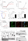
NAT6 is a host factor for EV71 infection in SK-N-SH cells. (A) SK-N-SH cells stably transduced with control or NAT6 sgRNA were infected with the EV71 virus (MOI = 5). After 24 h, cells were immunoblotted with NAT6 or EV71 VP1 antibody. GAPDH was used as a loading control. (B) vRNA from cells as described in panel A was quantified with q-RT-PCR. Values are normalized to control. ***P < 0.001. (C) Progeny viruses from the above cells were titrated. **P < 0.01 and ***P < 0.001. (D) Cells as described above were infected with EV71, and cell viability was determined at 24 h post-infection. Values are normalized to uninfected control cells. ***P < 0.001. (E) Control or NAT6 KO SK-N-SH cells were transduced with Flag-tagged sgRNA-resistant NAT6 and then infected with EV71. After 24 h, cells were immunoblotted with NAT6 or EV71 VP1 antibody. GAPDH was used as a loading control. (F) Cells as described in panel E were infected with EV71, and vRNA was quantified with q-RT-PCR. Values are normalized to control. ***P < 0.001. (G and H) SK-N-SH cells stably transduced with control or NAT6 sgRNA were infected with Echo7 virus (G, MOI = 5) or CVB5 virus (H, MOI = 1) for 24 h. Cells were then immunoblotted with Echo VP3 or CVB VP1 antibody. GAPDH was used as a loading control. (I and J) SK-N-SH cells were either mock infected or infected with EV71 (MOI = 5) for 24 h, and NAT6 or viral VP1 expression was determined by immunoblotting (I) or q-RT-PCR (J).
Fig 2
Fig 2
NAT6 is relevant to EV71 replication. (A and B) Control or NAT6 KO SK-N-SH cells were infected with EV71 (A) or EV71 with NanoLuc reporter (B), and viral titer (A) or luciferase activity (B) was determined at indicated time points. ns, not significant. *P < 0.05; **P < 0.01; and ***P < 0.001. (C) Schematic diagram of virus attachment and internalization assay. (D) Control, NAT6 KO, or SCARB2 KO SK-N-SH cells were treated with EV71 (MOI = 10) as described in panel C. Attached (left panel) or internalized (right panel) vRNA was quantified with q-RT-PCR. Values are normalized to control. ns, not significant. (E) Control or NAT6 KO SK-N-SH cells were transfected with EV71 SGR or EV71 SGR-GND RNA. Luciferase activity was determined at indicated time points. **P < 0.01 and ***P < 0.001. (F) SK-N-SH cells stably expressing NAT6-Flag were infected with EV71 (MOI = 5) for 24 h. Cells were then fixed and stained with antibodies against Flag (green) and dsRNA (red) with DAPI nuclear counterstaining (blue). Asterisk indicates infected cell, and arrowhead indicates uninfected cell. Bar, 10 µm. (G) 293T cells cotransfected with NAT6-Flag and green fluorescent protein (GFP)-tagged 2B, 2C, 3AB, 3C, 3D, or PFN2 plasmids were immunoprecipitated with anti-Flag antibody, and interactions between NAT6 and viral NS proteins were detected by anti-GFP antibody. (H) Uninfected or EV71-infected SK-N-SH cell homogenates (H) were centrifuged to prepare a “water-soluble” supernatant (WS) or “detergent-soluble” (DS) fraction. Detergent-resistant membranes were then fractionated on a density gradient. Fractions (numbered in order from light to heavy) were analyzed by immunoblotting for the indicated proteins.
Fig 3
Fig 3
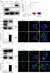
NAT6 is essential for Golgi integrity in SK-N-SH cells. (A and B) Control or NAT6 KO SK-N-SH cells were transduced with sgRNA-resistant catalytic-dead NAT6 mutant and then infected with EV71. After 24 h, viral VP1 expression was determined by immunoblotting (A) or q-RT-PCR (B). (C) Upper left: control or NAT6 KO SK-N-SH cells were immunoblotted with NAT6 or GM130 antibody. GAPDH was used as a loading control. Right panel: control or NAT6 KO SK-N-SH cells were immunostained with antibodies against GM130. Bar, 20 µm. Percentage of cells with fragmented Golgi was quantified and shown in the lower left panel. ***P < 0.001. (D) Upper left: control or NAT6 KO SK-N-SH cells were immunoblotted with NAT6 or TGN46 antibody. GAPDH was used as a loading control. Right panel: control or NAT6 KO SK-N-SH cells were immunostained with antibodies against TGN46. Bar, 20 µm. Percentage of cells with fragmented Golgi was quantified and shown in the lower left panel. ***P < 0.001.
Fig 4
Fig 4
NAT6 is dispensable for EV71 viral release. (A) Control or NAT6 KO SK-N-SH cells were immunostained with β-COP (green) and counterstained with DAPI (blue). Bar, 10 µm. (B) Control or NAT6 KO SK-N-SH cells were transfected with a plasmid encoding secreted NanoLuc, and luciferase from the supernatant was determined at indicated time points. *P < 0.05 and ***P < 0.001. (C) Schematic diagram of EV71 reinfection assay. Extracellular viruses from the supernatant and intracellular viruses from freeze-thawed cell lysate were used to inoculate naive SK-N-SH cells for vRNA quantitation or naive RD cells for viral titration. (D) Left panel: extracellular or intracellular vRNA was determined by q-RT-PCR, and values were normalized to control cells. ***P < 0.001. Right panel: extracellular or intracellular viral titers were determined and expressed as TCID50/mL. *P < 0.05 and **P < 0.01.
Fig 5
Fig 5
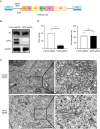
NAT6 supports viral RO biogenesis. (A) Schematic diagram of pTM1 (2A–3D). The EV71 NS encoding region was inserted into the pTM1 backbone under the control of a T7 promoter and EMCV IRES, followed by a T7 terminator. (B) Control or NAT6 KO cells stably expressing T7 RNA polymerase were transfected with pTM1 (2A–3D), and expression of viral 3D protein was determined 36 h post-transfection with immunoblotting. (C) Control or NAT6 KO cells as described in panel B were prepared for transmission electron microscope. Enlargements of the boxed areas are shown in the right panels. Bar, 1 µm. (D) Quantification of the number and diameter of vesicles in panel C. ns, not significant. ***P  <  0.001.
Fig 6
Fig 6
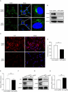
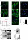
NAT6 regulates PI4KB and ACBD3 expression. (A) Control or NAT6 KO SK-N-SH cells were immunostained with PI4KB (green) and counterstained with DAPI (blue). Bar, 10 µm. (B) Control or NAT6 KO SK-N-SH cells were immunoblotted with indicated antibodies. (C) Control or NAT6 KO SK-N-SH cells were immunostained with an antibody against PI4P (red) and counterstained with DAPI (blue). Bar, 20 µm. (D) Quantitation of PI4P fluorescence from control or NAT6 KO SK-N-SH cells. ***P < 0.001. (E) PI4KB mRNA expression from control or NAT6 KO SK-N-SH cells was determined with q-RT-PCR. ns, not significant. (F and G) Control or NAT6 KO SK-N-SH cells were immunoblotted with Arf1 (F) or ACBD3 (G) antibody. (H) ACBD3 mRNA expression from control or NAT6 KO SK-N-SH cells was determined with q-RT-PCR.
Fig 7
Fig 7
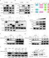
NAT6 interacts with ACBD3 and actin and stabilizes ACBD3 by suppressing the autophagy-lysosome degradation pathway. (A) SK-N-SH cells stably expressing NAT6-HA were immunoprecipitated using anti-ACBD3 antibody and immunoblotted with antibodies against ACBD3 or β-actin. (B) SK-N-SH cells stably expressing ACBD3-Flag were immunoprecipitated using anti-Flag antibodies and immunoblotted with antibodies against NAT6 or β-actin. (C) Schematic diagram of human ACBD3 domains and truncations. The amino acid residues are indicated on top. (D) HEK293T cells cotransfected with NAT6-GFP and ACBD3-Flag or truncated ACBD3-Flag were immunoprecipitated using anti-GFP antibody, and interactions between NAT6 and ACBD3 or mutants were detected with immunoblotting. (E) HEK293T cells cotransfected with a fixed amount of ACBD3-Flag, PI4KB, and an increasing amount of NAT6-HA plasmid (300, 600, and 900 ng) for 24 h before they were immunoblotted with indicated antibodies. (F) Mock or NAT6-HA were cotransfected with ACBD3-Flag into HEK293T cells for 18 h before they were treated with protein synthesis inhibitor cycloheximide (CHX) (100 µM). Cell lysates were collected at indicated time points and immunoblotted with indicated antibodies. The relative ACBD3 band density was normalized to that of GAPDH and shown on top. (G) SK-N-SH cells stably expressing NAT6-HA were treated with CHX (125 µM). Cell lysates were collected at indicated time points and immunoblotted with indicated antibodies. The relative ACBD3 band density was normalized to that of GAPDH and shown on top. (H) Control or NAT6 KO SK-N-SH cells were treated with DMSO, MG132 (10 µM), or bafilomycin A1 (BafA1) (10 nM) for 24 h. Cell lysates were collected at indicated time points and immunoblotted with indicated antibodies. The relative ACBD3 band density was normalized to that of GAPDH and shown on top. (I) Control or NAT6 KO SK-N-SH cells were immunoblotted with anti-LC3 and p62 antibodies. The relative amount of p62/GAPDH and LC3-II/LC3-I was shown below.
Fig 8
Fig 8
NAT6 supports Golgi integrity and EV71 infection by regulating actin dynamics. (A) Control or NAT6 KO SK-N-SH cells were stained with FITC-phalloidin (green) and counterstained with DAPI (blue). Bar, 10 µm. (B) SK-N-SH cells were treated with 200 or 500 nM Jpk for 1 h and then stained with FITC-phalloidin with nuclei counterstained with DAPI. Bar, 10 µm. (C) SK-N-SH cells were treated with 200 nM Jpk for 1 h and then stained with anti-GM130 with nuclei counterstained with DAPI. The percentage of cells with fragmented Golgi was quantified and shown below. Bar, 10 µm. (D) SK-N-SH cells infected with EV71-NanoLuc were treated with indicated amounts of Jpk for 12 h, and luciferase activity was determined. Cellular ATP content was determined to assess cytotoxicity. **P < 0.01. (E) SK-N-SH cells stably expressing NAT6-HA were immunoprecipitated with anti-HA antibodies and immunoblotted with the indicated antibodies. (F) Control or NAT6 KO SK-N-SH cells were analyzed by Phos-tag gel and immunoblotted with anti-GRASP55 antibody. Thapsigargin (TG) treatment was used as a positive control of phospho-GRASP55 induction.

Similar articles

Cited by

References

    1. Baggen J, Thibaut HJ, Strating JRPM, van Kuppeveld FJM. 2018. The life cycle of non-polio enteroviruses and how to target it. Nat Rev Microbiol 16:368–381. doi:10.1038/s41579-018-0022-3 - DOI - PubMed
    1. Solomon T, Lewthwaite P, Perera D, Cardosa MJ, McMinn P, Ooi MH. 2010. Virology, epidemiology, pathogenesis, and control of enterovirus 71. Lancet Infect Dis 10:778–790. doi:10.1016/S1473-3099(10)70194-8 - DOI - PubMed
    1. Rao CD. 2021. Enteroviruses in gastrointestinal diseases. Rev Med Virol 31:1–12. doi:10.1002/rmv.2148 - DOI - PubMed
    1. Yamayoshi S, Yamashita Y, Li J, Hanagata N, Minowa T, Takemura T, Koike S. 2009. Scavenger receptor B2 is a cellular receptor for enterovirus 71. Nat Med 15:798–801. doi:10.1038/nm.1992 - DOI - PubMed
    1. Hussain KM, Leong KLJ, Ng M-L, Chu JJH. 2011. The essential role of clathrin-mediated endocytosis in the infectious entry of human enterovirus 71. J Biol Chem 286:309–321. doi:10.1074/jbc.M110.168468 - DOI - PMC - PubMed

MeSH terms