. 2024 Jan 5;7(3):e202302258.
doi: 10.26508/lsa.202302258. Print 2024 Mar.

USP27X variants underlying X-linked intellectual disability disrupt protein function via distinct mechanisms

Affiliations

USP27X variants underlying X-linked intellectual disability disrupt protein function via distinct mechanisms

Intisar Koch et al. Life Sci Alliance. .

Abstract

Neurodevelopmental disorders with intellectual disability (ND/ID) are a heterogeneous group of diseases driving lifelong deficits in cognition and behavior with no definitive cure. X-linked intellectual disability disorder 105 (XLID105, #300984; OMIM) is a ND/ID driven by hemizygous variants in the USP27X gene encoding a protein deubiquitylase with a role in cell proliferation and neural development. Currently, only four genetically diagnosed individuals from two unrelated families have been described with limited clinical data. Furthermore, the mechanisms underlying the disorder are unknown. Here, we report 10 new XLID105 individuals from nine families and determine the impact of gene variants on USP27X protein function. Using a combination of clinical genetics, bioinformatics, biochemical, and cell biology approaches, we determined that XLID105 variants alter USP27X protein biology via distinct mechanisms including changes in developmentally relevant protein-protein interactions and deubiquitylating activity. Our data better define the phenotypic spectrum of XLID105 and suggest that XLID105 is driven by USP27X functional disruption. Understanding the pathogenic mechanisms of XLID105 variants will provide molecular insight into USP27X biology and may create the potential for therapy development.


Conflict of interest statement

MJ Guillen Sacoto is an employee of GeneDx, LLC. H Mor-Shaked is an employee of Geneyx Genomics. Other authors declare that they have no competing commercial interests.

Figures

Figure 1.
Figure 1.. Pedigrees suggestive of X-linked inheritance.
(A) Pedigrees for variant segregation in nine families with 10 affected individuals harboring USP27X variants drawn using QuickPed (Vigeland, 2022). The medical history of family members is described in the supplementary material. (B) Location of variants on a 2D schematic diagram of the USP27X protein. (C) Bar graph depicting the prevalence of the most commonly shared clinical manifestations. Human Phenotype Ontology (HP) terms are shown.
Figure S1.
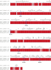
Figure S1.. XLID105 missense variants result in changes to conserved residues.
Protein sequence alignment of human, mouse, and rat USP27X. Conserved residues are highlighted in red and residues with missense variants in XLID105 are outlined in blue.
Figure 2.
Figure 2.. XLID105 variants do not affect USP27X localization or protein stability.
(A) Analysis of USP27X XLID105 mutant protein localization. Usp27x−/y ESCs were transfected with plasmids encoding the indicated HA-tagged USP27X mutants and their localization was analyzed via anti-HA (green) immunofluorescence and confocal microscopy. Actin staining (red) is shown as cytoskeleton marker and Hoechst (blue) was used as a nuclear marker. Scalebar: 20 μm, (n = 3). Quantification of the ratio between the nuclear and total HA-USP27X fluorescence intensity of 10 confocal image frames per variant is shown. No significant differences were found using one-way ANOVA analysis (data are presented as mean ± SEM). (B) Analysis of USP27X XLID105 mutant protein stability. Usp27x−/y ESCs were transfected with plasmids encoding the indicated HA-tagged USP27X constructs and cells were treated with cycloheximide for the indicated times. HA-tagged USP27X expression was analyzed via immunoblotting. ERK1 was used as a loading control. Quantification of relative HA-USP27X levels is displayed (data are presented as mean ± SEM, n = 3). No significant differences were found by t test analyses comparing each mutant with USP27X WT across four time points. Source data are available for this figure.
Figure 3.
Figure 3.. Structurally relevant USP27X residues are mutated in XLID105.
(A) USP27X predicted structure from the AlphaFold database. The fingers, palm, and thumb subdomains are colored pink, purple, and green, respectively. Insertions in the USP domain (>6 residue stretches with predicted Local Distance Difference Test scores <80) are colored gray. Residues found to be mutated in individuals with XLID105 are highlighted in boxes, with the confidence score (predicted Local Distance Difference Test) given. Hydrogen bonds are shown as dashed lines. (B) F313 and Y381 are predicted to be important to USP27X ubiquitin binding. The USP27X–ubiquitin complex predicted using ColabFold (three replicate computations shown). Close-up views of USP27X F313 and Y381 residues in contact with ubiquitin (yellow) are shown. Hydrogen bonds are depicted as dashed lines. (B, C) Analysis of fractional solvent accessible surface area of residues found to be mutated in XLID105 individuals, calculated from the AlphaFold structures from panel (B) (Data are presented as mean ± SEM).
Figure S2.
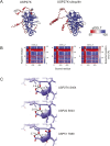
Figure S2.. Structural prediction of the USP27X–ubiquitin complex.
(A) AlphaFold pLDDT confidence scores of USP27X and the USP27X–ubiquitin complex. (B) Predicted Aligned Error plots of three replicate models of the USP27X–ubiquitin complex (A = ubiquitin, B=USP27X). The color scale is shown with blue indicating higher confidence and red lower confidence. (C) Close-up views of USP27X, USP22, and USP51 AlphaFold models around residue S404 of USP27X highlighting conservation of the local structure. Hydrogen bonds are depicted as dashed lines.
Figure S3.
Figure S3.. Activity-based probes measure USP27X ubiquitin recognition.
(A) Thermal shift assay of recombinant USP27X WT and XLID105 variants. The melt curve and melt peak graphs, and calculated melt temperature for these proteins are shown. (B) Recombinant GST-tagged USP2 or USP27X were incubated with a HA-tagged ubiquitin–PRG probe and USP2 and USP27X probe labelling was analyzed via SDS–PAGE and immunoblotting (n = 3). (C) WT USP27X activity-based probe labelling with ubiquitin–PRG was compared with a USP27X C87A mutant (n = 3). (D) A USP27X activity-based probe labelling assay with ubiquitin–PRG was performed in the presence of increasing concentrations of PR-619 (n = 3). USP27X probe labelling was analyzed via immunoblotting. A merged GST/HA (USP27X/Ub-PRG) image is displayed for visualization of the probe labelling. (#) non-specific band. Source data are available for this figure.
Figure 4.
Figure 4.. XLID105 variants disrupt USP27X substrate-independent deubiquitylating activity.
(A, B) GST-tagged WT or XLID105 mutant USP27X were incubated with K48 (A) or K63 (B) di-ubiquitin chains respectively. Data are presented as mean ± SEM, one-way ANOVA followed by Tukey’s analysis, K48: P = 0.0474 (*), P = 0.0080 (**), P < 0.0001 (****); K63: P = 0.0041 (**), P = 0.0001 (***), P < 0.0001 (****). (C) GST-tagged wild-type or XLID105 mutant USP27X was incubated with Ub-PRG probe and labelling was analyzed via immunoblotting. Labelling quantification of three replicates is shown. Data are presented as mean ± SEM, one-way ANOVA followed by Tukey’s analysis G76S: P = 0.0264(*), F313V: P = 0.0021 (**), Y381H: P = 0.0129 (*), and S404N: P < 0.0001 (****). A merged GST/HA (USP/Ub-PRG) image is displayed for visualization of the probe labelling. Source data are available for this figure.
Figure 5.
Figure 5.. USP27X XLID105 variants drive altered ATXN7L3 interaction.
(A) Detection of specific USP27X–ATXN7L3 protein interaction. Usp27x−/y ESCs were transfected with plasmids encoding HA-tagged USP27X and FLAG-tagged ATXN7L3. Lysates were subjected to anti-HA or anti-FLAG immunoprecipitation and immunoblotting, (n = 3). (B) Analysis of ATXN7L3 interaction with USP27X XLID105 variants. (A) Usp27x−/y ESCs were transfected with plasmids encoding FLAG-tagged ATXN7L3 or HA-tagged WT or the indicated XLID105 mutants and samples were subjected to anti-HA immunoprecipitation as in (A), (n = 3). (A, B) Co-immunoprecipitation and lysate control immunoblots are shown in (A, B). (A, B) ERK1 levels are shown in the lysates as loading control for (A, B). (B) A merged HA/FLAG image is displayed for the immunoprecipitates in (B). Quantification of ATXN7L3 enrichment by the individual USP27X mutants is displayed. Data are presented as mean ± SEM, One-way ANOVA followed by Tukey’s analysis P = 0.0003 (***), P > 0.0001 (****). Source data are available for this figure.

Similar articles

Cited by

References

    1. Adorno M, Sikandar S, Mitra SS, Kuo A, Nicolis Di Robilant B, Haro-Acosta V, Ouadah Y, Quarta M, Rodriguez J, Qian D, et al. (2013) Usp16 contributes to somatic stem-cell defects in Down’s syndrome. Nature 501: 380–384. 10.1038/nature12530 - DOI - PMC - PubMed
    1. Alam S, Zunic A, Venkat S, Feigin ME, Atanassov BS (2022) Regulation of cyclin D1 degradation by ubiquitin-specific protease 27X is critical for cancer cell proliferation and tumor growth. Mol Cancer Res 20: 1751–1762. 10.1158/1541-7786.MCR-22-0259 - DOI - PMC - PubMed
    1. Atanassov BS, Mohan RD, Lan X, Kuang X, Lu Y, Lin K, McIvor E, Li W, Zhang Y, Florens L, et al. (2016) ATXN7L3 and ENY2 coordinate activity of multiple H2B deubiquitinases important for cellular proliferation and tumor growth. Mol Cell 62: 558–571. 10.1016/j.molcel.2016.03.030 - DOI - PMC - PubMed
    1. Beck DB, Basar MA, Asmar AJ, Thompson JJ, Oda H, Uehara DT, Saida K, Pajusalu S, Talvik I, D’Souza P, et al. (2021) Linkage-specific deubiquitylation by OTUD5 defines an embryonic pathway intolerant to genomic variation. Sci Adv 7: eabe2116. 10.1126/sciadv.abe2116 - DOI - PMC - PubMed
    1. Bushby KMD, Cole T, Matthews JNS, Goodship JA (1992) Centiles for adult head circumference. Arch Dis Child 67: 1286–1287. 10.1136/adc.67.10.1286 - DOI - PMC - PubMed

Publication types

Substances

LinkOut - more resources