Review
. 2024 Mar;23(3):e14070.
doi: 10.1111/acel.14070. Epub 2024 Jan 5.

Microphysiological systems for human aging research

Affiliations
Review

Microphysiological systems for human aging research

Seungman Park et al. Aging Cell. 2024 Mar.

Abstract

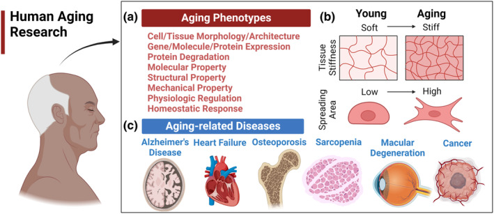
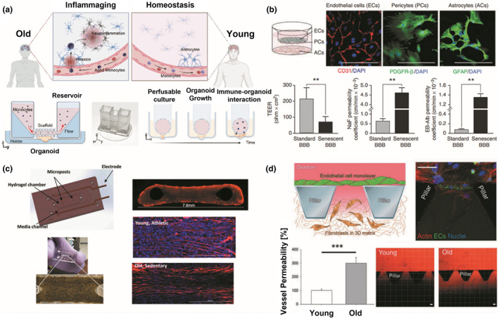
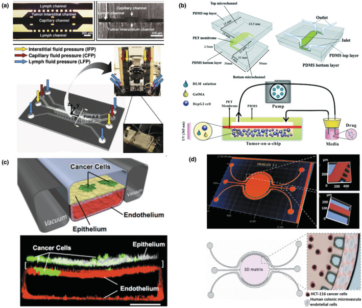
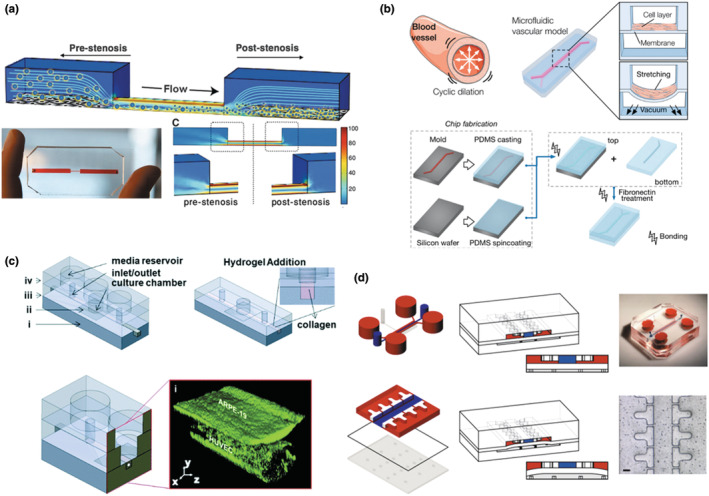
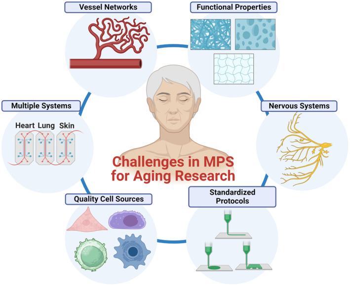
Recent advances in microphysiological systems (MPS), also known as organs-on-a-chip (OoC), enable the recapitulation of more complex organ and tissue functions on a smaller scale in vitro. MPS therefore provide the potential to better understand human diseases and physiology. To date, numerous MPS platforms have been developed for various tissues and organs, including the heart, liver, kidney, blood vessels, muscle, and adipose tissue. However, only a few studies have explored using MPS platforms to unravel the effects of aging on human physiology and the pathogenesis of age-related diseases. Age is one of the risk factors for many diseases, and enormous interest has been devoted to aging research. As such, a human MPS aging model could provide a more predictive tool to understand the molecular and cellular mechanisms underlying human aging and age-related diseases. These models can also be used to evaluate preclinical drugs for age-related diseases and translate them into clinical settings. Here, we provide a review on the application of MPS in aging research. First, we offer an overview of the molecular, cellular, and physiological changes with age in several tissues or organs. Next, we discuss previous aging models and the current state of MPS for studying human aging and age-related conditions. Lastly, we address the limitations of current MPS and present future directions on the potential of MPS platforms for human aging research.

Keywords: age-related changes; age-related diseases; aging; aging phenotypes; microphysiological systems.


Conflict of interest statement

D.‐H.K is a co‐founder and scientific advisory board member at Curi Bio, Inc.

Figures

FIGURE 1
FIGURE 1
Types of aging studies. (a) Age‐related phenotypes and age‐related diseases. Representative phenotypes of normal aging include changes in cell/tissue architecture, abnormal RNA or protein expression, a decline in various functional properties, such as molecular, structural, or mechanical properties, altered physiologic rhythms, loss of complexity, and homeostenosis. (b) Examples of age‐related changes in cellular and tissue levels. At the cellular level, cell morphology is changed in the aging process with a higher spreading area. At the tissue level, the stiffness of skin tissue is known to increase significantly during aging. (c) Age‐related diseases at the organ levels. Examples of age‐related diseases are Alzheimer's disease, cardiovascular disease, cancer, osteoporosis, and so on.
FIGURE 2
FIGURE 2
Microphysiological systems (MPS) for studying aging phenotypes. (a) MPS for immune‐driven brain aging. Primary monocytes from young and aged donors and human brain organoids were adopted and cultured in the 3D printed device to understand immune‐organoid interactions. Figure adapted with permission from (Ao et al., 2022), copyright Advanced Science. (b) In vitro triple co‐culture models of the blood–brain barrier (BBB). Endothelial cells (ECs) and pericytes (PCs) were cultured on the top and bottom sides of the semipermeable filters, while astrocytes were plated on the culture plate. Barrier functions of normal and aging BBB were quantified and compared through TEER measurement, and permeability coefficient for NaF and EB‐albumin. **p < 0.05. Figure adapted with permission from (Yamazaki et al., 2016), copyright Stroke. (c) Muscle myobundles in a microfluidic chip. Young/active and old/sedentary people‐derived muscle fibers were formed around the two posts in a microfluidic chip. The myobundles were fluorescently immunostained with myosin heavy chain antibody (MF‐20, red) and DAPI (blue). Figure adapted with permission from (Giza et al., 2022), copyright Aging Cell. (d) Microfluidic model of human endothelial cell aging. Aged endothelium was created in the microfluidic system containing pillars, and vessel permeability was measured using fluorescently‐labeled dextran. Results showed that vessel permeability in aged endothelium is substantially increased compared to that of young endothelium. ***p < 0.05. Figure adapted with permission from (Bersini et al., 2020), copyright Advanced Biosystems.
FIGURE 3
FIGURE 3
Design of diverse MPS for cancer research. (a) Mammary tumor‐on‐a‐chip. A functional unit of the mammary tumor (center channel), as well as capillary (top channel) and lymphatic vessels (two side channels), was emulated in a 3D microfluidic platform, which undergoes interstitial, capillary, and lymph fluid pressures. Figure adapted with permission from (Kwak et al., 2014), copyright Journal of Controlled Release. (b) Liver tumor‐on‐a‐chip. 3D cell culture microchip, composed of top and bottom microchannels and PET membrane, was integrated with several components derived from a decellularized liver matrix from a native liver with gelatin methacryloyl (GelMA). The hydrogel was created through photopolymerization by UV (ultraviolet) light (365 nm). Figure adapted with permission from (Lu et al., 2018), copyright Lab on a Chip. (c) Lung tumor‐on‐a‐chip. Human lung epithelial cells (white) and non‐small‐cell lung cancer cells (green) were plated on the upper surface of a porous membrane, and human microvascular endothelial cells (red) were cultured on the lower channel of the alveolus‐mimetic microfluidic chip (scale bar, 200 μm). Figure adapted with permission from (Hassell et al., 2017), copyright Cell Reports. (d) Colorectal tumor‐on‐a‐chip. Colon cancer cells (HCT‐116)‐laden Matrigel was embedded in the round central chamber (5 mm in diameter and 126 μm in depth), and human colonic microvascular endothelial cells were added in the side channels to mimic microvessels. Figure adapted with permission from (Carvalho et al., 2019), copyright Science Advances.
FIGURE 4
FIGURE 4
Schematics of diverse MPS for studying age‐related diseases. (a) Vascular‐on‐a‐chip to study thrombosis. A 3D microfluidic vascular stenosis model (90% lumen occlusion) with endothelial cells was fabricated to investigate shear‐induced dissociation by nanotherapeutics and nanoparticles, which are similar in size to platelets ranging from 1 to 5 μm in diameter. The shear rate in the region of the stenosis was increased to ~100,000 s−1 from 1000 s−1 of a physiological shear rate. Figure adapted with permission from (Korin et al., 2012), copyright Science. (b) Progeria‐on‐a‐chip. The microfluidic vascular model is comprised of two compartmentalized channels where senescent aortic smooth muscle cells are cultured on top of the deformable PDMS membrane in the upper channel and undergo cyclic strain mimicking pulsatile blood flow. Figure adapted with permission from (Ribas et al., 2017), copyright Small. (c) MPS for the outer blood‐retinal barrier to study macular degeneration. The microscale system contains (i) a bottom compartment, (ii) a polyester membrane with pores, (iii) an open‐top culture chamber, and (iv) a top compartment. The microvessel surrounded by collagen hydrogel I was created using blunt needles, and human endothelial cells and retinal pigment epithelial cells were cocultured. Figure adapted with permission from (Arik et al., 2021), copyright Lab on a Chip. (d) Cartilage‐on‐a‐chip model to study osteoarthritis. Two PDMS chambers were fabricated and separated by a PDMS membrane. The chondrocyte‐laden PEG polymer solution was injected into the central upper chamber (blue). By pressurizing the bottom chamber, mechanical compression can be generated by the deformation of the PDMS membrane (Scale bar, 100 μm). Figure adapted with permission from (Occhetta et al., 2019), copyright Nature Biomedical Engineering.
FIGURE 5
FIGURE 5
Challenges in MPS for aging research. Key challenges encompass the development of vascularization, integration of multi‐MPS platforms for various organs and tissues (e.g., human‐on‐a‐chip systems), procurement of high‐quality human cells representing healthy aging/aged states, establishment of standardizing protocols, replication of nervous system components, and recapitulation of functional properties including mechanical, structural, and transport properties.

Similar articles

Cited by

References

    1. Achberger, K. , Probst, C. , Haderspeck, J. C. , Bolz, S. , Rogal, J. , Chuchuy, J. , Nikolova, M. , Cora, V. , Antkowiak, L. , Haq, W. , Shen, N. , Schenke‐Layland, K. , Ueffing, M. , Liebau, S. , & Loskill, P. (2019). Merging organoid and organ‐on‐a‐chip technology to generate complex multi‐layer tissue models in a human retina‐on‐a‐chip platform. eLife, 8, e46188. - PMC - PubMed
    1. Ackert‐Bicknell, C. L. , Anderson, L. C. , Sheehan, S. , Hill, W. G. , Chang, B. , Churchill, G. A. , Chesler, E. J. , Korstanje, R. , & Peters, L. L. (2015). Aging research using mouse models. Current Protocols in Mouse Biology, 5, 95–133. - PMC - PubMed
    1. Acun, A. , Vural, D. C. , & Zorlutuna, P. (2017). A tissue engineered model of aging: Interdependence and cooperative effects in failing tissues. Scientific Reports, 7, 5051. - PMC - PubMed
    1. Agarwal, A. , Goss, J. A. , Cho, A. , McCain, M. L. , & Parker, K. K. (2013). Microfluidic heart on a chip for higher throughput pharmacological studies. Lab on a Chip, 13, 3599. - PMC - PubMed
    1. Aguzzi, A. , & O'Connor, T. (2010). Protein aggregation diseases: Pathogenicity and therapeutic perspectives. Nature Reviews. Drug Discovery, 9, 237–248. - PubMed

Publication types