Review
. 2024 Mar:99:101234.
doi: 10.1016/j.preteyeres.2023.101234. Epub 2024 Jan 2.

Squishy matters - Corneal mechanobiology in health and disease

Affiliations
Review

Squishy matters - Corneal mechanobiology in health and disease

Sara M Thomasy et al. Prog Retin Eye Res. 2024 Mar.

Abstract

The cornea, as a dynamic and responsive tissue, constantly interacts with mechanical forces in order to maintain its structural integrity, barrier function, transparency and refractive power. Cells within the cornea sense and respond to various mechanical forces that fundamentally regulate their morphology and fate in development, homeostasis and pathophysiology. Corneal cells also dynamically regulate their extracellular matrix (ECM) with ensuing cell-ECM crosstalk as the matrix serves as a dynamic signaling reservoir providing biophysical and biochemical cues to corneal cells. Here we provide an overview of mechanotransduction signaling pathways then delve into the recent advances in corneal mechanobiology, focusing on the interplay between mechanical forces and responses of the corneal epithelial, stromal, and endothelial cells. We also identify species-specific differences in corneal biomechanics and mechanotransduction to facilitate identification of optimal animal models to study corneal wound healing, disease, and novel therapeutic interventions. Finally, we identify key knowledge gaps and therapeutic opportunities in corneal mechanobiology that are pressing for the research community to address especially pertinent within the domains of limbal stem cell deficiency, keratoconus and Fuchs' endothelial corneal dystrophy. By furthering our understanding corneal mechanobiology, we can contextualize discoveries regarding corneal diseases as well as innovative treatments for them.

Keywords: Cell-matrix interactions; Cornea; Corneal wound healing; Mechanobiology; Mechanotransduction; Stiffness.


Conflict of interest statement

Declaration of competing interest

Employee: Novartis institutes for biomedical research (NIBR). Work described in the study were from VR’s time in academia and NIBR had no role in study design, execution, planning, funding, interpretation of results, manuscript preparation or decision to publish.

None for ST, BL, MG, and JS.

Figures

Fig. 1.
Fig. 1.. Cells maintain intimate connections via cell-to-cell junctions.
The three most common junctions between two cells are the adherens, tight, and desmosome junctions. Cell-to-cell junctions connect two cells but also join the two cells to the cytoskeleton via anchoring proteins on the cellular side of the cell membrane. Junction loss due to disruptions in the cellular monolayer will potentiate changes in signaling through these intracellular cytoskeletal networks.
Fig. 2.
Fig. 2.. Dynamic reciprocity exists between cells and their ECM.
Cells synthesize ECM proteins and deposit them into the extracellular space. The proteins assembled in the matrix present a rich set of topographic and stiffness cues to the cells. Integrins, syndecans, and other receptors mechanotransduce biophysical cues into the cells. Signaling molecules and the cytoskeleton convey these signals to the nucleus, where they influence cell behavior in many ways, including modulating changes in the expression of ECM genes and proteins. Adapted from Ali, M., Raghunathan V., Li, J.Y., Murphy, C.J., Thomasy, S.M., 2016. Biomechanical relationships between the corneal endothelium and Descemet’s membrane. Exp Eye Res 152, 57–70.
Fig. 3.
Fig. 3.. YAP/TAZ are the master conductors of mechanotransduction signaling.
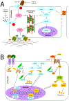
(A) Mechanical regulation of the Hippo pathway. Hippo is regulated by multiple signals generated by the biophysical (e.g., matrix stiffness) and biochemical (e.g., LPA, thrombin) environment. Importantly, many of these signals are modulated by tension in the actinomyosin cytoskeleton. Modulation of cytoskeletal mechanics through G-protein coupled receptors and matrix biophysics can likewise inhibit YAP/TAZ directly at the nuclear translocation stage or through activation of Hippo components. Note: This schematic is simplified to clarify the major components in the mechanical regulation of YAP/TAZ signaling. (B) Crosstalk between YAP/TAZ and TGFβ, and between YAP/TAZ and Wnt. TGFβ superfamily signaling is initiated by the binding of an extracellular ligand (e.g., TGFβ and BMP), which leads to the phosphorylation of SMADs and the formation of a complex with a Co-SMAD and 14-3-3s. After translocation to the nucleus, these complexes initiate the TGFβ/BMP transcriptional program. The SMAD complex also interacts with YAP/TAZ to initiate different transcriptional programs in the nucleus. Canonical Wnt is initiated by the binding of a Wnt ligand to the Fzd/LRP receptor complex. This induces the inhibitory behavior of Dvl on the Axin/APC/GSK3b complex, freeing β-catenin to translocate to the nucleus and initiate the Wnt transcriptional program. YAP/TAZ can inhibit Wnt signaling through inhibition of Dvl in the cytoplasm (TAZ) or in the nucleus (YAP) or cytoplasmic sequestration of β-catenin (YAP). Alternatively, YAP can encourage the transcriptional activity of β-catenin. Note: This schematic is simplified to clarify the intersections of YAP/TAZ and Wnt signaling. Reproduced from Ali, M., Raghunathan V., Li, J.Y., Murphy, C.J., Thomasy, S.M., 2016. Biomechanical relationships between the corneal endothelium and Descemet’s membrane. Exp Eye Res 152, 57–70.
Fig. 4.
Fig. 4.. Mechanotransduction feedback mechanisms can drive disease.
Mechanotransduction can be initiated in disease states through many different cell or matrix alterations including the deposition of new matrix, change to ECM composition, cell-to-cell junction alterations, cell-to-cell junction loss via cell dropout, and tissue fibrosis/scarring. Downstream propagation on intracellular signaling can occur through many different mechanisms, often converging into pathways mediated by YAP/TAZ, Wnt, TGFβ, and/or AKT. These signaling cascades ultimately yield alterations in gene transcription that can propagate cellular dysfunction.
Fig. 5.
Fig. 5.. Compartmentalized organization of stem cells in the ocular-surface epithelium.
(A) Schematic of the anatomy of the mammalian eye. The inset shows a magnification of the ocular anterior segment and the location of the cornea and limbus. (B) Cross-section diagram of the mouse cornea illustrating the components of the limbal stem cell niche. The limbus is located at the intersection between the conjunctival and corneal epithelia, which share a common surface. Stem cells and progenitors are located in the basal layer of the stratified epithelium. At least two distinct stem cell populations exist in the limbal niche. Mostly quiescent stem cells are located in the outer limbus. These stem cells do not contribute directly to the homeostasis of either the conjunctiva or the cornea but can be activated after large-scale injury to the corneal epithelium. Stem cells in the inner limbus are active and undergo mostly symmetric cell divisions to generate transient-amplified progenitors. These exit the limbus and drift centripetally while continuing to proliferate. Cells in the basal layer can commit to terminal differentiation by intrinsic or extrinsic cues, to replenish the cells that are shed from the epithelial surface. The probability of basal progenitors committing to terminal differentiation increases toward the center of the cornea. The stroma consists of cellular and noncellular components that can regulate and influence the activity and fate of stem cells. (C) A 3D model of the cornea showing the organization of the limbal compartments and the clonal behavior of the stem cells within. Reprinted with permission from Lee, V., Rompolas, P., 2022. Corneal regeneration: insights in epithelial stem cell heterogeneity and dynamics. Curr Opin Genetic Dev 77, 101981.
Fig. 6.
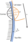
Fig. 6.. Schematic model of lid – cornea interaction with lid velocity (v), lid pressure against the cornea (p), and tear film thickness (h).
Figure and figure legend adapted from Pult, H., Tosatti, S.G., Spencer, N.D., Asfour, J.M., Ebenhoch, M., Murphy, P.J., 2015. Spontaneous Blinking from a Tribological Viewpoint. Ocul Surf 13, 236–249.
Fig. 7.
Fig. 7.. A schematic depicting the layers of the human cornea and the corresponding elastic modulus values obtained from atomic force microscopy:
Epithelium, anterior basement membrane (7.5 kPa), Bowman’s layer (110 kPa), anterior stroma (16 kPa), posterior stroma (2.5 kPa), Descemet’s membrane (50 kPa) and the endothelium. Data from (Last et al., 2012; Leonard et al., 2019a). Figure and figure legend adapted from Last, J.A., Thomasy, S.M., Croasdale, C.R., Russell, P., Murphy, C.J., 2012. Compliance profile of the human cornea as measured by atomic force microscopy. Micron 43, 1293–1298.
Fig. 8.
Fig. 8.. Scanning electron microscopy images of human corneal epithelial cells.
(A) Cell cultured on a silicon oxide substrate patterned with 70 nm wide ridges, on a 400 nm pitch. The groove depth was 600 nm. (B) Cell on a smooth silicon oxide substrate. Reprinted with permission from Teixeira, A.I., Abrams, G.A., Bertics, P.J., Murphy, C.J., Nealey, P.F., 2003. Epithelial contact guidance on well-defined micro- and nanostructured substrates. J Cell Sci 116, 1881–1892.
Fig. 9.
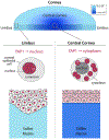
Fig. 9.. Biomechanical properties of the corneal matrix maintain stem cell populations at the limbus and promote differentiation towards the central cornea.
At the limbus, the extracellular matrix (ECM) is softer (light blue) and YAP1 (red circles) is localized to the nucleus of the limbal epithelial stem cells. When moving from the limbal biomechanical niche to the central cornea, there is a transition to a stiffer ECM with cytoplasmic localization of YAP1. The nuclear localization of YAP1 has been shown to maintain the stemness of the limbal corneal epithelial cells, whereas cytoplasmic localization results in the differentiation to more mature corneal epithelial cells (Bhattacharya et al., 2023).
Fig. 10.
Fig. 10.. Vertebrates demonstrate diverse corneal shape, lamellar collagen branching and orientation as well as stromal stiffness.
Aquatic species such as shark and sturgeon have flat corneas with no refractive power while semi-terrestrial and terrestrial species utilize a curved cornea as a refractive lens. Cross-sectional high-resolution macroscopic images of corneas demonstrate nearly absent branching in the shark and sturgeon, intermediate branching in bullfrog and alligator, and extensive branching in the peregrine falcon. In non-mammalian corneas, nearly perpendicular orthogonal layering of collagen sheets (fish, amphibians and reptiles or ribbons) or ribbons (birds) is observed, which leads to a clockwise chiral-nematic arrangement reminiscent of a cholesteric liquid crystal. By contrast, mammalian corneas demonstrate a random orientation of collagen fiber bundling with diverse interweaving ranging from just anterior (dog) to all but the most posterior cornea (human). In all species, the anterior stroma is stiffer than the posterior stroma. Adapted from Winkler, M., Shoa, G., Tran, S.T., Xie, Y., Thomasy, S., Raghunathan, V. K., Murphy, C., Brown, D.J., Jester, J.V., 2015. A Comparative Study of Vertebrate Corneal Structure: The Evolution of a Refractive Lens. Invest Ophthalmol Vis Sci 56, 2764–2772, copyright Association for Research in Vision and Ophthalmology.
Fig. 11.
Fig. 11.. Mammalian corneas exhibit dramatic differences in their stromal collagen intertwining and stiffness.
Note the dramatic differences in collagen fiber structure between the human, dog, and rabbit. A Bowman’s layer is present in humans and absent in domestic mammals like dogs and rabbits. These differences are likely responsible for the differences in elastic modulus observed between these three species in the anterior (A) and posterior (P) stroma and likely the reason why an ex vivo corneoscleral button harvested from a human maintains its shape while the cornea in a rabbit or dog donor button collapses. Data from (Leonard et al., 2019a; Thomasy et al., 2014, 2016b).
Fig. 12.
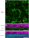
Fig. 12.. Two-photon and second-harmonic imaging facilitates monitoring of corneal cells and extracellular matrix in vivo.
High magnification en face image of keratocytes in a normal mouse cornea at 80 μm depth (A). A cross-sectional view of the epithelium, stroma and endothelium are shown with keratocytes visible in green (B) or stromal extracellular matrix in cyan (C). A globally expressed, membrane-localized fluorescent reporter (Rosa26-mTmG) was used to visualize cell morphology in vivo. The stromal ECM is visualized by Second-Harmonic Generation and depicted in cyan. Scale bar represents 100 μm. Image courtesy of Dr. Panteleimon Rompolas, Departments of Dermatology and Ophthalmology, University of Pennsylvania Perelman School of Medicine.
Fig. 13.
Fig. 13.. Dynamic interactions between the matrix and cellular stiffness during corneal wound healing.
We have demonstrated that the intrinsic stiffness of the corneal stroma undergoes dynamic changes in stiffness throughout wound healing in a rabbit PTK model and that transformation of quiescent keratocytes to contractile myofibroblasts is facilitated by both soluble factors (e.g. TGFβ) and substratum stiffness in vitro (Dreier et al., 2013; Raghunathan et al., 2017). By contrast, 17AAG can revert fibroblasts and myofibroblasts to a keratocyte phenotype in vitro (Raghunathan et al., 2021). Furthermore, we have demonstrated that crosslinking stiffens the stroma resulting in myofibroblast persistence and increased stromal haze in a rabbit model in vivo (Moore et al., 2023). Novel therapeutics that target stromal softening may facilitate return to a more homeostatic corneal matrix and cell population.
Fig. 14.
Fig. 14.. Compliant substrates limit the transformation to the myofibroblast phenotype and modulate stromal cell stiffness.
Rabbit corneal fibroblasts were cultured in the absence or presence of TGFβ1 on substrates with varying compliance then fixed and immunohistochemical stains performed for α-smooth muscle actin (αSMA, green), F-actin (red), and 40,6-diamidino-2-phenylindole (DAPI; blue). Most cells on TCP displayed strong staining for αSMA when exposed to TGFβ1, consistent with a myofibroblast phenotype. By contrast, few cells (on 28-kPa gels) or no cells (on 4-kPa gels) stained positive for αSMA, even with TGFβ1 treatment; F-actin demonstrated increased stress fiber formation on TCP versus the compliant substrates (A). Rabbit (B) and human (C) corneal stromal cells were stiffer on substrates mimicking the elastic modulus of native stroma in health and disease; TGFβ1 treatment further increased cell stiffness. Adapted from Dreier, B., Thomasy, S.M., Mendonsa, R., Raghunathan, V.K., Russell, P., Murphy, C.J., 2013. Substratum compliance modulates corneal fibroblast to myofibroblast transformation. Invest Ophthalmol Vis Sci 54, 5901–5907, copyright Association for Research in Vision and Ophthalmology and Raghunathan, V.K., Thomasy, S.M., Strøm, P., Yañez-Soto, B., Garland, S.P., Sermeno, J., Reilly, C.M., Murphy, C.J., 2017. Tissue and cellular biomechanics during corneal wound injury and repair. Acta Biomater 58, 291–301.
Fig. 15.
Fig. 15.. Growth factors and their subsequent deprivation initiate distinct cell morphologies, behaviors, and matrix secretion in keratocytes.
Cells treated with IGF demonstrated similar morphology to control keratocytes with low, peripheral traction force, low fibrillar fibronectin (Fn) and collagen deposition, absent Fn fiber strain, low expression of transglutaminase 2 (TG2), high expression of keratocan and ALDH1a1, and low metabolic activity. Withdrawal of IGF did not alter keratocyte morphology, force, fibronectin fiber strain and metabolic activity. By contrast, keratocytes treated with PDGF or FGF demonstrated a proliferative, metabolically active, low contractility phenotype with contact-guided migration and formation of a fibrillar fibronectin matrix. The FBS- or TGFβ1-treated keratocytes displayed extensive stress fibers, an actin cap, and high traction force consistent with a (myo)fibroblast phenotype and deposited the densest Fn and collagen matrix with marked contraction and extensive reorganization of their surrounding ECM. Withdrawal of PDGF, FGF, FBS or TGFβ1 returned the corneal cells to a keratocyte-like phenotype with low, peripheral traction force, low to decreased fibrillar Fn and collagen deposition, and absent to low Fn fiber strain, and low metabolic activity. Reprinted with permission from Pot, S.A., Lin, Z., Shiu, J., Benn, M.C., Vogel, V., 2023. Growth factors and mechano-regulated reciprocal crosstalk with extracellular matrix tune the keratocyte-fibroblast/myofibroblast transition. Sci Rep 13, 11350, CC04.0.
Fig. 16.
Fig. 16.. Intimate fibrosis and remodeling feedback loop between corneal stromal cells and their matrix.
The damaged corneal epithelium induces release of soluble factors, notably PDGF and TGFβ1, leading to keratocyte activation. (A) Activated keratocytes (from left to right: control keratocyte, PDGF-primed fibroblast, FBS-primed fibroblast, TGFβ1-primed myofibroblast) show changes in cell morphology, increased metabolic activity, ECM assembly, contractility, and fibronectin (Fn) fiber unfolding. (B) Growth factor removal from cell culture was used to mimic decreasing stromal growth factor amounts following epithelial and basement membrane healing. Substrate type contributed to the completeness of cell phenotype reversal (compare B to A), such that substrate sensing by cells facilitates their phenotype development. (C) Fluorescence Resonance Energy Transfer (FRET) experiments demonstrated that forces generated by cells translate into changes in mechanical matrix strain (Fn unfolding). The most contractile phenotypes, TGFβ1-and FBS and (myo)fibroblasts, stretched Fn fibers within the ECM such that partial secondary/tertiary Fn protein structure loss occurred. This Fn fiber unfolding exposes cryptic Fn-Fn self-assembly sites, accelerating Fn fibrillogenesis, cross-linking and fiber bundling, and stabilization of the early Fn matrix. This Fn fiber unfolding exposes a cryptic Toll-like receptor (TLR) 4 activating site on Fn’s ED-A domain, resulting in TLR activation, and subsequent TGFβ, tenascin-C (TNC), Fn, and collagen-1 gene expression. Cell generated force induced Fn fiber stretching promotes Fn fibrillogenesis with subsequent assembly of a collagen matrix. Increased tissue stresses and tension from cell proliferation, contractility, and matrix assembly drives profibrotic gene expression (incl. αSMA, collagen, TNC), and thus myofibroblast transition and the deposition of a contracted, dense, crosslinked collagen-1-rich matrix (DF), in a self-amplifying manner. Finally, decreasing PDGF and TGFβ1 concentrations following epithelial and basement membrane healing, together with normalizing ECM properties, including tissue specific matrix topography, stiffness, and collagen fiber composition, regulates the disappearance of myofibroblasts from wound sites. Thus, an ECM niche supportive of homeostasis and regenerative remodeling is created. Experimental results (bold) from Pot et al., (2023) with published literature (italics). Reprinted with permission from Pot, S.A., Lin, Z., Shiu, J., Benn, M.C., Vogel, V., 2023. Growth factors and mechano-regulated reciprocal crosstalk with extracellular matrix tune the keratocyte-fibroblast/myofibroblast transition. Sci Rep 13, 11350, CC-BY-NC-ND 4.0.
Fig. 17.
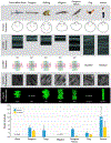
Fig. 17.. Differential responses of primary human corneal stromal fibroblasts to exogenous drug treatments.
Primary human corneal stromal fibroblasts were cultured either untreated or with 10 μM ROCK inhibitor (Y27632), or 10 μM Wnt inhibitors (KY20111 or LGK974) for 3 days in growth media. (A) Cytoskeletal structure imaged by AFM demonstrates presence of stress fibers in control cells, but this was inconspicuously absent in Y-27632 treated cells. Fluorescent labelling with phalloidin (F-actin) and DAPI (nucleus) corroborates these findings. (B) Elastic moduli of cells were measured at the tallest region (over the nucleus) by AFM. While ROCK inhibition softened the cells, Wnt inhibition had no significant effects on moduli compared with untreated control cells. *p < 0.05, ANOVA followed by multiple comparison test against control.
Fig. 18.
Fig. 18.
(A) Modulation of mechanical properties through crosslinking of cell-derived extracellular matrices. Primary human and/or rabbit corneal stromal fibroblasts (HCFs or RCFs, respectively) were cultured for 4 weeks in DMEM media supplemented with 10% FBS, penn/strep and 50 mg/ml ascorbic acid, with media changes performed twice weekly. At the end of the 4 weeks, cells were removed by successive incubation with a buffer containing 20 mM NH4OH, 0.05% Triton X-100 in water. Decellularization was confirmed by detecting the presence/absence of fluorescent labelling for F-actin and nucleus (data not shown). Elastic moduli of decellularized cell-derived matrices were then either measured with no subsequent treatment or after crosslinking with either 0.05% riboflavin/UV-A for 15 min, or with EDC/NHS for 90 min. Both Riboflavin/UV-A and EDC/NHS crosslinking treatments potently increased matrix stiffness. (BD) Impact of decellularization on potency of crosslinking in human cornea. (B) Elastic moduli of human cornea prior and after decellularization was comparable with no significant differences noted. Decellularization was achieved by using an antigen removal method without the use of sodium dodecyl sulfate (SDS). Decellularization was confirmed by H&E staining (data not shown) (C) Elastic moduli of the anterior stroma of naïve human corneas with cells intact were measured with no treatment or after treatment with Riboflavin/UV-A following epi-Off protocol. Significant stiffening of the stroma was observed as expected. ***P < 0.001, Mann-Whitney U test. (D) Human corneas were decellularized using the antigen retrieval method referenced above. Elastic moduli of decellularized corneas were measured with either no subsequent treatment or after crosslinking with riboflavin/UV-A or EDC/NHS. No significant differences in elastic moduli were observed between the groups by ANOVA.
Fig. 19.
Fig. 19.. Collagen crosslinking (CXL) prior to or at the same time as corneal wounding with a phototherapeutic keratectomy (PTK) increased stromal haze relative to PTK alone in a rabbit model.
(A) Stromal haze thickness (%) of the central cornea was significantly higher in the CXL groups than controls; CXL + PTK and CXL then PTK groups did not significantly differ (B) Representative color photographs and OCT images of a single rabbit in each group at baseline and days 0, 7, 21, and 90. Marked stromal haze was observed from day 7 until day 90 in the simultaneous CXL + PTK group. (C) The elastic modulus of corneas with simultaneous CXL and PTK was significantly higher than CXL then PTK corneas at day 21, but no significant differences were observed by day 90. In addition, no significant differences were observed between the CXL then PTK and the debride then PTK groups. *P < 0.05, repeated measure two-way ANOVA followed by Tukey’s multiple comparisons test. Reproduced from Moore, B.A., Jalilian, I., Kim, S., Mizutani, M., Mukai, M., Chang, C., Entringer, A.M., Dhamodaran, K., Raghunathan, V.K., Teixeira, L.B., Murphy, C.J., Thomasy, S.M., 2023. Collagen crosslinking impacts stromal wound healing and haze formation in a rabbit phototherapeutic keratectomy model Mol Vis 29, 101–115.
Fig. 20.
Fig. 20.. Administration of cysteamine hydrochloride, a TGM2 inhibitor, does not affect corneal scarring or stromal stiffness.
Representative clinical, slit beam, and OCT images illustrating the area of corneal haze at final day 42 and graphic representation of clinical haze scores (A). Corneal haze was present but not significantly different between control and CH treated eyes (n = 8 eyes per group to day 28, n = 4 eyes per group to day 42, P > 0.05 for all timepoints). Corneal fibrosis, as scored based on H&E histology sections, was not significantly different between treated and control eyes (n = 7 per group, P = 0.3473, B). Immunohistochemical α-SMA expression showed no differences between treated and control eyes, suggesting no appreciable change in KFM transformation (n = 7 per group, P = 0.2821, C). Corneal thickness as measured by OCT, was not different between control and treated eyes suggesting no difference in amount of scarring (n = 8 eyes per group to day 28, n = 4 eyes per group to day 42, P > 0.05 for all timepoints, D). Atomic force microscopy of sections from the area of wounded cornea showed no difference in stiffness between control and treated eyes (n = 4 eyes per time point, P = 0.99 at Day 28, P = 0.96 at Day 42. E). Reprinted from Minella, A.L., Casanova, M.I., Chokshi, T.J., Kang, J., Cosert, K., Gragg, M.M., Bowman, M.A., McCorkell, M.E., Daley, N.L., Leonard, B.C., Murphy, C.J., Raghunathan, V.K., Thomasy, S.M., 2023. The TGM2 inhibitor cysteamine hydrochloride does not impact corneal epithelial and stromal wound healing in vitro and in vivo. Exp Eye Res 226, 109338.
Fig. 21.
Fig. 21.. Variation in corneal endothelial proliferative capacity among species.
Following corneal endothelial injury, rabbits demonstrate rapid and robust mitosis. The corneal endothelial cells of rodents also demonstrate some mitotic capacity although it is slower than in rabbits. Dogs, particularly young ones, also exhibit a proliferative corneal endothelium post-injury but it is slower than that of rodents. By contrast, cats, non-human primates (NHPs), and humans demonstrate little mitotic ability consistent with that of humans. Adapted from Park, S., Leonard, B.C., Raghunathan, V.K., Kim, S., Li, J.Y., Mannis, M.J., Murphy, C.J., Thomasy, S.M., 2021. Animal models of corneal endothelial dysfunction to facilitate development of novel therapies. Ann Transl Med 9, 1271, CC BY-NC-ND 4.0.
Fig. 22.
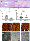
Fig. 22.. Stiffness and topography of corneal Descemet’s membranes (DM) differed between normal and guttae-affected donors.
Elastic moduli of DM were significantly higher in normal versus guttae-affected donors whereas elastic moduli of the endothelium were similar. Representative histologic images of a decellularized healthy DM from a 65-year-old female (A) and a decellularized guttae-affected DM from a 69-year-old male (B) with three large guttae (excrescences of extracellular matrix on DM, arrows) observed. Scale bar equivalent to 20 μm. Elastic modulus of DM (C) and corneal endothelial cells (D) in healthy donors (n = 10) and guttae-affected donors (n = 6). Data points represents single atomic force microscopy (AFM) measurements from all individuals within the healthy or guttae-affected patients. Horizontal lines indicate mean and error bars represent SD. The P values were determined by a Mann-Whitney rank sum test (DM) and Student’s t-test (endothelium), **P < 0.01. ns: not significant. The DM of normal corneas and those with guttae were critical point dried and imaged by AFM (EG). Tissues were glued to AFM compatible dishes on their epithelial side and were then scanned at a rate of 0.1 μm/s under PeakForce Tapping mode using a AC-240TS cantilever. Each image was taken in 1024 × 1024 pixel and the height channel was used to show the details of the surface of normal and guttae-affected DMs. The DMs severely affected with guttae demonstrate collagen structures on their surface (G, blue arrows) which were not observed in controls (E) and corneas moderately affected with guttae (F). Scale bar, 1 μm. Subsequent to imaging by AFM, samples were sputter coated with gold and imaged using a scanning electron microscope (HJ) which validated dysregulated collagen banding in samples severely affected with guttae (J) which was absent in control (H) and corneas moderately affected with guttae (I). Figure and legend adapted from Leonard, B.C., Park, S., Kim, S., Young, L.J., Jalilian, I., Cosert, K., Zhang, X., Skeie, J.M., Shevalye, H., Echeverria, N., Rozo, V., Gong, X., Xing, C., Murphy, C.J., Greiner, M.A., Mootha, V.V., Raghunathan, V.K., Thomasy, S.M., 2023. Mice Deficient in TAZ (Wwtr1) Demonstrate Clinical Features of Late-Onset Fuchs’ Endothelial Corneal Dystrophy. Invest Ophthalmol Vis Sci 64, 22 with unpublished data.
Fig. 23.
Fig. 23.. TAZ-deficient mice exhibit reduced corneal endothelial cell density (ECD), abnormal morphology, softer DM, and delayed endothelial wound healing when compared with WT controls.
(AB) In vivo confocal microscopy revealed reduced ECD in both Wwtr1+/ and Wwtr1−/−mice at 2 months compared with WT mice. Despite age-related corneal endothelial cell loss, TAZ-deficient mice continued to have further declines in ECD at 6 and 12 months of age. Additionally, endothelial cells from TAZ-deficient mice demonstrated abnormal morphology (lower percentage of hexagonal cells, higher shape variability, altered reflectivity) when compared with WT mice. Scale bar equivalent to 50 μm. (B) Each dot represents a single eye (n ≥ 9 eyes in each group); horizonal line represents mean of the group and error bars reflect standard deviation. (C) Atomic force microscopy (AFM) performed on DM revealed decreased elastic modulus (softer) in TAZ-deficient mice, with Wwtr1/ mice having the lowest measurement, followed by Wwtr1+/ mice and finally WT mice (stiffest). Each dot represents a single eye (n ≥ 9 eyes in each group), horizonal line represents mean of the group and error bars reflect standard deviation. A two-factor ANOVA was performed to detect statistical differences, *, **, ***, **** represent P < 0.05, P < 0.01, P < 0.001, P < 0.0001, respectively. Wwtr1 deficient mice demonstrated impaired corneal endothelial cell regeneration after corneal cryoinjury (DF). A cryoinjury wound was created with a 2 mm diameter steel probe immersed in liquid nitrogen for 3 min (−196 °C) and subsequently applied to the cornea for 10 s in WT (n = 8), Wwtr1+/ (n = 8), and Wwrt1/ (n = 5) mice. On day 2, animals were euthanized, and the right eye was stained with Alizarin red to calculate total denuded area (D) and the left eye was stained with EdU to assess cell proliferation (EF). On day 2, there was a significantly larger denuded area in Wwtr1+/ mice compared with WT mice (E). Additionally, Wwtr1/ mice had a trend towards larger denuded areas compared with WT mice (E). There were no differences in EdU staining, indicating that the proliferative capacity of the corneal endothelial cells was equal across groups (F). Both Alizarin red and EdU staining were analyzed using Kruskal-Wallis tests, **P < 0.01. Alizarin red scale bars equivalent to 500 μm and EdU staining scale bars equivalent to 20 μm (inset). Figure and legend adapted from Leonard, B.C., Park, S., Kim, S., Young, L.J., Jalilian, I., Cosert, K., Zhang, X., Skeie, J.M., Shevalye, H., Echeverria, N., Rozo, V., Gong, X., Xing, C., Murphy, C.J., Greiner, M.A., Mootha, V.V., Raghunathan, V.K., Thomasy, S.M., 2023. Mice Deficient in TAZ (Wwtr1) Demonstrate Clinical Features of Late-Onset Fuchs’ Endothelial Corneal Dystrophy. Invest Ophthalmol Vis Sci 64, 22.
Fig. 24.
Fig. 24.. ECM pathology in DM impairs corneal endothelial cell function in FECD.
We hypothesize that abnormal ECM deposition leads to YAP/TAZ upregulation and nuclear translocation (Hippo pathway inactivation) and thus contributes to corneal endothelial cell death. Cell death, in turn, drives further dysfunction and abnormal ECM deposition of surrounding corneal endothelial cells that leads to an escalating cycle of pathology in the posterior cornea in FECD.
Fig. 25.
Fig. 25.. TAZ and YAP localize to the nucleus of corneal endothelial cells surrounding large guttae in FECD surgical explants compared to non-FECD healthy controls.
Compared to healthy donor corneal endothelial tissues (A, B, F, G) isolated <18h after donor death and prior to storage in media, protein expression of both TAZ (green) and YAP (green) in age-matched FECD surgical samples (N = 7 and 3, respectively; C-E, H-J) is greater in all surgical tissues compared to controls. Expression of TAZ is greatest in the nucleus of corneal endothelial cells of FECD tissues in the central cornea adjacent to large guttae (E) compared to cells in the midperipheral cornea adjacent to smaller guttae (D); expression of TAZ is greatest in the cytoplasm in the peripheral cornea where there are fewer guttae (C). Similarly, expression of YAP is greatest in the nucleus of corneal endothelial cells in the central cornea adjacent to large guttae (J), but in contrast, YAP nuclear expression is also increased in the midperiphery (I) and periphery (H) of FECD tissues. In aggregate, this pattern indicates translocation of TAZ and YAP from the cytosol to the nucleus (e.g., Hippo pathway is “off”) when corneal endothelial cells lose confluency or grow on top of or next to guttae, consistent with aberrant Hippo pathway mediated mechanotransduction in FECD progression. Images are representative samples. Nuclear counter stain = DAPI (blue). Scale bars are 50 μm. Figure and legend adapted from Leonard, B.C., Park, S., Kim, S., Young, L.J., Jalilian, I., Cosert, K., Zhang, X., Skeie, J.M., Shevalye, H., Echeverria, N., Rozo, V., Gong, X., Xing, C., Murphy, C.J., Greiner, M.A., Mootha, V.V., Raghunathan, V.K., Thomasy, S.M., 2023. Mice Deficient in TAZ (Wwtr1) Demonstrate Clinical Features of Late-Onset Fuchs’ Endothelial Corneal Dystrophy. Invest Ophthalmol Vis Sci 64, 22 with unpublished data.
Fig. 26.
Fig. 26.. Extracellular matrix protein alterations are prevalent in diabetic donor corneas that mechanically alter DM and the corneal endothelium.
Colocalization of adhesive glycoproteins with advanced glycation end products (AGEs) at the DM–stroma (St) interface in diabetic and normal control corneas. (A–L) Immunohistochemical double-labeling experiments showing differential staining patterns of the DM–St interface region (arrows) for AGEs and vitronectin (A, B), fibronectin (C, D), amyloid P (E, F), TGFBI (G, H), tenascin-C (I, J), and collagen IV (K, L). Nuclei are counterstained with DAPI (blue). EN, endothelium. (M, N) Immunogold double-labeling for AGEs (10-nm gold particles) and vitronectin (M) or fibronectin (N) (20-nm gold particles) in the IFM of diabetic corneas. ABL, anterior banded layer of the DM. These results indicate that AGE residues are localized specifically to the adhesive matrix proteins in the interfacial matrix of diabetic tissues. (O, P) The DM and endothelium were markedly stiffer in diabetic versus control corneas as determined by atomic force microscopy. The elastic modulus was measured in six diabetic and six age-matched control corneas and was significantly greater at 16.77 ± 5.01 and 8.95 ± 4.07 kPa for the DM and 1.29 ± 0.48 and 0.70 ± 0.31 kPa for the endothelium, respectively (P < 0.0001). Box plots depict the median (solid line) and 25th and 75th percentiles, and whiskers show the 10th and 90th percentiles. Black circles indicate individual measurements. Figure and legend adapted from Kingsbury, K.D., Skeie, J.M., Cosert, K., Schmidt, G.A., Aldrich, B. T., Sales, C.S., Weller, J., Kruse, F., Thomasy, S.M., Schlötzer-Schrehardt, U., Greiner, M.A., 2023. Type II Diabetes Mellitus Causes Extracellular Matrix Alterations in the Posterior Cornea That Increase Graft Thickness and Rigidity. Invest Ophthalmol Vis Sci 64, 26.
Fig. 27.
Fig. 27.. Response to topical ripasudil four times daily was variable in CED-affected dogs with 62% demonstrating stable disease or improvement while 38% had disease progression.
Representative digital photographs and IVCM images of CED-affected eyes at baseline and 12-months post treatment (A) An 8-year-old male castrated Boston terrier demonstrated improved corneal edema (A1 to A2) and improved endothelial cell density (ECD, A3 to A4), increasing from 1565 ± 150 cells/mm2 at baseline to 1733 ± 224 cells/mm2 at 12 months. (B) A 5-year-old male castrated chihuahua mix demonstrated progressive corneal edema (B1 to B2) and decreased ECD (B3 to B4) from 1324 ± 138 cells/mm2 at baseline to 881 ± 17 cells/mm2 at 12 months. (C) A 12-year-old male castrated shih tzu demonstrated stable corneal edema (C1 to C2) and stable ECD (C3 to C4) from 1674 ± 199 cells/mm2 at baseline to 1588 ± 30 cells/mm2 at 12 months. The ECD is reported as mean ± SD. Kaplan-Meier curves demonstrated that CED-affected corneas treated with topical ripasudil progressed slower than historical controls (D). Tick marks indicate censored subjects, and the shaded area indicates 95% confidence intervals. Median time from the initial visit to meeting any of the progression criteria in historical controls was 223 days. Median time to progression was not reached in ripasudil-treated eyes. Log-rank test identified a significant difference in distribution of time to progression between eyes receiving topical ripasudil and untreated historical controls (P = 0.023). Seventeen ripasudil-treated eyes and 17 untreated, age- and breed/size-matched historical controls were included in the analysis. Adapted from Michalak, S.R., Kim, S., Park, S., Casanova, M.I., Bowman, M.A.W., Ferneding, M., Leonard, B.C., Good, K.L., Li, J.Y., Thomasy, S.M., 2022. Topical Ripasudil for the Treatment of Primary Corneal Endothelial Degeneration in Dogs. Transl Vis Sci Technol 11, 2.

Similar articles

Cited by

References

    1. Ablamowicz AF, Nichols JJ, 2016. Ocular surface membrane-associated mucins. Ocul. Surf 14, 331–341. - PubMed
    1. Abrams GA, Bentley E, Nealey PF, Murphy CJ, 2002. Electron microscopy of the canine corneal basement membranes. Cells Tissues Organs 170, 251–257. - PubMed
    1. Abrams GA, Goodman SL, Nealey PF, Franco M, Murphy CJ, 2000a. Nanoscale topography of the basement membrane underlying the corneal epithelium of the rhesus macaque. Cell Tissue Res 299, 39–46. - PubMed
    1. Abrams GA, Schaus SS, Goodman SL, Nealey PF, Murphy CJ, 2000b. Nanoscale topography of the corneal epithelial basement membrane and Descemet’s membrane of the human. Cornea 19, 57–64. - PubMed
    1. Afshari NA, Igo RP Jr., Morris NJ, Stambolian D, Sharma S, Pulagam VL, Dunn S, Stamler JF, Truitt BJ, Rimmler J, Kuot A, Croasdale CR, Qin X, Burdon KP, Riazuddin SA, Mills R, Klebe S, Minear MA, Zhao J, Balajonda E, Rosenwasser GO, Baratz KH, Mootha VV, Patel SV, Gregory SG, Bailey-Wilson JE, Price MO, Price FW Jr., Craig JE, Fingert JH, Gottsch JD, Aldave AJ, Klintworth GK, Lass JH, Li YJ, Iyengar SK, 2017. Genome-wide association study identifies three novel loci in Fuchs endothelial corneal dystrophy. Nat. Commun 8, 14898. - PMC - PubMed