Review
. 2024 Jan 3;9(1):9.
doi: 10.1038/s41392-023-01723-x.

Lymphatic vessel: origin, heterogeneity, biological functions, and therapeutic targets

Affiliations
Review

Lymphatic vessel: origin, heterogeneity, biological functions, and therapeutic targets

Zhaoliang Hu et al. Signal Transduct Target Ther. .

Abstract

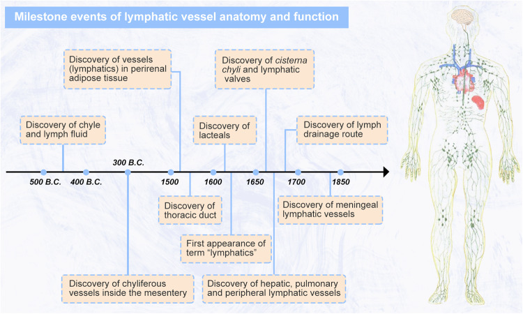
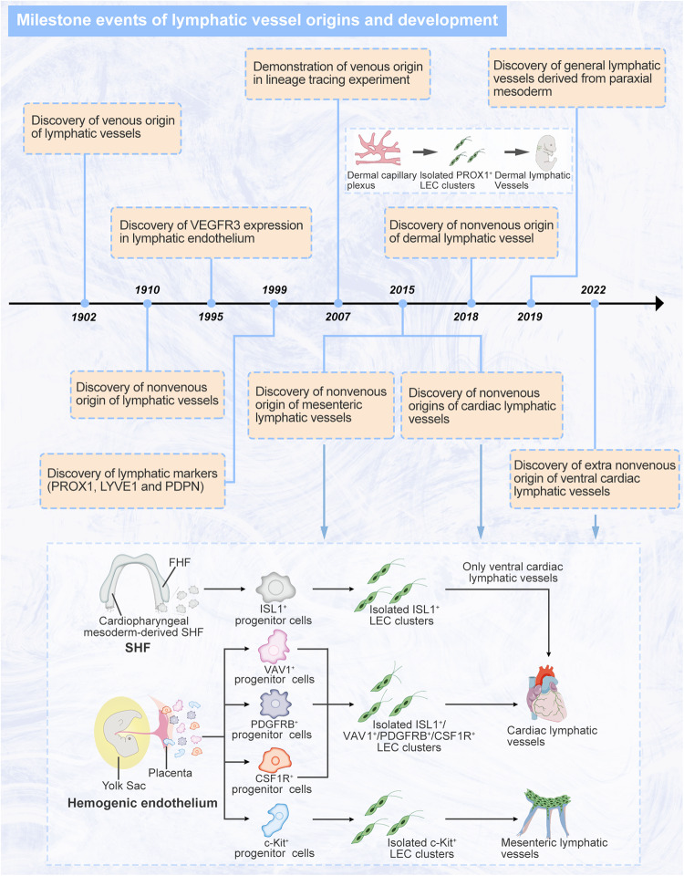
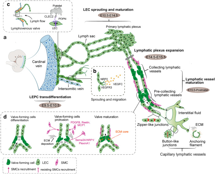
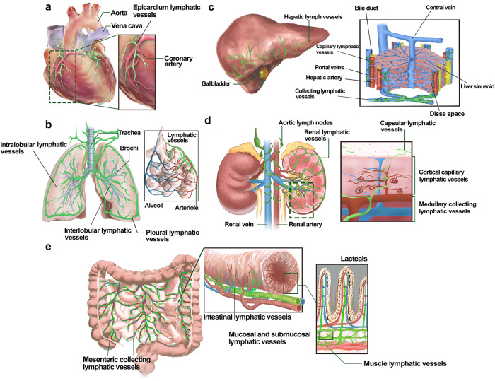
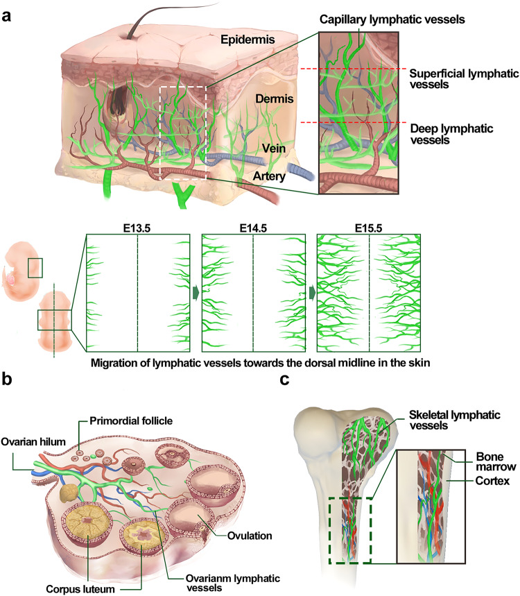
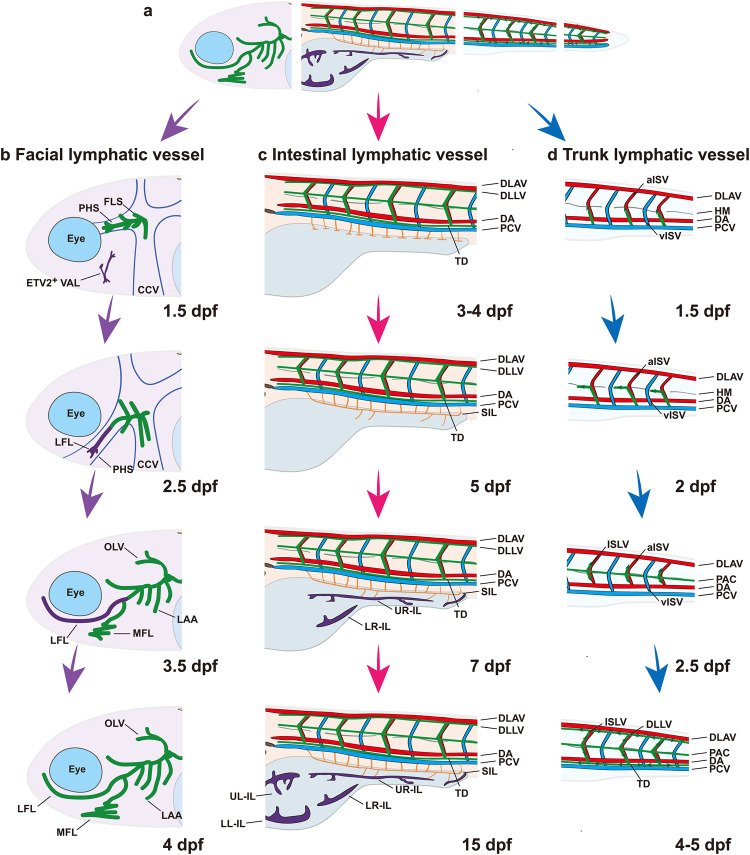
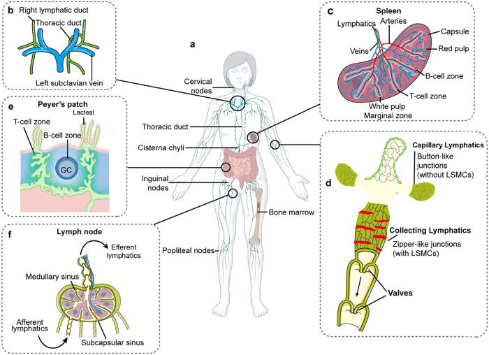
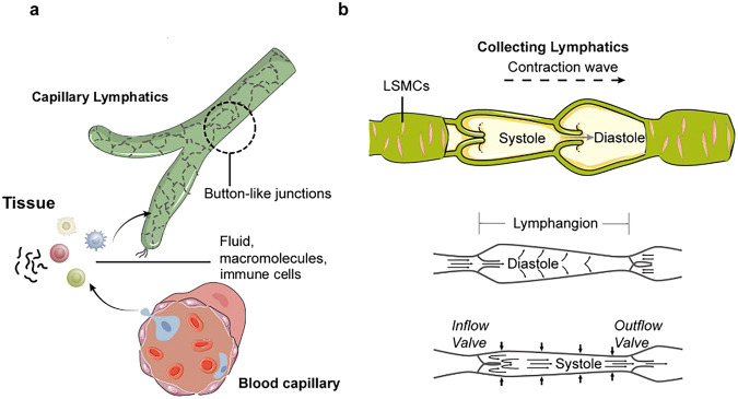
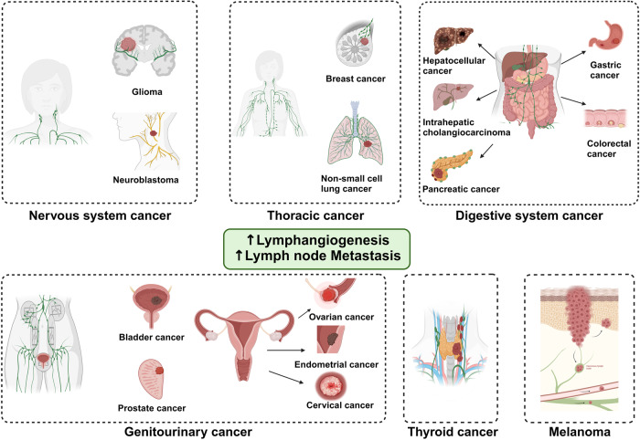
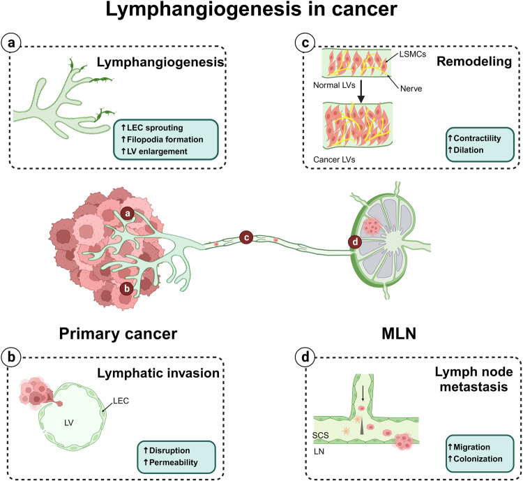
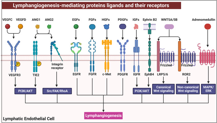
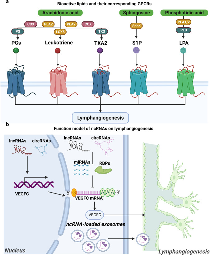
Lymphatic vessels, comprising the secondary circulatory system in human body, play a multifaceted role in maintaining homeostasis among various tissues and organs. They are tasked with a serious of responsibilities, including the regulation of lymph absorption and transport, the orchestration of immune surveillance and responses. Lymphatic vessel development undergoes a series of sophisticated regulatory signaling pathways governing heterogeneous-origin cell populations stepwise to assemble into the highly specialized lymphatic vessel networks. Lymphangiogenesis, as defined by new lymphatic vessels sprouting from preexisting lymphatic vessels/embryonic veins, is the main developmental mechanism underlying the formation and expansion of lymphatic vessel networks in an embryo. However, abnormal lymphangiogenesis could be observed in many pathological conditions and has a close relationship with the development and progression of various diseases. Mechanistic studies have revealed a set of lymphangiogenic factors and cascades that may serve as the potential targets for regulating abnormal lymphangiogenesis, to further modulate the progression of diseases. Actually, an increasing number of clinical trials have demonstrated the promising interventions and showed the feasibility of currently available treatments for future clinical translation. Targeting lymphangiogenic promoters or inhibitors not only directly regulates abnormal lymphangiogenesis, but improves the efficacy of diverse treatments. In conclusion, we present a comprehensive overview of lymphatic vessel development and physiological functions, and describe the critical involvement of abnormal lymphangiogenesis in multiple diseases. Moreover, we summarize the targeting therapeutic values of abnormal lymphangiogenesis, providing novel perspectives for treatment strategy of multiple human diseases.


Conflict of interest statement

The authors declare no competing interests.

Figures

Fig. 1
Fig. 1
Milestone events of lymphatic vessel anatomy and function. Since the first discovery of chyle and lymph fluid at 5th B.C., some milestone findings have gradually revealed lymphatic vessel network and function in mammals. Created with Adobe Illustrator
Fig. 2
Fig. 2
Milestone events of lymphatic vessels origins and development. In modern research (from 1902 to now), numerous researchers have gradually discovered the diverse origins and molecules of the lymphatic vessel development. These studies have doubtless initiated the understanding of heterogeneous development processes and regulatory mechanism of lymphatic vessels. VEGFR3 vascular endothelial growth factor receptor 3, PROX1 prospero homeobox protein 1, LYVE1 lymphatic vessel endothelial hyaluronan receptor 1, PDPN podoplanin, LEC lymphatic endothelial cell, FHF first heart field, SHF second heart field, ISL1 Islet 1, VAV1 vav guanine nucleotide exchange factor 1, PDGFRB platelet-derived growth factor receptor B, CSF1R colony-stimulating factor 1 receptor. Created with Adobe Illustrator
Fig. 3
Fig. 3
The schematic diagram of embryonic lymphatic vessel development. a Beginning at E9.5, VeECs, located at CV and ISVs, transdifferentiate into LEPCs. During E10.5-E15.5, the lymphatic plexus continues to sprouting and migrating, and expanding the primary lymphatic vessel network. Continuing from E15.5 until the early postnatal stage, the primary lymphatic plexus undergoes the maturation events to remodel into the hierarchical lymphatic vessels, comprising of capillary lymphatic vessels, pre-collecting lymphatic vessels, and collecting lymphatic vessels. Capillary lymphatic vessels sense interstitial pressure changes by anchoring filaments to control the opening of button-like junctions. The formation of pre-collecting and collecting lymphatic vessels requires for lymphatic valves morphogenesis and SMCs recruitment to drive lymph drainage; b At E10.5, upregulated VEGFR3 and NRP2 mediate LEPCs sprouting and LECs migration in response to VEGFC stimulation. VEGFC/VEGFR3 is an irreplaceable signaling regulates lymphatic vessel development; c The lymphovenous valve serves as the only connection of the lymphatic-venous system to prevent blood backflow. Platelet dynamically regulated lymphovenous hemostasis via interacting with LECs to activate CLEC2/PDPN signaling pathway to maintain platelet aggregation; d Under the stimulation of OSS, the differentiation of valve-forming cells prepares to proliferation, elongation, and protrusion. Moreover, ECM deposition and selective SMCs coverage further promote lymphatic vessel maturation. Ex embryonic day x, VeECs venous endothelial cells, CV cardinal vessel, ISVs intersomitic veins, LECs lymphatic endothelial cells, LEPCs lymphatic endothelial progenitor cells, VEGFC vascular endothelial growth factor C, VEGFR3 vascular endothelial growth factor receptor 3, NRP1/2 neuropilin 1/2, CLEC2 c-type lectin-like receptor 2, PDPN podoplanin, OSS oscillatory shear stress, MCP1 monocyte chemotactic protein 1, PDGFB platelet-derived growth factor B, ECM extracellular matrix, SMCs smooth muscle cells. Created with Adobe Illustrator
Fig. 4
Fig. 4
The lymphatic vessels in meninge and eyes. a The meningeal lymphatic vessels are mainly located at the dural region abut to the cranium, and developing along the cerebral vessels and nerves. Meningeal lymphatic vessels enter the nasal submucosal interstitium traveling through cribriform plate and form nasal lymphatic vessels extracranially, partly participating in the extracranial CSF drainage; b The meningeal lymphatic vessels exchange CSF with glymphatic system at subarachnoid space and eventually drain it into dcLNs; c SC abuts juxtacanalicular region of the TM, consisting of inner and outer wall constituted by heterogeneous endothelial cells. The inner wall could sense flow and transport aqueous humor into the SC and further drain to downstream episcleral veins; d The ocular surface lymphatic vessels originate from the nasal canthus and encircle laterally along the corneal limbus and the bulbar conjunctiva. CSF cerebrospinal fluid, dcLNs deep cervical lymph nodes, SC Schlemm’s canal, TM trabecular meshwork. Created with Adobe Illustrator
Fig. 5
Fig. 5
The lymphatic vessels in heart, lung, liver, kidney, and intestine. a Cardiac lymphatic vessels develop from the extracardiac region and follow the basal-to-tip manner along the developing coronary arteries to the ventricles; b Pulmonary lymphatic vessels consists of interlobular, intralobular and pleural lymphatic vessels, and develop surround airway, blood vessels and developing alveoli; c Capillary lymphatic vessels of the portal tract region mainly absorb the lymph secreted into the Disse space and eventually drain through collecting lymphatic vessels into thoracic duct; d Capsular lymphatic vessels are located near the renal surface. Cortical capillary lymphatic vessels accompany the renal tubules, glomeruli and small arteries and run along the medullary collecting lymphatic network and are finally excluded from the kidney via the hilar lymphatic vessels; e The intestinal lymphatic vessels consist of mesenteric collecting lymphatic vessels, mucosal, submucosal, muscle lymphatic vessels, and lacteals. Created with Adobe Illustrator
Fig. 6
Fig. 6
The lymphatic vessels in skin, ovary, and bone. a Skin lymphatic vessels, consisting of superficial and deep lymphatic vessel networks, are mainly located in the dermis and partly accompanied by dermal blood vessels. During E13.5 to E15.5, the primitive lymphatic plexus of the dorsally cervical region of skin begins to develop from the bilateral sides toward the midline of the back; b Ovarian lymphatic vessels develop along established blood vessels in the interstitium, which happened during the period of the first wave of follicular development; c Skeletal lymphatic vessels could develop at the sternum, femur, and tibia. And this part shows the skeletal lymphatic vessels go along the bone marrow of the long bones. Ex embryonic day x. Created with Adobe Illustrator
Fig. 7
Fig. 7
The schematic of lymphatic vessels development in zebrafish. a Zebrafish possess an extensive lymphatic vessel network throughout the body. Specialized lymphatic vessels development in zebrafish includes facial lymphatic vessel development (b), intestinal lymphatic vessel development (c), and trunk lymphatic vessel development (d); b The facial lymphatic vessels of zebrafish undergo a step-wise assembly from 1.5 dpf to 4 dpf. The FLS are derived from the CCV beginning to form along the PHS. Subsequently, a group of ETV2-expressing cells, known as VAL, begin to fuse with the lymphatic sprouts to form the LFL along the PHS. The LFL then begins to bud out to form a complex facial lymphatic vessels including the OLV, LAA, and MFL; c The development of intestinal lymphatic vessels proceeds from 3 dpf–15 dpf. At 3 dpf-4 dpf, LECs sprout from the PCV to the ventral and bilateral sides, respectively to form segmental lymphatic vessels, which subsequently interconnect to form L-SIL, R-SIL. The SILs first bud toward the right side of the abdomen along the vascular network to form UR-IL and IR-IL. Finally, the lymphatic vessel network continues to expand follow the left vascular track to form UL-IL and LL-IL and take up intestinal lymph; d The formation of trunk lymphatic vessels is the earliest event of embryonic lymphatic vessel development in zebrafish, budding from the PCV to form the ISLV along the trajectory of the ISVs, and subsequently sprouting ventrally and dorsally to form the DLLV and TD, respectively. Dpf day postfertilization, CCV common cardinal vein, PHS primary head sinus, FLS facial lymphatic sprouting, VAL ventral aorta lymphangioblast, LFL lateral facial lymphatic vessel, OLV otolithic lymphatic vessel, ETV2 ETS variant transcription factor 2, LAA lymphatic branchial arches, MFL medial facial lymphatic vessels, PCV posterior cardinal vein, L-SIL the left supraintestinal vessel, R-SIL the right supraintestinal vessels, UR-IL upper-right intestinal lymphatics, LR-IL lower-right intestinal lymphatics, UL-IL upper-left intestinal lymphatics, LL-IL lower-left intestinal lymphatics, ISLV intersegmental lymphatic vessel, aISV arterial intersegmental vessels, vISV venous intersegmental vessel, PAC parachordal line, DA dorsal aorta, DLAV dorsal longitudinal anastomotic vessel, HM horizontal myoseptum, DLLV dorsal longitudinal lymphatic vessel, TD thoracic duct. Created with Adobe Illustrator
Fig. 8
Fig. 8
Anatomy of the lymphatic system. a The lymphatic system includes the primary and secondary lymphoid organs and lymphatic vessels, providing a one-way drainage route from all tissues back ultimately to the blood circulation via the great veins in the neck. In the primary lymphoid organs (bone marrow and thymus), immune cell production and maturation takes place, whereas secondary lymphoid organs (lymph nodes, spleen, and mucosa-associated lymphoid organs such as Peyer’s patch, tonsils, and adenoids) are the sites for lymphocyte activation; b The thoracic duct is responsible for the lymph drainage coming from most of the body with the exception of the right side of the head and neck, the right side of the thorax and the right upper limb where drain lymph primarily into the right lymphatic duct; ce The spleen, the Peyer’s patch and lymph nodes are highly organized structures with segregated B-cell and T-cell zones to optimize the induction of adaptive immune responses; f The capillary lymphatics drain downstream into the collecting lymphatics. Capillary LECs are interconnected via discontinuous junctions allowing the fluid to enter the system passively. Collecting LECs present with continuous junctions. Collecting lymphatics differ from capillary lymphatics by possessing intraluminal valves, LSMCs and a continuous basement membrane. LSMCs lymphatic smooth muscle cells, LECs lymphatic endothelial cells, GC germinal center. Created with Adobe Illustrator
Fig. 9
Fig. 9
Lymph absorption and transport. a Capillary lymphatics comprise a single layer of loosely connected LECs lacking a continuous basement membrane and perivascular mural cells. LECs within capillary lymphatics are interconnected through discontinuous button-like junctions that facilitate the uptake of interstitial fluid, macromolecules and immune cells which are released by the blood capillary; b Collecting lymphatics have a period of brisk contraction (systole) and a period of relaxation (diastole) between each phasic contraction. Each lymphangion, defined as the segment between two valves, can typically exhibit systole and diastole. When a lymphangion is relaxed, the inflow (or upstream) valve will open (given sufficient inflow pressure). During systole, the phasic contraction pushes the lymph, but the inflow valve closes, so that lymph is forced forward through the outflow valve. LECs lymphatic endothelial cells. Created with Adobe Illustrator
Fig. 10
Fig. 10
Compartmentalized functions of LN LECs. a Decoy CCL21 receptor CCRL1 produced by cLECs creates a CCL21 gradient and regulates intranodal migration of CCR7-expressing DCs; b The expression of CSF1 in LECs is maintained by RANK expressed on LECs, which is activated by RANKL produced by MRCs; c Medullary LN LECs express CD209 to retain neutrophils, which may be important in clearing lymph-borne pathogens. cLEC ceiling lymphatic endothelial cell, fLEC floor lymphatic endothelial cell, LN lymph node, LEC lymphatic endothelial cell, CCL21 chemokine (C-C motif) ligand 21, CCRL1 chemokine (C-C motif) receptor like 1, CSF1 colony-stimulating factor 1, RANK receptor activator of nuclear factor-kappaB, RANKL receptor activator of nuclear factor-kappaB ligand, DCs dendritic cells, S1P sphingosine 1 phosphate, SCS subcapsular sinus, SSM subcapsular sinus macrophage, MM medullary macrophage, MRC marginal reticular cell. Created with Adobe Illustrator
Fig. 11
Fig. 11
Lymphangiogenesis in cancers. Lymphangiogenesis plays a crucial role in lymph node metastasis, which is associated with poor prognosis and overall survival in a range of malignancies. The molecular mechanisms underlying lymphangiogenesis exhibit diversity across different cancer contexts, potentially suggesting targeted therapeutic strategies for cancers. Created with BioRender.com
Fig. 12
Fig. 12
Lymphangiogenesis and lymph node metastasis in cancer. a Lymphatic vessels undergo sprouting, filopodia formation, and lymphatic vessel enlargement; b The disruption of lymphatic vessels and augmented permeability contribute to the intravasation of cancer cells into lymphatic vessels; c An increased coverage by LSMCs and a higher innervation present in the dilated collecting lymphatic vessels, which coordinately enhances collecting lymphatic vessel contractility and pumping frequency; d LECs forming the boundaries of the SCS create and maintain chemokine gradients that direct cancer cells to arrive in the SCS of MLN. Furthermore, LECs within the MLN upregulate adhesion molecules, that further support cancer cell colonization. LV lymphatic vessel, LSMC lymphatic smooth muscle cell, LEC lymphatic endothelial cell, SCS subcapsular sinus, MLN metastatic lymph node. Created with BioRender.com
Fig. 13
Fig. 13
Lymphangiogenesis-mediating proteins ligands and their receptors. Schematic diagram showing the main promoters of lymphangiogenesis with soluble ligands or interacting proteins present outside the cell and the transmembrane receptors expressed by lymphatic endothelial cells (LECs) at 7the cell surface. VEGFC vascular endothelial growth factor C, VEGFD vascular endothelial growth factor D, VEGFR3 vascular endothelial growth factor receptors 3, ANG angiopoietin, TIE, tunica interna endothelial cell kinase, EGF epidermal growth factor, EGFR epidermal growth factor receptor, FGF fibroblast growth factor, FGFR fibroblast growth factor receptor, HGF hepatocyte growth factor, PDGF platelet-derived growth factor, PDGFR platelet-derived growth factor receptor, IGF insulin-like growth factor, IGFR insulin-like growth factor receptor, AM adrenomedullin. Created with BioRender.com
Fig. 14
Fig. 14
The role of bioactive lipids and ncRNAs on lymphangiogenesis. a The bioactive lipids derived from the metabolism of arachidonic acid, Sphingosine, phosphatidic acid could regulate lymphangiogenesis through binding their specific GPCRs; b The function model of ncRNAs on lymphangiogenesis. Some ncRNAs were reported to have evident role on lyphangiogenesis, especially in tumor associated-lymphangiogenesis. These ncRNAs could be potential therapeutic targets for controlling abnormal lymphangiogenesis in cancer. GPCRs G Protein-Coupled Receptors, COX cyclooxygenase, PLA2 phospholipase A2, PS prostanoid synthases, TXA thromboxane, LOX5 5-lipoxygenase, S1P sphingosine 1-phosphate, LPA lysophosphatidic acid, ncRNAs noncoding RNAs, lncRNAs long noncoding RNAs, circRNAs circular RNAs, RBPs, RNA binding proteins. Created with BioRender.com

Similar articles

Cited by

References

    1. Breslin, J. W. et al. Lymphatic vessel network structure and physiology. Compr. Physiol.9, 207–299 (2018). - PMC - PubMed
    1. Oliver, G., Kipnis, J., Randolph, G. J. & Harvey, N. L. The lymphatic vasculature in the 21st century: novel functional roles in homeostasis and disease. Cell182, 270–296 (2020). - PMC - PubMed
    1. Natale, G., Bocci, G. & Ribatti, D. Scholars and scientists in the history of the lymphatic system. J. Anat.231, 417–429 (2017). - PMC - PubMed
    1. Irschick, R., Siemon, C. & Brenner, E. The history of anatomical research of lymphatics—from the ancient times to the end of the European Renaissance. Ann. Anat.223, 49–69 (2019). - PubMed
    1. Kanter, M. A. The lymphatic system: an historical perspective. Plast. Reconstr. Surg.79, 131–139 (1987). - PubMed

MeSH terms