. 2024 Mar 1;35(3):ar38.
doi: 10.1091/mbc.E23-09-0345. Epub 2024 Jan 3.

The ubiquitin ligases Cbl and Cbl-b regulate macrophage growth by controlling CSF-1R import into macropinosomes

Affiliations

The ubiquitin ligases Cbl and Cbl-b regulate macrophage growth by controlling CSF-1R import into macropinosomes

Lu Huang et al. Mol Biol Cell. .

Abstract

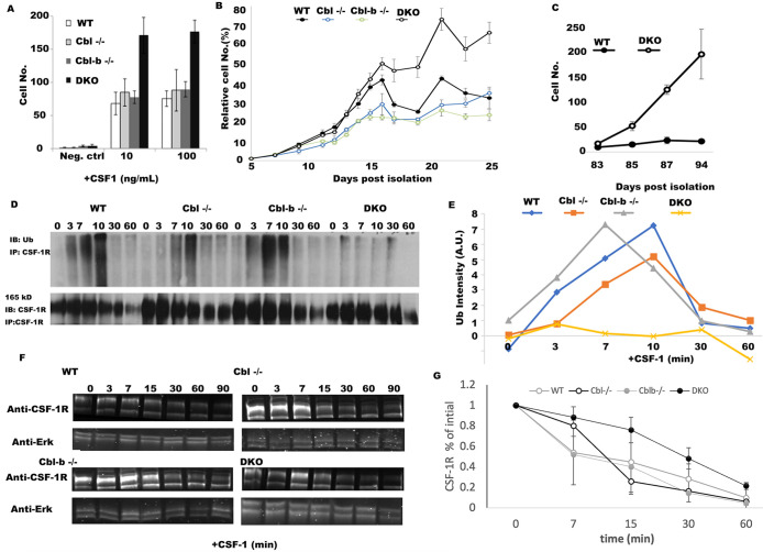
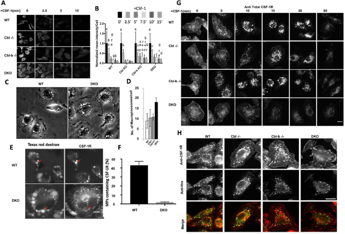
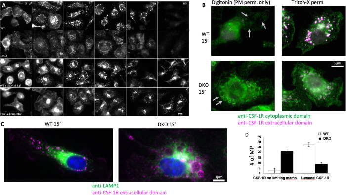
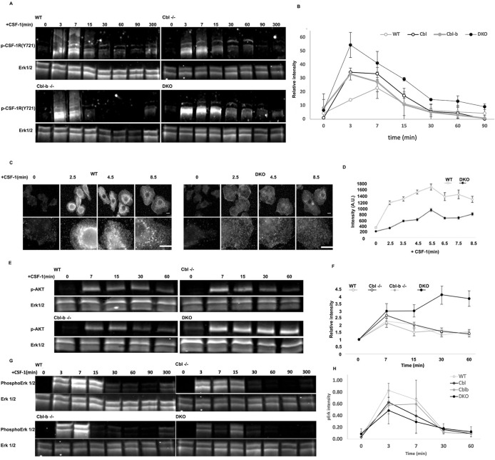
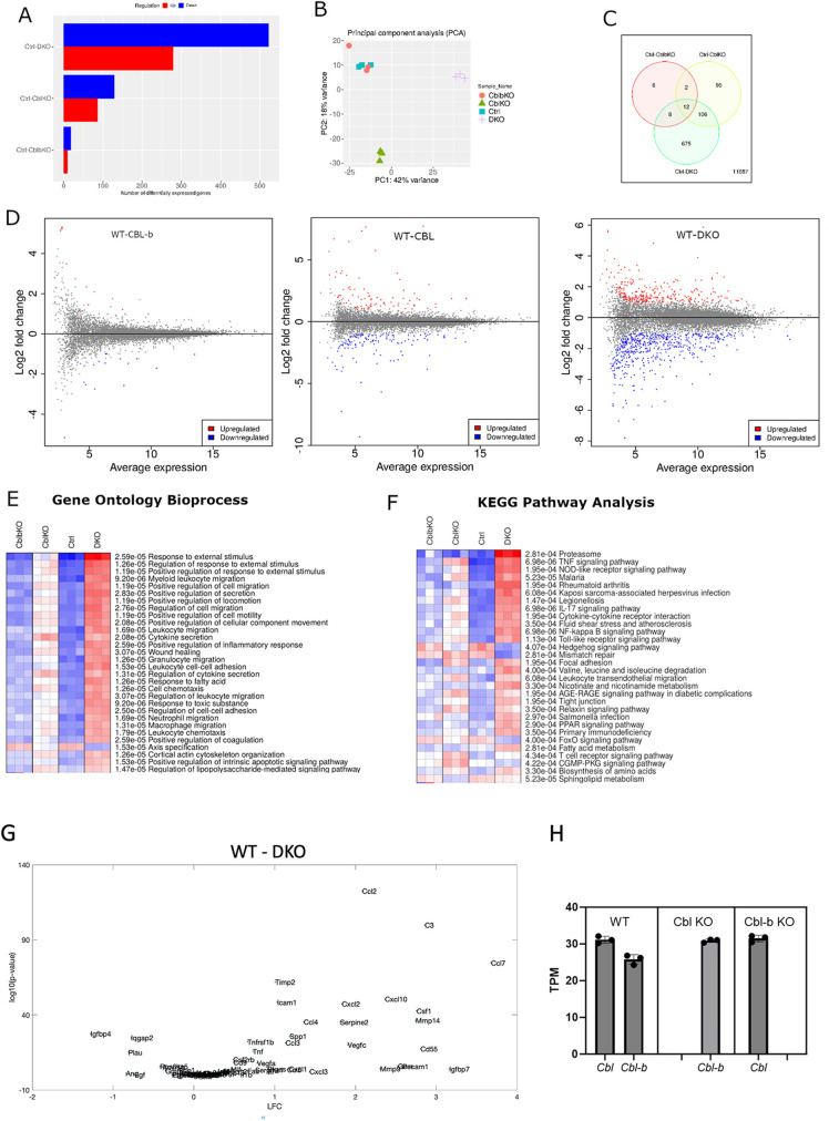
The ubiquitination of transmembrane receptors regulates endocytosis, intracellular traffic, and signal transduction. Bone marrow-derived macrophages from myeloid Cbl-/- and Cbl-b-/- double knockout (DKO) mice display sustained proliferation mirroring the myeloproliferative disease that these mice succumb to. Here, we found that the ubiquitin ligases Cbl and Cbl-b have overlapping functions for controlling the endocytosis and intracellular traffic of the CSF-1R. DKO macrophages displayed complete loss of ubiquitination of the CSF-1R whereas partial ubiquitination was observed for either single Cbl-/- or Cbl-b-/- macrophages. Unlike wild type, DKO macrophages were immortal and displayed slower CSF-1R internalization, elevated AKT signaling, and a failure to transport the CSF-1R into the lumen of nascent macropinosomes, leaving its cytoplasmic region available for signaling. CSF-1R degradation depended upon lysosomal vATPase activity in both WT and DKO macrophages, with this degradation confined to macropinosomes in WT but occurring in distributed/tubular lysosomes in DKO cells. RNA-sequencing comparison of Cbl-/-, Cbl-b-/- and DKO macrophages indicated that while the overall macrophage transcriptional program remained intact, DKO macrophages had alterations in gene expression associated with growth factor signaling, cell cycle, inflammation and senescence. Cbl-b-/- had minimal effect on the transcriptional program whereas Cbl-/- led to more alternations but only DKO macrophages demonstrated substantial changes in the transcriptome, suggesting overlapping but unique functions for the two Cbl-family members. Thus, Cbl/Cbl-b-mediated ubiquitination of CSF-1R regulates its endocytic fate, constrains inflammatory gene expression, and regulates signaling for macrophage proliferation.


Figures

FIGURE 1:
FIGURE 1:
Cbl and Cbl-b DKO macrophages evade exit from the cell cycle and have minimal CSF-1R ubiquitination. (A) BMDM growth measured at day 6 postisolation (n = 5 fields of view and n = 3 replicates, error bar = std. dev.). (B) Growth curves, measured by alamar blue for WT, Cbl–/–, Cbl-b–/– and DKO BMDMs measured over 25 d from 1,000 cells starting at day 5 postisolation. CSF-1−containing media was added every other day followed by replating to a density of 1,000 cells/well (indicated by arrows, n = 6 replicates, error bars = std. dev.). (C) The growth rates between day 83 to day 94 of DKO and WT cells (n = 3 replicates, error bars are std. dev.). (D) CSF-1R immunoprecipitation and ubiquitin blot across BMDM of different genotypes following stimulation with CSF-1 (minutes after stimulation). (E) Quantification of D (data is representative of 3 independent experiments). F. CSF-1R western blot and quantification in BMDM stimulated with CSF-1 (after 0 min). Erk1/2 was used as a loading control. G. Quantification of CSF-1R degradation across three experiments (mean +/- std. dev.).
FIGURE 2:
FIGURE 2:
Cbl and Cbl-b facilitate CSF-1R internalization and translocation to the macropinosome lumen. (A) Immunofluorescence of cell surface CSF-1R in WT and DKO macrophages at indicated CSF-1 stimulation period. (B) Quantification of surface CSF-1R at different time points, scale bars are SD with n = 100, same letters are not significantly different (p< 0.05) by 2-way ANOVA followed by Tukey HSD comparison of means. (C) Phase contrast images illustrating the number of macropinosomes in DKO BMDM. (D) Quantified number of macropinosomes per cell (error bars = std. dev., n = 100 macrophages across two experiments). (E) Delivery of the CSF-1R to the macropinosome visualized labeling by sequential imaging of Texas-red dextran (macropinosome) and CSF-1R immunofluorescence following permeabilization (scale bar = 5 μm). (F) Quantification of the percentage of macropinosomes that contain luminal CSF-1R (error bar = std. dev. with n = 50 from two independent experiments). (G) CSF-1Rimmunofluorescence delivery to macropinosomes and degradation (scale bar = 10 μm). (H). Colocalization of CSF-1R and Hrs 10 min after CSF-1 exposure (scale bar = 10 μm).
FIGURE 3:
FIGURE 3:
Cbl and Cbl-b are essential for sequestration of the CSF-1R and subsequent degradation within macropinosomes. (A) Immunofluorescence of the CSF-1R with and without inhibition of the vacuolar ATPase with 30 min pretreatment with bafilomycin A1 followed by CSF-1 exposure time course (images are identically window leveled and were collected under identical conditions). (B) Immunofluorescence staining of the intracellular and extracellular epitopes of the CSF-1R with selective permeabilization of the plasma membrane with digitonin or complete membrane permeabilization with Triton-x 100 at 15 min post CSF-1 stimulation. Gray arrows denote locations of macropinosomes. Green staining in during digitonin indicates cytosolic accessibility of the CSF-1R. (C) Distribution of LAMP1-positive lysosomes in relation to the CSF-1R contained with the lumen (WT) or limiting membrane of the macropinosome (DKO) at 15 min post CSF-1 stimulation. (D) Quantification of the CSF-1R localization from 30 macropinosomes for each WT and DKO based on morphology in the Triton-X 100 staining condition.
FIGURE 4:
FIGURE 4:
Cbl and Cbl-b limit CSF-1R and AKT phosphorylation while supporting Erk1/2 and broad tyrosine phosphorylation. (A) Immunoblot of CSF-1R phosphorylation (pY721) following CSF-1 stimulation for 0−300 min. Note the higher molecular weight products consistent with ubiquitination observed in all blots except DKO. (B) Quantification of the phospho-CSF-1R signal intensity from three immunoblots (all molecular weights of CSF-1R), normalized to total Erk1/2 (mean+/– std. dev.) (C and D) Immunofluorescence and quantification of pan phosphotyrosine in WT and DKO BMDM following CSF-1 stimulation, illustrated broadly impaired tyrosine phosphorylation in the DKO. Error bar is standard error of mean, n = 50 cells per genotype. (E) AKT(S473) phosphorylation following CSF-1 stimulation. (F) Quantification of pAKT(S473), mean+/-std. dev. from two independent experiments. (G) Western blot of Erk1/2 phosphorylation following CSF-1 stimulation. (H) Quantification of pErk (mean +/- std. dev., n = 3 experiments).
FIGURE 5:
FIGURE 5:
Differential gene expression analysis of Cbl, Cbl-b, and DKO BMDMs. (A) Differential genes expression (DESeq2) across genotypes as compared WT(ctrl) (FDR = 0.05). (B) Principal component analysis indicated that gene expression in Cbl-b closely matched WT, whereas Cbl and DKO displayed more unique variations in gene expression. (C) Venn diagram illustrating the number of differentially expressed genes shared and unique to each genotype comparison. (D) MA plots comparing each mutant with WT. Statistically significant expression differences are color coded. (E and F) Gene ontology bioprocess and KEGG pathway analysis of genes with FDR of < 0.05. G. DQseq2 analysis comparing WT and DKO genes contained within the mouse Saul_Sen_Mayo GSEA molecular signatures database.

Similar articles

References

    1. Andoniou CE, Lill NL, Thien CB, Lupher ML, Ota S, Bowtell DD, Scaife RM, Langdon WY, Band H (2000). The Cbl proto-oncogene product negatively regulates the Src-family tyrosine kinase Fyn by enhancing its degradation. Mol Cell Biol 20, 851–867. - PMC - PubMed
    1. An W, Nadeau SA, Mohapatra BC, Feng D, Zutshi N, Storck MD, Arya P, Talmadge JE, Meza JL, Band V, Band H (2015). Loss of Cbl and Cbl-b ubiquitin ligases abrogates hematopoietic stem cell quiescence and sensitizes leukemic disease to chemotherapy. Oncotarget 6, 10498–10509. - PMC - PubMed
    1. An W, Mohapatra BC, Zutshi N, Bielecki TA, Goez BT, Luan H, Iseka F, Mushtaq I, Storck MD, Band V, Band H (2016). VAV1-Cre mediated hematopoietic deletion of CBL and CBL-B leads to JMML-like aggressive early-neonatal myeloproliferative disease. Oncotarget 7, 59006–59016. - PMC - PubMed
    1. Cannarile MA, Weisser M, Jacob W, Jegg A-M, Ries CH, Rüttinger D (2017). Colony-stimulating factor 1 receptor (CSF1R) inhibitors in cancer therapy. J Immunother Cancer 5, 53. - PMC - PubMed
    1. Chitu V, Stanley ER (2017). Regulation of Embryonic and Postnatal Development by the CSF-1 Receptor. Curr Top Dev Biol 123, 229–275. - PMC - PubMed

MeSH terms

Substances

LinkOut - more resources