. 2024 Mar 1;35(3):ar35.
doi: 10.1091/mbc.E23-07-0294. Epub 2024 Jan 3.

Tonic ErbB signaling underlies TGFβ-induced activation of ERK and is required for lens cell epithelial to myofibroblast transition

Affiliations

Tonic ErbB signaling underlies TGFβ-induced activation of ERK and is required for lens cell epithelial to myofibroblast transition

Judy K VanSlyke et al. Mol Biol Cell. .

Abstract

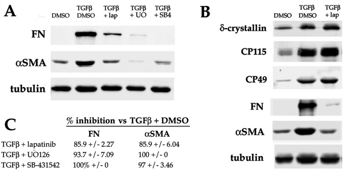
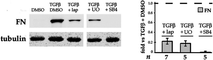
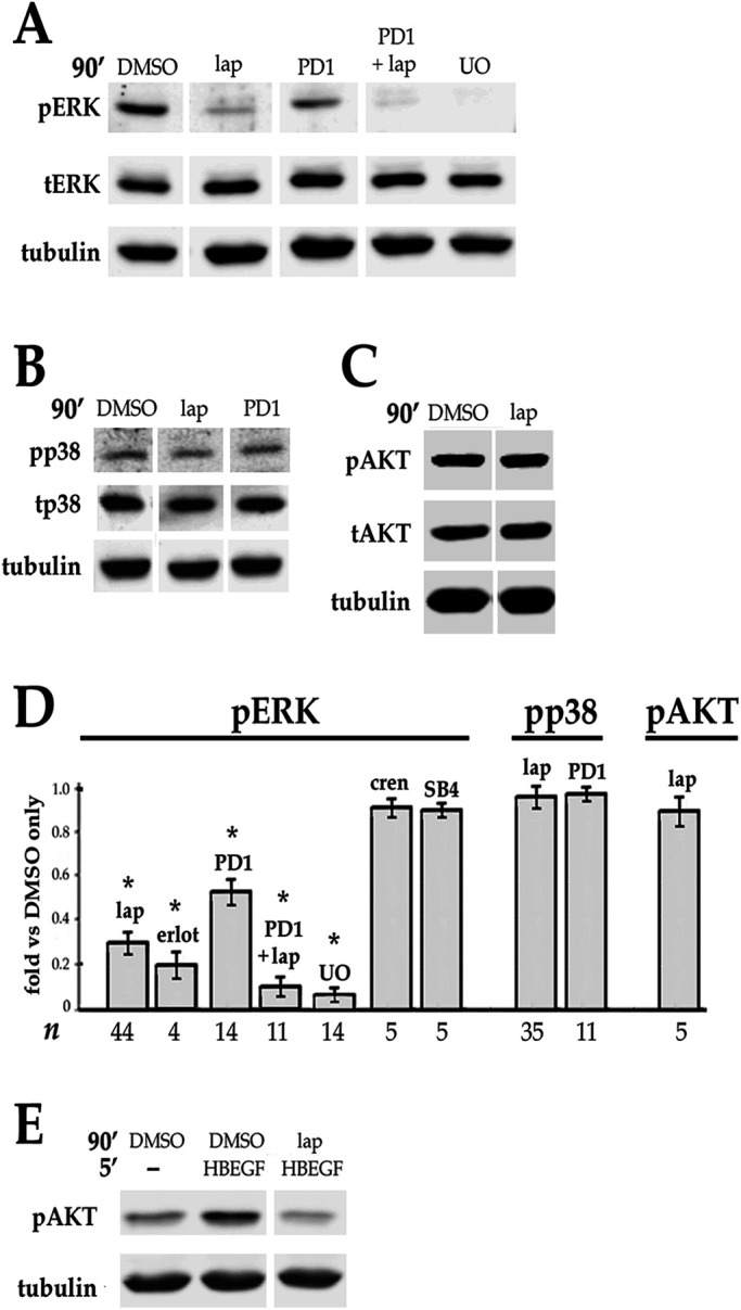
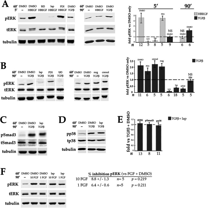
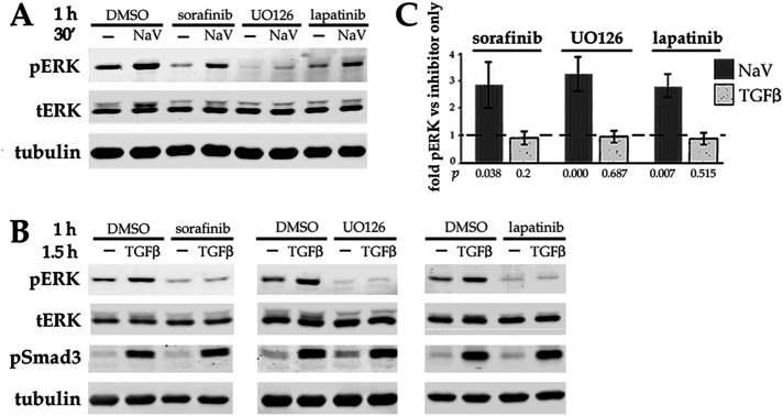
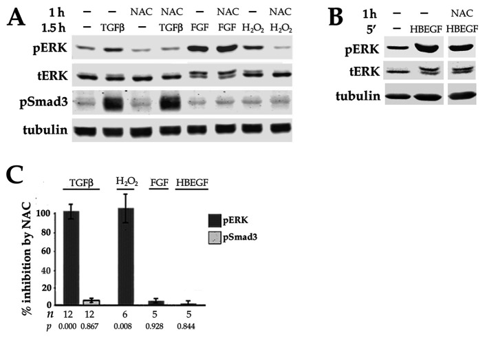
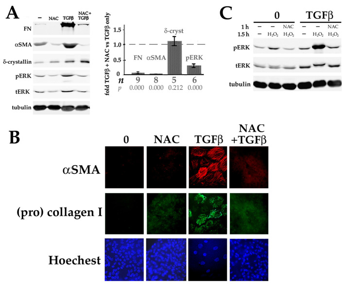
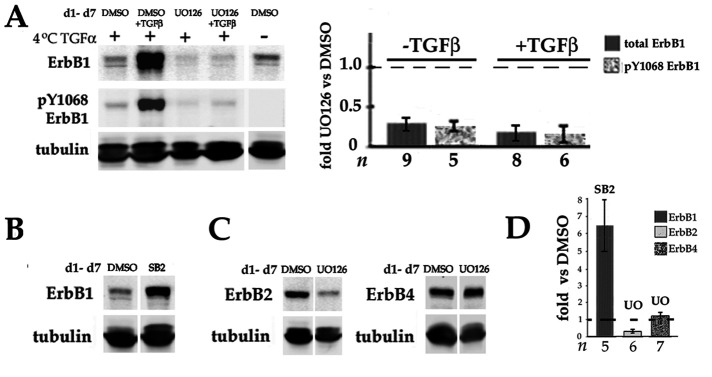
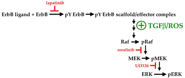
Fibrosis is a major, but incompletely understood, component of many diseases. The most common vision-disrupting complication of cataract surgery involves differentiation of residual lens cells into myofibroblasts. In serum-free primary cultures of lens epithelial cells (DCDMLs), inhibitors of either ERK or of ErbB signaling prevent TGFβ from upregulating both early (fibronectin) and late (αSMA) markers of myofibroblast differentiation. TGFβ stimulates ERK in DCDMLs within 1.5 h. Kinase inhibitors of ErbBs, but not of several other growth factor receptors in lens cells, reduce phospho ERK to below basal levels in the absence or presence of TGFβ. This effect is attributable to constitutive ErbB activity playing a major role in regulating the basal levels pERK. Additional studies support a model in which TGFβ-generated reactive oxygen species serve to indirectly amplify ERK signaling downstream of tonically active ErbBs to mediate myofibroblast differentiation. ERK activity is in turn essential for expression of ErbB1 and ErbB2, major inducers of ERK signaling. By mechanistically linking TGFβ, ErbB, and ERK signaling to myofibroblast differentiation, our data elucidate a new role for ErbBs in fibrosis and reveal a novel mode by which TGFβ directs lens cell fate.


Figures

FIGURE 1:
FIGURE 1:
Effect of inhibition of ERK or ErbB on TGFβ stimulation of myofibroblast differentiation in DCDMLs. DCDML primary lens epithelial cells were treated starting on d 1 of culture with or without 4 ng/ml TGFβ1, in the presence or absence of the following kinase inhibitors: lapatinib (lap), UO126 (UO), or SB-431542 (SB4). Cells were lysed on d 7 and whole cell lysates analyzed for the EMyT markers FN and αSMA (A), or for FN, αSMA, and the lens fiber cell markers δ-crystallin, CP115, and CP49 (B) by Western blot. (C) Quantitation of EMyT marker data, expressed as the percent inhibition induced by lapatinib, UO126, or SB-431542 relative to cells treated with TGFβ and vehicle (0.1% DMSO) only. All data are normalized to tubulin in the same sample. For all, n ≥ 6; p = 0.000.
FIGURE 2:
FIGURE 2:
Both ERK and ErbB activity are required for TGFβ to induce the early stage of EMyT. DCDMLs were cultured from d 1–3 in the presence or absence of TGFβ with or without lapatinib, UO126, or SB-431542 as indicated. Cells were lysed on d 3 and whole cell lysates analyzed for expression of FN. For all, p ≤ 0.004.
FIGURE 3:
FIGURE 3:
Effect of blocking ErbB and/or other growth factor receptors on the basal level of phospho ERK, p38, and AKT in DCDMLs. (A–C) DCDMLs were incubated for 90 min with DMSO, lapatinib, the selective FGFR inhibitor PD173074, lapatinib plus PD173074, or UO126 before Western blot analysis of phospho and total forms of ERK, p38, AKT, or of tubulin as indicated. (D) Results graphed as fold controls treated with DMSO only. Also included are data demonstrating that the ErbB inhibitor erlotinib (erlot), but not inhibitors of the receptors for TGFβ (SB-431542) or PDGF (crenolanib), reduces basal levels of pERK. Asterisks indicate p ≤ 0.001; for all other conditions, p ≥ 0.337. (E) DCDMLs were incubated with DMSO or lapatinib and lysed after a 5 min treatment with 10 ng/ml HB-EGF or medium only to demonstrate that the anti-pAKT antibody recognizes activated (pS475) AKT in DCDMLs.
FIGURE 4:
FIGURE 4:
ErbB and TGFβ signaling in lens cells. DCDMLs were pretreated for 1 h with DMSO or the indicated inhibitors before incubation for 5 or 90 min with either 10 ng/ml HB-EGF, 4 ng/ml TGFβ, 1 or 10 ng/ml FGF, or an equal volume of M199 medium (-). Whole cell lysates were analyzed for phospho and total forms of ERK, Smad3, and p38, or for tubulin by Western blot. (A) ErbB ligand induces a rapid and transient activation of ERK compared with TGFβ. Results graphed as fold pERK relative to DMSO only control. p < 0.001, except NS (p >0.325). Activation by phosphorylation decreases the apparent mobility of total (t) ERK(2) on SDS–PAGE, resulting in a doublet under conditions in which ERK is partially activated. (B) Inhibitor sensitivity of activation of ERK by TGFβ. Results graphed as fold pERK relative to DMSO only (no TGFβ) control. p < 0.001, except NS (p = 0.22). Compared to cells treated with TGFβ plus DMSO, the level of pERK in cells treated with TGFβ plus PD173074 is significantly lower (p = 0.000), whereas crenolanib (0.5 μM) and noggin (1 μg/ml) have no effect on activation of pERK by TGFβ (p > 0.628). Also graphed are data demonstrating that the ErbB inhibitor erlotinib and the TGFβR inhibitor SB-431542 reduce upregulation of pERK by TGFβ. (C) Lapatinib does not block activation of Smad3 by TGFβ. (D) Lapatinib does not block activation of p38 by TGFβ. (E) Quantitation of the data as compared with treatment with TGFβ and DMSO. In all cases, p ≥ 0.133. (F) Lapatinib does not block activation of pERK by FGF, as quantitated as percent inhibition of pERK in cells treated with FGF + lapatinib compared with cells treated with FGF + DMSO.
FIGURE 5:
FIGURE 5:
Effect of long-term exposure to FGF or TGFβ on ERK. (A) TGFβ continues to stimulate ERK in DCDMLs chronically exposed to physiological levels of FGF. DCDMLs were cultured for 6 d in either the absence or presence of 1 ng/ml FGF2. Cells were then incubated for 60 min with DMSO or lapatinib, followed by a 90 min treatment with no additions (–), 1 ng/ml FGF2, or 4 ng/ml TGFβ. Whole cell lysates were assessed for phospho ERK, total ERK, or tubulin by Western blot. All results shown are from the same blot of a single experiment. Levels of pERK obtained under the indicated conditions are graphed relative to controls treated with DMSO without inhibitor or growth factor. NS, p = 0.593; for all other conditions, p < 0.012. (B) Prolonged exposure to TGFβ increases both phospho and total levels of ERK. DCDMLs were cultured for 6 d in either the absence or presence of TGFβ. Whole cell lysates were assessed for pERK, tERK, or tubulin by Western blot. pERK and tERK values were normalized to tubulin in the same sample.
FIGURE 6:
FIGURE 6:
TGFβ does not stimulate the release of HB-EGF from DCDMLs. (A) DCDMLs were transiently transfected with plasmids encoding either a full-length, transmembrane proform of HB-EGF or an irrelevant control transmembrane protein (E208K Cx32; VanSlyke et al., 2023; Ctl) and cultured for 48 h in either the absence or presence of TGFβ. Untransfected HEK293 cells were then incubated at 4°C for 15 min with either 10–0 ng/ml mature, soluble recombinant HB-EGF (lanes 1–4), or the indicated DCDML transfectant conditioned medium (lanes 5–9). HEK whole cell lysates were analyzed by Western blot using antibodies against phosphotyrosine. To confirm the identity of the bracketed 170 kD pY band as autophosphorylated ErbB, recipient HEK cells were incubated at 37°C for 1 h with lapatinib before addition of conditioned medium (lanes 8 and 9); asterisk indicates a nonspecific band. Uniform expression of ErbB1, a major endogenous ErbB in HEKs, is also shown. All data shown are from the same blot of a single experiment. Phospho-Y values (in arbitrary units, normalized to β actin in the same samples) for the HB-EGF concentration curve are provided. (B) ErbB-associated 4G10 signal from three independent experiments was normalized to β actin and showed that conditioned medium from proHB-EGF transfectants cultured with TGFβ induced 0.893 ± 0.115 times the ErbB-associated 4G10 signal as was elicited by conditioned medium from cells cultured in the absence of TGFβ. Individual data points are plotted as black squares.
FIGURE 7:
FIGURE 7:
Short-term treatment with TGFβ does not enhance activation of ErbBs in DCDMLs. (A) The level of tyrosine phosphorylated ErbBs on the cell surface is unaffected by TGFβ. DCDMLs were incubated at 37°C for 1 h with DMSO or lapatinib, or for 5, 45, or 90 min with or without TGFβ as indicated. In lanes 1 and 2, cells were then incubated at 4°C for 15 min with 100 ng/ml HB-EGF. All cultures were then subjected to cell surface biotinylation at 4°C, and plasma membrane pools of autoactivated ErbB analyzed by Western blotting using the antiphosphotyrosine 4G10 antibody. To demonstrate equal loading, biotinylated samples were reprobed with ErbB1 antibody and whole cell lysates were analyzed for β actin. (B) TGFβ does not increase autophosphorylation of ErbB1. DCDMLs were incubated at 37°C for 1 h with DMSO or lapatinib, or for 5, 45, or 90 min with or without TGFβ as indicated. In lanes 13-16, cells were then treated at 37°C for 5 min with 0.1, 1, or 10 ng/ml HB-EGF. All cultures were then lysed. Western blotting of whole cell lysates with a rabbit antibody specific to pY1068ErbB1 demonstrated that treatment with HB-EGF, but not with TGFβ, increased the activation of ErbB1. Blots were reprobed with a rat antibody against total ErbB1. (C). Results from (A) and (B) were graphed as fold value from cells incubated with TGFβ versus without TGFβ for the indicated time. In all cases, p > 0.2. Individual data points are plotted from experiments in which n = 3.
FIGURE 8:
FIGURE 8:
Stimulation of ERK by TGFβ requires the canonical ErbB-to-ERK pathway and is not phenocopied by a tyrosine phosphatase inhibitor. DCDMLs were preincubated for 1 h with either DMSO, 10 uM sorafenib, UO126, or lapatinib before exposure to either the general tyrosine phosphatase inhibitor sodium orthovanadate (NaV; 0.2 mM) for 30 min (A), or to TGFβ for 1.5 h (B). The levels of pERK, tERK, pSmad3, and tubulin were assessed by Western blot. (C) The level of pERK in cells treated with either sodium orthovanadate or TGFβ in the presence of each kinase inhibitor was graphed relative to pERK in cells exposed to kinase inhibitor only. n = 5.
FIGURE 9:
FIGURE 9:
TGFβ upregulation of pERK is specifically inhibited by the antioxidant NAC. DCDMLs were preincubated for 1 h with or without 20 mM NAC before treatment with: (A) 4 ng/ml TGFβ, 1 ng/ml FGF, or 250 μM H2O2 for 90 min, or (B) 10 ng/ml HB-EGF for 5 min. Whole cell lysates were then analyzed for pERK, tERK, pSmad3, and tubulin. (C) Results were graphed as percent inhibition by NAC of upregulation of pERK or pSmad3 by the indicated treatment. Similar results were obtained with 10 mM NAC.
FIGURE 10:
FIGURE 10:
Oxidative stress is required for upregulation of EMyT by TGFβ and is enhanced by TGFβ. (A) EMyT in response to TGFβ is blocked by the antioxidant NAC. DCDMLs were cultured for 6 d with no additions (-), 20 mM NAC, TGFβ, or both (NAC added 1 h before TGFβ). Whole cell lysates were then analyzed by Western blot for FN, αSMA, δ-crystallin, phospho and total ERK, and tubulin. Results graphed as fold values obtained in cells cultured with NAC plus TGFβ relative to values from cells cultured with TGFβ only. (B) DCDMLs cultured as in (A) were fixed and immunostained to demonstrate the lack of αSMA-containing stress fibers in NAC treated cells. NAC also prevented TGFβ from inducing the expression of the fibrotic marker collagen I as assessed by an antibody specific to procollagen I. (C) Multi-day exposure to TGFβ increases pERK levels in response to oxidative stress. DCDMLs cultured for 6 d in the presence or absence of TGFβ were preincubated with or without 20 mM NAC for 1 h before a 1.5 h incubation in the absence or presence of 250 μM H2O2. Cell lysates were analyzed for phospho and total ERK, or tubulin. NAC inhibited the activation of ERK by H2O2 by 100% ± 0 in cells cultured in the absence of TGFβ, and by 84% ±10.8 in cells cultured with TGFβ (n = 9; p = 0.00).
FIGURE 11:
FIGURE 11:
ERK is required for expression of ErbB1 and ErbB2 protein. (A) DCDMLs were cultured for 6 d with DMSO, UO126, TGFβ, or both (DMSO or UO126 added 1 h before TGFβ). The cells were then incubated at 4°C for 15 min in either the absence or presence of 60 ng/ml TGFα as indicated. Whole cell lysates were then analyzed by Western blot with mouse anti-pY1068ErbB1 antibodies, followed by reprobing for total ErbB1. Data are graphed as fold obtained in cultures treated with DMSO instead of UO126 in the same experiment. For all conditions, p = 0.000. Note that the anti-pY1068ErbB1 antibody (#2236) employed in these experiments is not as sensitive as the rabbit reagent used in Figure 7B. (B) DCDMLs were cultured for 6 d with DMSO or the p38 inhibitor SB203580 before analysis of whole cell lysates for total ErbB1. (C) DCDMLs cultured for 6 d with DMSO or UO126 were lysed and analyzed for ErbB2 or ErbB4. (D) Effect of SB203580 on the level of ErbB1 (p = 0.012), or UO126 on the levels of ErbB2 (p = 0.000) and ErbB4 (p = 0.123) detected by Western blot expressed as fold DMSO only cultures in the same experiment.
FIGURE 12:
FIGURE 12:
Kinases in the core canonical ErbB-to-ERK pathway, and proposed step at which TGFβ/ROS acts to stimulate ERK in lens epithelial cells in an ErbB-dependent manner.

Similar articles

Cited by

References

    1. Albeck JG, Mills GB, Brugge JS (2013). Frequency-modulated pulses of ERK activity transmit quantitative proliferation signals. Mol Cell 49, 249–261. - PMC - PubMed
    1. Apple DJ, Ram J, Foster A, Peng Q (2000). Elimination of cataract blindness: a global perspective entering the new millenium. Surv Ophthalmol 45(Suppl 1), S1–S196. - PubMed
    1. Awasthi N, Guo S, Wagner BJ (2009). Posterior capsular opacification: a problem reduced but not yet eradicated. Arch Ophthalmol 127, 555–562. - PubMed
    1. Boswell BA, Korol A, West-Mays JA, Musil LS (2017). Dual function of TGFβ in lens epithelial cell fate: implications for secondary cataract. Mol Biol Cell 28, 907–921. - PMC - PubMed
    1. Boswell BA, Le AC, Musil LS (2009). Upregulation and maintenance of gap junctional communication in lens cells. Exp Eye Res 88, 919–927. - PMC - PubMed

Substances