. 2023 Dec 27;19(12):e1011089.
doi: 10.1371/journal.pgen.1011089. eCollection 2023 Dec.

Rhotekin regulates axon regeneration through the talin-Vinculin-Vinexin axis in Caenorhabditis elegans

Affiliations

Rhotekin regulates axon regeneration through the talin-Vinculin-Vinexin axis in Caenorhabditis elegans

Yoshiki Sakai et al. PLoS Genet. .

Abstract

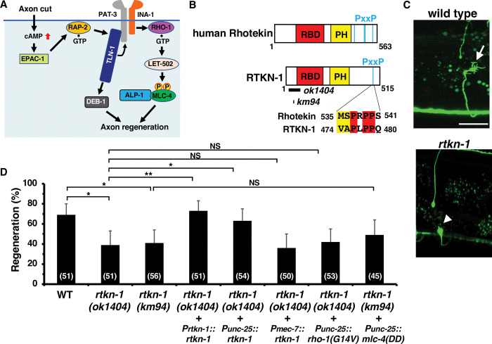
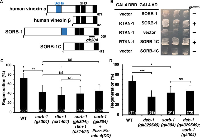
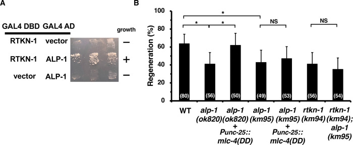
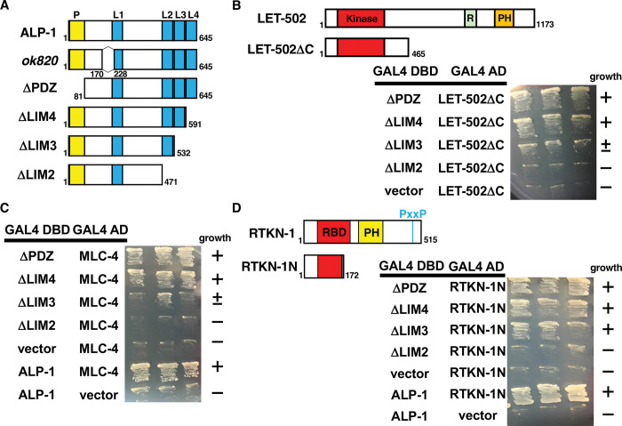
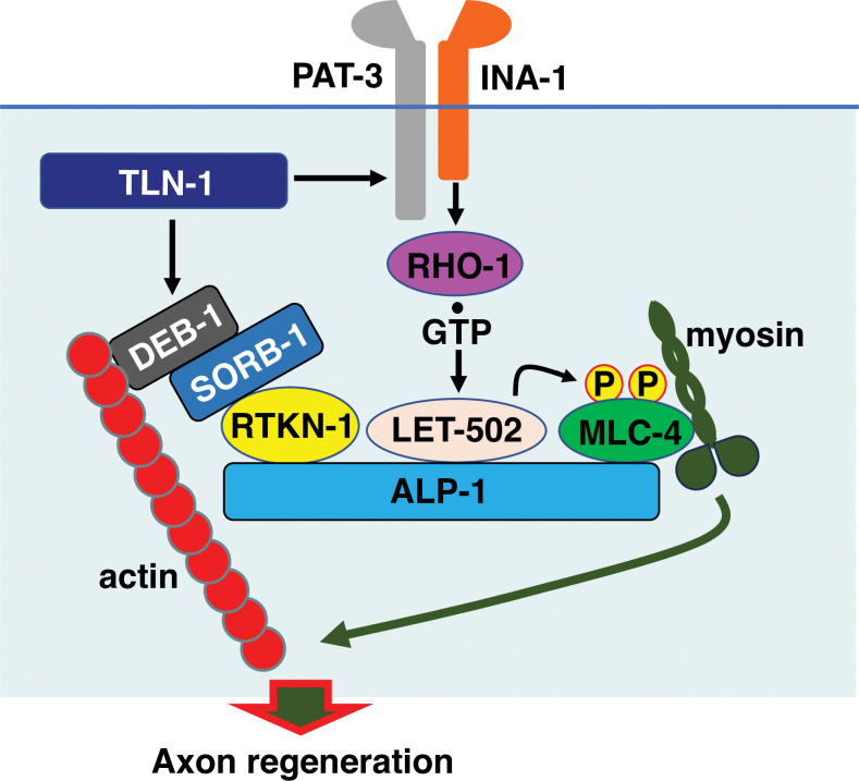
Axon regeneration requires actomyosin interaction, which generates contractile force and pulls the regenerating axon forward. In Caenorhabditis elegans, TLN-1/talin promotes axon regeneration through multiple down-stream events. One is the activation of the PAT-3/integrin-RHO-1/RhoA GTPase-LET-502/ROCK (Rho-associated coiled-coil kinase)-regulatory non-muscle myosin light-chain (MLC) phosphorylation signaling pathway, which is dependent on the MLC scaffolding protein ALP-1/ALP-Enigma. The other is mediated by the F-actin-binding protein DEB-1/vinculin and is independent of the MLC phosphorylation pathway. In this study, we identified the svh-7/rtkn-1 gene, encoding a homolog of the RhoA-binding protein Rhotekin, as a regulator of axon regeneration in motor neurons. However, we found that RTKN-1 does not function in the RhoA-ROCK-MLC phosphorylation pathway in the regulation of axon regeneration. We show that RTKN-1 interacts with ALP-1 and the vinculin-binding protein SORB-1/vinexin, and that SORB-1 acts with DEB-1 to promote axon regeneration. Thus, RTKN-1 links the DEB-1-SORB-1 complex to ALP-1 and physically connects phosphorylated MLC on ALP-1 to the actin cytoskeleton. These results suggest that TLN-1 signaling pathways coordinate MLC phosphorylation and recruitment of phosphorylated MLC to the actin cytoskeleton during axon regeneration.


Conflict of interest statement

The authors have declared that no competing interests exist.

Figures

Fig 1
Fig 1. SVH-7/RTKN-1 is required for axon regeneration.
(A) Regulation of axon regeneration by the TLN-1/talin signaling network. In response to axon injury, cAMP levels are elevated, resulting in the activation of EPAC-1, which then activates RAP-2. GTP-bound RAP-2 interacts with and activates TLN-1, which then activates PAT-3 and DEB-1. Activation of PAT-3 leads to phosphorylation of MLC-4 via the RHO-1–LET-502 pathway. ALP-1 acts as a platform for LET-502-mediated phosphorylation of MLC-4. DEB-1 acts independently of the MLC-4 phosphorylation pathway. (B) Domain structure of RTKN-1 and human Rhotekin. The Rho binding domain (RBD) and pleckstrin homology (PH) domains are shown in red and yellow, respectively. The Pro-rich motif (PxxP) is also shown. Identical and similar residues are highlighted in red and yellow shading, respectively. The bold lines below the RTKN-1 diagram indicate the extent of the deleted regions in the ok1404 and km94 mutants. (C) Representative D-type motor neurons in wild-type and rtkn-1(ok1404) mutant animals 24 h after laser surgery. Yellow and white arrowheads indicate regenerating and non-regenerating growth cones, respectively. Scale bar, 10 μm. (D) Percentage of axons that initiated regeneration 24 h after laser surgery at the young adult stage. The number of axons examined is shown. The error bar represents the 95% confidence interval (CI). *P < 0.05, **P < 0.01, according to Fisher’s exact test. NS, not significant.
Fig 2
Fig 2. SORB-1/Vinexin is required for axon regeneration.
(A) Domain structure of SORB-1 and human vinexin. The sorbin homology (SoHo) and Src homology 3 (SH3) domains are shown in blue and black, respectively. The bold line below the SORB-1 diagram indicates the extent of the deleted region in the gk304 mutant. (B) Yeast two-hybrid assays for RTKN-1 interaction with SORB-1 and SORB-1C. The reporter strain PJ69-4A was co-transformed with the expression vectors encoding GAL4 DBD-RTKN-1, GAL4 AD-SORB-1 and GAL4 AD-SORB-1C as indicated. Yeasts carrying the indicated plasmids were grown on selective plates lacking histidine and containing 10 mM 5-aminotriazole for 4 days. (C and D) Percentage of axons that initiated regeneration 24 h after laser surgery at the young adult stage. The number of axons examined is shown. The error bar represents the 95% confidence interval (CI). *P < 0.05, **P < 0.01, ***P < 0.001, according to Fisher’s exact test. NS, not significant.
Fig 3
Fig 3. Relationship between RTKN-1 and ALP-1.
(A) Yeast two-hybrid assays for RTKN-1 interaction with ALP-1. The reporter strain PJ69-4A was co-transformed with expression vectors encoding GAL4 DBD-RTKN-1 and GAL4 AD-ALP-1 as indicated. Yeasts carrying the indicated plasmids were grown on selective plates lacking histidine and containing 10 mM 5-aminotriazole for 4 days. (B) Percentage of axons that initiated regeneration 24 h after laser surgery in the young adult stage. The number of axons examined is shown. The error bar represents the 95% confidence interval (CI). *P < 0.05, according to Fisher’s exact test. NS, not significant.
Fig 4
Fig 4. Interaction of ALP-1 with LET-502, MLC-4 and RTKN-1.
(A) Structure of ALP-1. Schematic diagrams of ALP-1 are shown. The PDZ (P) and LIM domains are shown in yellow and blue, respectively. The ALP-1(Δ171–227) protein is missing 57 amino acids before the LIM1 domain. (B–D) Yeast two-hybrid assays for ALP-1 interaction with LET-502ΔC (B), MLC-4 (C) and RTKN-1N (D). Schematic diagrams of LET-502 and LET-502ΔC are shown (B). The kinase, RBD (R) and PH domains are shown in red, green and orange, respectively. Schematic diagrams of RTKN-1 and RTKN-1N are shown (D). The RBD and PH domains are shown in red and yellow, respectively. The PxxP is also shown. Yeasts carrying the indicated plasmids were grown on selective plates lacking histidine and containing 10 mM (B and D) or 5 mM (C) 5-aminotriazole for 4 days.
Fig 5
Fig 5. The A2534T mutation is responsible for tln-1(e259) phenotypes.
(A) Domain structure of TLN-1 and human talin-1. The tln-1(e259) allele harbors the A2534T mutation in the C-terminal dimerization domain (DD). Identical and similar residues are highlighted in red and yellow shading, respectively. The L2509P mutation in human talin-1 is shown. The FERM (F) domain (F0–F3) and the 13 talin rod domains R1–R13 are shown. (B) Percentage of axons that initiated regeneration 24 h after laser surgery at the young adult stage. The number of axons examined is shown. The error bar represents the 95% confidence interval (CI). *P < 0.05, **P < 0.01, according to Fisher’s exact test. (C) Representative trajectories of wild-type and tln-1 mutants. Wild-type and tln-1(e259; T2534A) mutant animals moved in a smooth sinusoidal pattern, whereas tln-1(e259) and tln-1(A2534T) animals displayed uncoordinated movement. Scale bar, 500 μm.
Fig 6
Fig 6. Schematic model of axon regeneration regulated by the TLN-1–PAT-3 and TLN-1–DEB-1 pathways.
TLN-1 promotes axon regeneration through the PAT-3 and DEB-1 signaling pathways. The PAT-3 pathway leads to the activation of RHO-1. GTP-bound active RHO-1 activates LET-502, which binds to ALP-1 and phosphorylates MLC-4 on ALP-1. Activated DEB-1 interacts with SORB-1, leading to the binding of SORB-1 and RTKN-1. RTKN-1 then interacts with ALP-1 and recruits the DEB-1–SORB-1 complex to ALP-1. On the ALP-1 platform, phosphorylated MLC-4 binds to the adhesion-associated actin cytoskeleton and promotes actomyosin-dependent force generation that facilitates axon regeneration.

Similar articles

References

    1. Franze K. Integrating chemistry and mechanics: the forces driving axon growth. Annu Rev Cell Dev Biol. 2020; 36: 61–83. doi: 10.1146/annurev-cellbio-100818-125157 - DOI - PubMed
    1. Myers JP, Santiago-Medina M, Gomez TM. Regulation of axonal outgrowth and pathfinding by integrin-ECM interactions. Dev Neurobiol. 2011; 71: 901–923. doi: 10.1002/dneu.20931 - DOI - PMC - PubMed
    1. Sweeney HL, Stull JT. Alteration of cross-bridge kinetics by myosin light chain phosphorylation in rabbit skeletal muscle: implications for regulation of actin-myosin interaction. Proc Natl Acad Sci USA. 1990; 87: 414–418. doi: 10.1073/pnas.87.1.414 - DOI - PMC - PubMed
    1. Kamm KE, Stull JT. The function of myosin and myosin light chain kinase phosphorylation in smooth muscle. Annu Rev Pharmacol Toxicol. 1985; 25: 593–620. doi: 10.1146/annurev.pa.25.040185.003113 - DOI - PubMed
    1. Amano M, Ito M, Kimura K, Fukata Y, Chihara K, Nakano T, et al.. Phosphorylation and activation of myosin by Rho-associated kinase (Rho-kinase). J Biol Chem. 1996; 271: 20246–20249. doi: 10.1074/jbc.271.34.20246 - DOI - PubMed

MeSH terms