. 2024 Feb;33(2):e4885.
doi: 10.1002/pro.4885.

Structural and functional validation of a highly specific Smurf2 inhibitor

Affiliations

Structural and functional validation of a highly specific Smurf2 inhibitor

Tanner M Tessier et al. Protein Sci. 2024 Feb.

Abstract

Smurf1 and Smurf2 are two closely related member of the HECT (homologous to E6AP carboxy terminus) E3 ubiquitin ligase family and play important roles in the regulation of various cellular processes. Both were initially identified to regulate transforming growth factor-β and bone morphogenetic protein signaling pathways through regulating Smad protein stability and are now implicated in various pathological processes. Generally, E3 ligases, of which over 800 exist in humans, are ideal targets for inhibition as they determine substrate specificity; however, there are few inhibitors with the ability to precisely target a particular E3 ligase of interest. In this work, we explored a panel of ubiquitin variants (UbVs) that were previously identified to bind Smurf1 or Smurf2. In vitro binding and ubiquitination assays identified a highly specific Smurf2 inhibitor, UbV S2.4, which was able to inhibit ligase activity with high potency in the low nanomolar range. Orthologous cellular assays further demonstrated high specificity of UbV S2.4 toward Smurf2 and no cross-reactivity toward Smurf1. Structural analysis of UbV S2.4 in complex with Smurf2 revealed its mechanism of inhibition was through targeting the E2 binding site. In summary, we investigated several protein-based inhibitors of Smurf1 and Smurf2 and identified a highly specific Smurf2 inhibitor that disrupts the E2-E3 protein interaction interface.

Keywords: E3 ligases; HECT domain; crystal structure; inhibitor; phage display; protein engineering; ubiquitin variants.


Figures

FIGURE 1
FIGURE 1
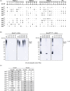
Ubiquitin variant (UbV) selection and specificity analysis. (a) UbVs generated against Smurf1 (UbV S1.1–5) and Smurf2 (UbV S2.1–5) by phage display. Regions 1, 2 and 3 correspond to regions of wild‐type ubiquitin that were subjected to diversification. Sequence differences, relative to wild‐type ubiquitin, for each UbV are indicated. (b) Specificity of UbVs assessed by in vitro auto‐ubiquitination assays using purified full‐length Smurf1 (left) or Smurf2 lacking the autoinhibitory C2 domain (Smurf2FLΔC2; right). (c) Affinities of UbVs for full‐length Smurf1 and Smurf2 proteins were determined by biolayer interferometry assays.
FIGURE 2
FIGURE 2
Specificity analysis of UbV S2.4 in cells. (a) Co‐immunoprecipitation of UbV S2.4 with Smurf1 and Smurf2. HEK293 cells were transfected with either FLAG‐Smurf1 or FLAG‐Smurf2 and V5‐tagged UbV S2.4. Immunoprecipitations using FLAG were performed and coimmunoprecipitation of UbV S2.4 was determined using anti‐V5 antibody. (b) Affinity purification‐mass spectrometry analysis of UbV S2.4 interactors. HEK293T cells were transfected with FLAG‐tagged UbV S2.4, FLAG‐EGFP, or an empty vector control. Spectral counts (SPC) were used to identify significant interactors relative to the control sample using SAINTexpress. Proteins with a Bayesian false‐discovery rate (BFDR) ≤0.05 are deemed significant compared to the empty vector control. (c) Smurf2 auto‐ubiquitination assay. HEK293 cells were transfected with FLAG‐tagged Smurf2 and HA‐tagged wild‐type ubiquitin and a 0.5:1 (+), 1:1 (++) or 2:1 (+++) ratio of V5‐UbV S2.4 to Smurf2. Cells were treated with 40 μM MG‐132 prior to anti‐FLAG immunoprecipitation to enrich Smurf1 or Smurf2. Immunoblotting with anti‐HA antibody was used to determine the level of auto‐ubiquitinated Smurf2 protein. See Figure S1A for Smurf1 auto‐ubiquitination. (d) Quantitative western blotting of UbV S2.4 mediated inhibition of Smurf1 and Smurf2. Auto‐ubiquitination assays were performed as outlined in panel (c) using 2:1 ratio of V5‐UbV S2.4 to Smurf1 and Smurf2. Student's t test was used to determine statistical significance. n = 3. See Figure S1B for western blots used for quantification. (e) UbV S2.4 mediated inhibition of Smad1 ubiquitination. HEK293 cells were transfected with Smad1‐HA, FLAG‐Smurf2, and V5‐UbV S2.4, treated with 10 μM MG132 and immunoprecipitated using anti‐HA. Smad1 ubiquitination levels were assessed using anti‐ubiquitin antibody against endogenous ubiquitin.
FIGURE 3
FIGURE 3
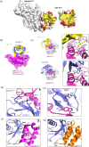
Structural analysis of the Smurf2 HECT domain in complex with UbV S2.4. (a) Open book view of the complex with the HECT domain of Smurf2 (left panel) and UbV S2.4 (right panel) shown as molecular surfaces. Contacting residues are colored yellow, non‐contacting residues are colored white and weakly bound (<20% buried upon formation of the complex) are colored red. (b, c) Structural comparison of UbV S2.4 bound to the Smurf2 HECT domain and UbV P1.1 bound to the WWP1 HECT domain (PDB ID: 5HPS), with UbV P1.1 and S2.4 colored yellow and blue, respectively. HECT residues are denoted with an asterisk to distinguish them from UbV residues. (b) Position of UbVs following superposition of the E2‐binding sites of the two HECT domains. The E2‐binding domain of Smurf2 (residues 516–593) is shown as a transparent surface and HECT E2‐binding region and superimposed UbVs are shown as cartoons. (c) Two views of the hydrophobic pocket of the WWP1 (top panels) and Smurf2 (bottom panels) HECT domains in complex with their UbVs. In the left panels, the HECT domains are shown as transparent molecular surfaces, with residues contacting L8 and W6 colored pink and shown as sticks, and L8 of WWP1 and W6 and F8 shown as sticks are highlighted with a circle. In the right panels, a closeup of L8 of UbV P1.1 (top), and W6 of UbV S2.4 (bottom), and their contacting residues are shown. (d, e) Closeup of two additional regions. (d) Interactions of the UbV with a loop in Smurf2 (residues 544–547), in which a number of hydrogen bonds and a salt bridge are observed (yellow dashes represent hydrogen bonds, the blue dash represents the salt bridge between R72 of the UbV and D546 of Smurf2). (e) UbV S2.4 residues contacting Smurf2 W535. (f, g) Basis for the specificity of UbV S2.4 for Smurf2 versus Smurf1. (f) The three residues in Smurf2 that interact with UbV S2.4 and differ from Smurf1 are shown and labeled, as well as their contacting residues in UbV S2.4. (g) A structural model of the Smurf1 HECT domain (orange) in complex with UbV S2.4 built using SWISS‐MODELLER. The three residues in Smurf1 that interact with UbV S2.4 and differ from Smurf2 are shown and labeled, as well as their putative contacting residues in UbV S2.4.

Similar articles

References

    1. Agirre J, Atanasova M, Bagdonas H, Ballard CB, Baslé A, Beilsten‐Edmands J, et al. The CCP4 suite: integrative software for macromolecular crystallography. Acta Crystallogr D Struct Biol. 2023;79(Pt 6):449–461. 10.1107/S2059798323003595 - DOI - PMC - PubMed
    1. Al‐Hakim A, Escribano‐Diaz C, Landry M‐C, O'Donnell L, Panier S, Szilard RK, et al. The ubiquitous role of ubiquitin in the DNA damage response. DNA Repair. 2010;9(12):1229–1240. 10.1016/j.dnarep.2010.09.011 - DOI - PMC - PubMed
    1. Chen C, Matesic LE. The Nedd4‐like family of E3 ubiquitin ligases and cancer. Cancer Metastasis Rev. 2007;26(3–4):587–604. 10.1007/s10555-007-9091-x - DOI - PubMed
    1. Damgaard RB. The ubiquitin system: from cell signalling to disease biology and new therapeutic opportunities. Cell Death Differ. 2021;28:423–426. 10.1038/s41418-020-00703-w - DOI - PMC - PubMed
    1. David D, Nair SA, Pillai MR. Smurf E3 ubiquitin ligases at the cross roads of oncogenesis and tumor suppression. Biochim Biophys Acta. 2013;1835(1):119–128. 10.1016/j.bbcan.2012.11.003 - DOI - PubMed

MeSH terms

LinkOut - more resources