Review
. 2023 Dec 10;24(24):17325.
doi: 10.3390/ijms242417325.

The Nexus of Inflammation-Induced Epithelial-Mesenchymal Transition and Lung Cancer Progression: A Roadmap to Pentacyclic Triterpenoid-Based Therapies

Affiliations
Review

The Nexus of Inflammation-Induced Epithelial-Mesenchymal Transition and Lung Cancer Progression: A Roadmap to Pentacyclic Triterpenoid-Based Therapies

Kirill V Odarenko et al. Int J Mol Sci. .

Abstract

Lung cancer is the leading cause of cancer-related death worldwide. Its high mortality is partly due to chronic inflammation that accompanies the disease and stimulates cancer progression. In this review, we analyzed recent studies and highlighted the role of the epithelial-mesenchymal transition (EMT) as a link between inflammation and lung cancer. In the inflammatory tumor microenvironment (iTME), fibroblasts, macrophages, granulocytes, and lymphocytes produce inflammatory mediators, some of which can induce EMT. This leads to increased invasiveness of tumor cells and self-renewal of cancer stem cells (CSCs), which are associated with metastasis and tumor recurrence, respectively. Based on published data, we propose that inflammation-induced EMT may be a potential therapeutic target for the treatment of lung cancer. This prospect is partially realized in the development of EMT inhibitors based on pentacyclic triterpenoids (PTs), described in the second part of our study. PTs reduce the metastatic potential and stemness of tumor cells, making PTs promising candidates for lung cancer therapy. We emphasize that the high diversity of molecular mechanisms underlying inflammation-induced EMT far exceeds those that have been implicated in drug development. Therefore, analysis of information on the relationship between the iTME and EMT is of great interest and may provide ideas for novel treatment approaches for lung cancer.

Keywords: aggressiveness; epithelial-to-mesenchymal transition; inflammation; mechanism of action; natural products; pulmonary malignancy; tumor stem cells.


Conflict of interest statement

The authors declare no conflict of interest.

Figures

Figure 1
Figure 1
Key molecular events of EMT. Epithelial tumor cells receive EMT-inducing signals from the tumor microenvironment, such as growth factors, low pH, hypoxia, and ECM modifications. EMT-inducing factors trigger various intracellular signaling pathways and activate EMT-TFs, which then downregulate and upregulate epithelial and mesenchymal markers, respectively. EMT is further sustained by autocrine loops: for example, inhibition of E-cadherin leads to its dissociation from β-catenin, which translocates to the nucleus and transactivates EMT-associated genes. Later stages of EMT involve transformation of both intra- and extracellular compartments. The formation of actin stress fibers and vimentin intermediate filaments provides the mechanical force for migration. The cells that have undergone EMT have multidirectional effects on the ECM: they disrupt the basement membrane by degrading collagen IV with matrix metalloproteinases (MMPs) 2 and 9, but at the same time produce other ECM components such as collagen I and fibronectin, which further maintain EMT. Downward (↓) and upward (↑) arrows indicate downregulation and upregulation of expression, respectively.
Figure 2
Figure 2
iTME as a driving source of EMT in lung cancer. A number of cell types from iTME, including cancer-associated fibroblasts (CAFs), tumor-associated macrophages (TAMs), neutrophils, eosinophils, mast cells, T cells (T helper 17 (Th17) and regulatory T (Tregs) cells), and B cells, induce EMT in lung cancer cells by producing various cytokines. Among them, transforming growth factor (TGF-β) is the most studied, but EMT-inducing activity has also been reported for anti-inflammatory cytokines (interleukin 4 (IL-4), IL-10), pro-inflammatory cytokines (IL-1β, IL-6, IL-17, tumor necrosis factor α (TNF-α)), chemokines (C-C Motif Chemokine Ligand 2 (CCL2), C-X-C Motif Chemokine Ligand 12 (CXCL12), IL-8), prostaglandins D2 and E2 (PGD2, PGE2), and neutrophil extracellular traps (NETs). Along with secretion, some EMT-inducing factors are transported to tumor cells via extracellular vesicles (TGF-β, Snail, microRNA-210 (miR-210)). In addition, lung tumor cells produce PGE2 to stimulate their EMT in an autocrine manner.
Figure 3
Figure 3
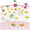
Contribution of CAFs to inflammation-induced EMT in lung cancer CAFs secrete a variety of EMT-inducing factors, including insulin-like growth factor 1 (IGF-1), hepatocyte growth factor (HGF), high mobility group box 1 (HMGB1), CXCL12, TGF-β, and extracellular vesicles (EVs). The IGF-1 and HGF signaling pathways are dependent on annexin A2 (ANXA2) phosphorylation. Autophagy-induced secretion of HMGB1 by CAFs induces EMT via the nuclear factor kappa B (NF-κB) pathway. CXCL12 triggers CXCR4/β-catenin to upregulate EMT-associated genes, among which peroxisome proliferator activated receptor delta (PPARδ) plays a specific role. CAF-derived EVs carry the EMT-TF Snail and miR-210, which induces the PTEN/PI3K/AKT pathway through UPF1 inhibition.
Figure 4
Figure 4
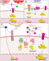
Immune component of inflammation-induced EMT in lung cancer. (A) Granulocytes, namely neutrophils and eosinophils, induce EMT through TGF-β/Smad signaling. CysLTs increase TGF-β production in eosinophils in an autocrine manner. (B) TAMs induce EMT in lung cancer by releasing IL-6, CCL2, and TGF-β. IL-6 and CCL2 share the same downstream JAK2/STAT3 signaling and mutually upregulate each other to induce EMT. (C) Mast cells release chymase, which reduces cell–cell adhesion by cleaving E-cadherin. Inhibition of the p52 tumor suppressor can be mentioned as another process induced by chymase in lung cancer cells. Mast cells secrete TGF-β-coated EVs that induce the classical Smad-dependent pathway. However, EVs contain other EMT-inducing molecules as they activate the phosphorylation of many proteins in lung cancer cells involved in PI3K/AKT, JAK/STAT, NF-κB, and HIF-1 signaling pathways, as well as the formation of focal adhesions and tight junctions.
Figure 5
Figure 5
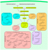
Biosynthetic pathway of PTs. In plant cells, the mevalonate and methylerythritol phosphate pathways provide isoprene units in the form of isopentenyl pyrophosphate (IPP) and dimethylallyl pyrophosphate (DMAPP), which are used as building blocks for the production of squalene. Oxidation of squalene yields 2,3-oxidosqualene, which serves as a precursor for the synthesis of all types of PT scaffolds, including lupane, ursane, oleanane, and friedelane. For simplicity, enzymes and most synthesis steps are omitted.
Figure 6
Figure 6
Inhibition of inflammation-induced EMT by PTs. Lupane-, oleanane-, and ursane-type PTs exhibit a variety of EMT inhibition mechanisms. PTs inhibit TGF-β signaling pathways (UA, AA, celastrol, pristimerin, C DDO-Me) and the downstream SMAD (UA, AA, celastrol, β-peltoboykinolic acid, glycyrrhizin) and ERK (UA, UNA, OA) signaling axes. PT-induced disruption of integrin signaling involves integrin αVβ5 expression inhibition (UA) and activation of FAK and AKT (BA, SYK023). The blockade of F-actin polymerization by the lupane-type PTs BA and SYK023 is mediated by the downregulation of Synpo, which presumably activates Smurf1-dependent ubiquitination of RhoA. In contrast, BA disrupts the Skp2-SCF E3 ligase complex, thereby protecting E-cadherin from degradation. The ursane-type PTs UA and AA inhibit β-catenin through a GSK-β-dependent mechanism. The effect of UA on EMT is also associated with repression of the AEG-1 oncogene, which regulates several steps in the NF-κB signaling pathway, and inhibition of the EGFR signaling pathway. Glycyrrhizin inhibits EMT induced by HMGB1, a nuclear protein released from tumor cells (TLR4 and RAGE are shown as possible HMGB1 receptors due to uncertainty in the downstream pathway). Some triterpenoids have an unknown mechanism of action but regulate EMT-associated genes (SM, evoditrilone A, pristimerin). Downward (↓) and upward (↑) arrows indicate downregulation and upregulation of expression, respectively.
Figure 7
Figure 7
Inhibition of stemness by PTs. Lupane-type PT BA inhibits stem cell proliferation by interfering with Skp2-SCF-dependent degradation of the cyclin-dependent kinase inhibitor p27. Oleanane-type PT β-escin suppresses the activity of ALDH1A1 and the downstream AKT pathway, which is thought to regulate the CSC transcriptome through the β-catenin pathway. The anti-stem cell activity of ursane-type PT UA is mediated through the EGFR pathway. Downward (↓) and upward (↑) arrows indicate downregulation and upregulation of expression, respectively.

Similar articles

Cited by

References

    1. Sung H., Ferlay J., Siegel R.L., Laversanne M., Soerjomataram I., Jemal A., Bray F. Global Cancer Statistics 2020: GLOBOCAN Estimates of Incidence and Mortality Worldwide for 36 Cancers in 185 Countries. CA Cancer J. Clin. 2021;71:209–249. doi: 10.3322/caac.21660. - DOI - PubMed
    1. Arseniev A.I., Nefedov A.O., Novikov S.N., Barchuk A.A., Tarkov S.A., Kostitsin K.A., Nefedova A.V., Aristidov N.Y., Semiletova Y.V., Ryazankina A.A. Algorithms of non-invasive, minimally-invasive and invasive diagnostics for lung cancer (review) Prev. Clin. Med. 2021;2:69–77. (In Russian)
    1. Ribatti D., Tamma R., Annese T. Epithelial-Mesenchymal Transition in Cancer: A Historical Overview. Transl. Oncol. 2020;13:100773. doi: 10.1016/j.tranon.2020.100773. - DOI - PMC - PubMed
    1. Menju T., Date H. Lung cancer and epithelial-mesenchymal transition. Gen. Thorac. Cardiovasc. Surg. 2021;69:781–789. doi: 10.1007/s11748-021-01595-4. - DOI - PubMed
    1. Tsoukalas N., Aravantinou-Fatorou E., Tolia M., Giaginis C., Galanopoulos M., Kiakou M., Kostakis I.D., Dana E., Vamvakaris I., Korogiannos A., et al. Epithelial-Mesenchymal Transition in Non Small-cell Lung Cancer. Anticancer Res. 2017;37:1773–1778. doi: 10.21873/anticanres.11510. - DOI - PubMed

MeSH terms