. 2024 Jan 9;19(1):54-67.
doi: 10.1016/j.stemcr.2023.11.009. Epub 2023 Dec 21.

Interspecies chimerism with human embryonic stem cells generates functional human dopamine neurons at low efficiency

Affiliations

Interspecies chimerism with human embryonic stem cells generates functional human dopamine neurons at low efficiency

Hu Wang et al. Stem Cell Reports. .

Abstract

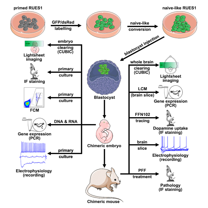
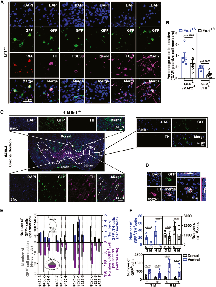
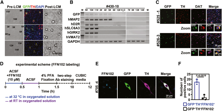
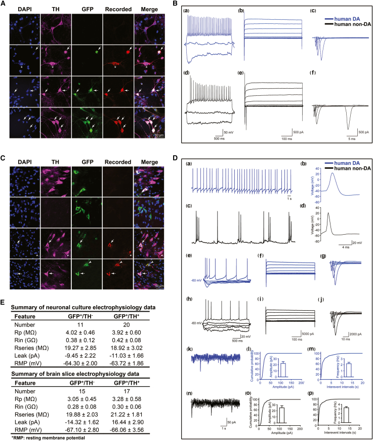
Interspecies chimeras offer great potential for regenerative medicine and the creation of human disease models. Whether human pluripotent stem cell-derived neurons in an interspecies chimera can differentiate into functional neurons and integrate into host neural circuity is not known. Here, we show, using Engrailed 1 (En1) as a development niche, that human naive-like embryonic stem cells (ESCs) can incorporate into embryonic and adult mouse brains. Human-derived neurons including tyrosine hydroxylase (TH)+ neurons integrate into the mouse brain at low efficiency. These TH+ neurons have electrophysiologic properties consistent with their human origin. In addition, these human-derived neurons in the mouse brain accumulate pathologic phosphorylated α-synuclein in response to α-synuclein preformed fibrils. Optimization of human/mouse chimeras could be used to study human neuronal differentiation and human brain disorders.


Conflict of interest statement

Declaration of interests The authors declare no competing interests.

Figures

None
Graphical abstract
Figure 1
Figure 1
Chimeric contribution of human ES RUES1-derived cells in En1 knockout mouse brain (A) Summary of blastocyst injected chimeric embryos and offspring. (B) Representative bright-field image of mouse embryo at E10.5, derived from blastocyst with RUES1 injection. (C) Representative integration of RUES1-derived cells into E10.5 embryonic midbrain (En1−/−) region. (D) Representative confocal immunofluorescence images showing the integration of RUES1-derived cells into an E10.5 embryo. Anti- hNA antibody was co-stained with anti-GFP. (E) Quantification of GFP+ fluorescence intensity in the midbrain in En1+/+ (n = 3 biological replicates), En1+/−(n = 3 biological replicates), and En1−/− (n = 1 biological replicate) genotypes. All data are represented as median with standard errors. (F) Representative images showing the integration of RUES1-derived cells in chimeric mouse forepaw at E10.5. (G) Flow cytometry analysis of DA neurons dissociated from chimeric embryos at E18. (H) Representative images (bright view and GFP channel) of neuronal culture from E16 embryos.
Figure 2
Figure 2
Chimeric contribution of human ES RUES1-derived cells in 23-day-old En1 knockout mouse brain (A) Representative dorsal and ventral views of brain from chimeric mice. (B) Left: Horizontal view in WT embryos without hES injection and En1−/− embryos with hES injection. Right: Representative sagittal light sheet confocal images of RUES1-derived neurons in En1−/− (23-day-old) chimeric midbrain region. cx, cortex; cb, cerebellum; mb, midbrain. (C) Representative immunofluorescence images showing the integration of RUES1-derived cells in a 23-day-old chimeric brain. The same age of mouse brain (without hESC blastocyst injection) was used as control; anti-hNA antibody was co-stained with anti-GFP.
Figure 3
Figure 3
Characterization of human ES RUES1-derived neurons in En1+/− mouse brain (A) Representative images showing the GFP+ neuron in midbrain (E18 mouse embryos derived from blastocyst injection of naive-like RUES1-GFP) culture. (B) Quantification of GFP+ MAP2 or TH+ cells in neuronal culture (n = 3 biological replicates). All data are represented as median with standard errors. (C) Representative images showing the integration of RUES1-derived neurons in the SNc, SNR, and RMC regions of a 4-month-old chimeric mouse (En1+/−). RMC, red nucleus, magnocellular part. VTA, ventral tegmental area. (D) Colocalization of GFP in TH+ neuron from chimeric mouse midbrain. (E) Summary of RUES1-GFP-derived neurons from 3-month-old (numbers 430-3, 430-5, 431-1, 430-7, 430-8, 431-9) and 6-month-old (numbers 525-2, 525-4, 525-5, 525-3, 525-6, 525-7) EN1+/+ (numbers 430-7, 430-8, 431-9, 525-3, 525-6, 525-7) and EN1+/− chimeric mice (numbers 430-3, 430-5, 431-1, 525-2, 525-4, 525-5). Schematic diagrams indicate the region of analysis for the quantification of RUES1-derived neurons in the bar graph. (F) Top: Quantification of (TH+ and TH) RUES1-GFP-derived neurons in 3- and 6-month-old EN1+/+ (n = 3 biological replicates) and EN1+/− (n = 3 biological replicates) chimeric mice. Bottom: Number of GFP+ cells in the ventral versus dorsal midbrain in 3- and 6-month-old EN1+/+ and EN1+/− chimeric mice.
Figure 4
Figure 4
Characterization of human ES RUES1-derived DA neurons in En1+/− mouse brain (A) LCM of GFP and TH positive and negative neurons in 4-month-old chimeric mouse brain stained with anti-GFP and anti-TH antibodies. (B) PCR analysis of DA neuron–related marker gene expression in neurons captured by LCM. The number of samples corresponds to the number shown in (A). (C) Representative images of RUES1-derived GFP+/TH+ neurons expressing DAT. (D) Schematic experimental procedure of FFN102-based DA tracing. (E) DA tracing in hESC-derived EN1+/− GFP+/TH+ cells using FFN102. (F) Quantification of FFN102-traced GFP+/TH+ cells (n = 3 biological replicates). All data are represented as median with standard errors.
Figure 5
Figure 5
Electrophysiological analysis of RUES1-derived neurons in neuronal culture and brain slices (A) Postimmunostaining showing recorded neurons in neuronal culture. Alexa Fluor 555 was patched as a marker of recording. (B) Electrophysiological properties of human DA neurons and human non-DA neurons. (a) Evoked AP of GFP+/TH+ DA neurons. (b) Sodium current and potassium current of GFP+/TH+ DA neurons. (c) Sodium current of GFP+/TH+ DA neurons. (d) Evoked AP of GFP+ non-DA neurons. (e) Sodium current and potassium current of GFP+ non-DA neurons. (f) Sodium current of GFP+ non-DA neurons. (C) Postimmunostaining showing recorded neurons in midbrain slice. Alexa Fluor 555 was patched as a marker of recording. (D) Electrophysiological properties of RUES1-derived DA neurons and RUES1-derived non-DA neurons. (a) Spontaneous AP of GFP+/TH+ DA neurons. (b) Single wave of spontaneous AP. (c) Spontaneous AP of GFP+ non-DA neuron. (d) Single wave of spontaneous AP of non-DA neuron. (e) Evoked AP of GFP+/TH+ DA neurons. (f) Sodium current and potassium current of GFP+/TH+ DA neurons. (g) Sodium current of GFP+/TH+ DA neurons. (h) Evoked AP of GFP+ non-DA neurons. (i) Sodium current and potassium current of GFP+ non-DA neurons. (j) Sodium current of GFP+ non-DA neurons. (k) mEPSCs of GFP+/TH+ DA neurons. (l and m) Amplitude and frequency of GFP+/TH+ DA neurons. (n) mEPSCs of GFP+ non-DA neurons. (o and p) Amplitude and frequency of mEPSCs in GFP+ non-DA neurons. (E) Summary of electrophysiological data from neuronal cultures and brain slices. Rp, pipette resistance; Rin, input resistance; R-series, series resistance; Leak, leak current; RMP, rest membrane potential.
Figure 6
Figure 6
RUES1-derived neurons can respond to α-syn PFFs (A) Schematic diagram of mPFF or hPFF injection in RUES1-GFP En1+/− chimeric mice. Chimeric mice were euthanized 30 days after the intrastriatal injection. (B) Representative images of ThS staining in SNc of En1+/− chimeric mice. (C) Representative images of pSyn+ immunostaining in SNc region of En1+/− chimeric mice injected with mPFF or hPFF. (D) Quantification of α-Syn pathology in the midbrain of mPFF- (n = 3 biological replicates) and hPFF- (n = 3 biological replicates) injected En1+/− chimeric mice. All data are represented as median with standard errors. (E) Representative images of pSyn and ubiquitin coimmunostaining in the SNc region of En1+/− chimeric mice injected with mPFF or hPFF.

Similar articles

Cited by

References

    1. Alves dos Santos M.T., Smidt M.P. En1 and Wnt signaling in midbrain dopaminergic neuronal development. Neural Dev. 2011;6:23. - PMC - PubMed
    1. Das S., Koyano-Nakagawa N., Gafni O., Maeng G., Singh B.N., Rasmussen T., Pan X., Choi K.D., Mickelson D., Gong W., et al. Generation of human endothelium in pig embryos deficient in ETV2. Nat. Biotechnol. 2020;38:297–302. - PubMed
    1. Funakoshi K., Bagheri M., Zhou M., Suzuki R., Abe H., Akashi H. Highly sensitive and specific Alu-based quantification of human cells among rodent cells. Sci. Rep. 2017;7:13202. - PMC - PubMed
    1. Gherbassi D., Simon H.H. The engrailed transcription factors and the mesencephalic dopaminergic neurons. J. Neural. Transm. Suppl. 2006;1:47–55. - PubMed
    1. Hanks M., Wurst W., Anson-Cartwright L., Auerbach A.B., Joyner A.L. Rescue of the En-1 mutant phenotype by replacement of En-1 with En-2. Science. 1995;269:679–682. - PubMed

Substances

LinkOut - more resources