Moyamoya-Like Vasculopathy and Orbital Trauma: An Association
- PMID: 38130810
- PMCID: PMC10732633
- DOI: 10.1080/01658107.2023.2212756
Moyamoya-Like Vasculopathy and Orbital Trauma: An Association
Abstract
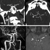
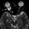
An Asian man in his 20s developed asymptomatic ipsilateral moyamoya-like vascular changes following orbital and head trauma. An ipsilateral traumatic optic neuropathy with extensive optic cupping ensued. The complex embryology of the ocular vascular development is reviewed as having a potential causative role in the intracranial carotid vasculopathy.
Keywords: Moyamoya; ocular embryology; optic neuropathy; vasculopathy.
© 2023 Taylor & Francis Group, LLC.
Conflict of interest statement
No potential conflict of interest was reported by the authors.
Figures


Similar articles
-
Successful Surgical Management of Traumatic Intracranial Hemorrhaging After Revascularization Surgery for Moyamoya Vasculopathy: A Case Report and Review of Literature.World Neurosurg. 2020 May;137:24-28. doi: 10.1016/j.wneu.2020.01.184. Epub 2020 Jan 31. World Neurosurg. 2020. PMID: 32014547 Review.
-
Indirect optic nerve injury in two-wheeler riders in northeast India.Indian J Ophthalmol. 2008 Nov-Dec;56(6):475-80. doi: 10.4103/0301-4738.43367. Indian J Ophthalmol. 2008. PMID: 18974518 Free PMC article.
-
Acute Thyrotoxicosis of Graves Disease Associated with Moyamoya Vasculopathy and Stroke in Latin American Women: A Case Series and Review of the Literature.World Neurosurg. 2016 Aug;92:95-107. doi: 10.1016/j.wneu.2016.04.122. Epub 2016 May 7. World Neurosurg. 2016. PMID: 27163552 Review.
-
Moyamoya Vasculopathy in Indian Children: Our Experience.J Pediatr Neurosci. 2017 Oct-Dec;12(4):320-327. doi: 10.4103/jpn.JPN_65_17. J Pediatr Neurosci. 2017. PMID: 29675069 Free PMC article.
-
Unilateral supraclinoid internal carotid artery stenosis with moyamoya-like vasculopathy. Noninvasive assessments.J Neuroimaging. 1994 Jan;4(1):11-6. doi: 10.1111/jon19944111. J Neuroimaging. 1994. PMID: 8136574
References
Publication types
Grants and funding
LinkOut - more resources
Full Text Sources
