. 2023 Nov 20;14(12):1647-1655.
doi: 10.1021/acsmedchemlett.3c00154. eCollection 2023 Dec 14.

Conformationally Restricted Analogues of α-Galactosylceramide as Adjuvant in COVID-19 Subunit Vaccine

Affiliations

Conformationally Restricted Analogues of α-Galactosylceramide as Adjuvant in COVID-19 Subunit Vaccine

Xing Hu et al. ACS Med Chem Lett. .

Abstract

iNKT cells are a type of T lymphocyte that recognizes glycolipid antigens presented by CD1d protein. αGC is an agonistic glycolipid that activates iNKT cells and triggers immune modulatory cytokine responses, making it a promising vaccine adjuvant. To find more potent immunostimulating glycolipids, we prepared 4,6-O-galactosyl conformationally restricted analogues of αGC. Mice vaccinated with the SARS-CoV-2 RBD-Fc vaccine adjuvanted with these newly developed glycolipids produced robust anti-RBD antibody responses, comparable to those achieved with αGC. Importantly, we also found that omitting αGC, α-C-GalCer (Th1-type agonist), or C20:2 (Th2-type agonist) from the booster vaccine had negligible impact on antibody and cellular responses, potentially reducing the frequency of adjuvant use required to maintain potent immune responses.


Conflict of interest statement

The authors declare no competing financial interest.

Figures
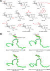
Figure 1
Figure 1
(A) Structures of αGC and its analogues. (B) Conformation of αGC, α-C-GC, and GCK127 as found in the crystal structure of the iNKT TCR-CD1d-glycolipid ligand. green, carbon; red, oxygen; blue, nitrogen.
Scheme 1
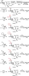
Scheme 1. αGC Analogues with Conformationally Restricted 4,6-O-Galactosyl Moieties
Figure 2
Figure 2
Synthesis of conformationally restricted analogues of αGC.
Figure 3
Figure 3
Synthesis of PhC-αGC.
Figure 4
Figure 4
Omission of αGC from the booster vaccine has little impact on anti-RBD IgG responses. Female BALB/c mice (n = 5 per group) were intraperitoneally immunized on days 1 and 15. The specific composition of each vaccine administered to the mice can be found in Supporting Information, Tables S1. Sera were collected on day 28 and tested for titers of SARS-CoV-2 RBD-specific IgG, IgG1, and IgG2a antibodies. (A–C) Anti-RBD IgG, IgG1, and IgG2a end point titers on day 28. Antibody titer below the limit of detection was assigned a value of 1. Data are representative of two similar experiments. Only p-values equal to or less than 0.05 are displayed.
Figure 5
Figure 5
Omission of α-C-GC (Th1-type agonist) and C20:2 (Th2-type agonist) from the booster vaccine. Female BALB/c mice (n = 5 per group) were intraperitoneally immunized on days 1 and 15. The specific composition of each vaccine administered to the mice can be found in Supporting Information, Table S2. Sera were collected on day 28 and tested for titers of SARS-CoV-2 RBD-specific IgG, IgG1, and IgG2a antibodies. Splenocytes were also collected on day 28 after the second immunization and processed for evaluating SARS-CoV-2 RBD-specific T cell-mediated responses. (A–C) Anti-RBD IgG, IgG1, and IgG2a end point titers on day 28. Data are representative of two similar experiments. (D) ELISPOT analysis of IFN-γ secretion in immunized mice. Antibody titer below the limit of detection was assigned a value of 1. Only p-values equal to or less than 0.05 are displayed.
Figure 6
Figure 6
Immunization protocol used for the evaluation of conformationally restricted analogues of αGC. Female BALB/c mice (n = 5 per group) were intraperitoneally immunized on day 1 with 30 μg of RBD-Fc protein admixed with 2 nmol of αGC analogues and on day 15 with 30 μg of RBD-Fc protein combined with no adjuvant. IFN-γ and IL-4 were analyzed in serum at 2 and 24 h after first immunization. Sera were collected on days 14 and 28 and tested for titers of SARS-CoV-2 RBD-specific IgG, IgG subtypes (IgG1, IgG2a, IgG2b, and IgG3), and pseudovirus neutralization activity. Splenocytes were also collected on day 28 and processed for evaluating SARS-CoV-2 RBD-specific T cell-mediated responses.
Figure 7
Figure 7
Humoral responses elicited by conformationally restricted analogues of αGC. (A) Anti-RBD IgG end point titers on days 14 and 28. (B–E) Anti-RBD IgG subclasses (IgG1, IgG2a, IgG2b, and IgG3) on day 28. Antibody titer below the limit of detection was set to 1. (F) IC50 titer of spike pseudotyped virus neutralization on day 28. The average IC50 values are labeled on the plots. Asterisks directly over a group denote a significant difference from the corresponding nonadjuvanted RBD-Fc group (labeled as “No adjuvant”). Data are representative of two similar experiments. Only p-values equal to or less than 0.05 are displayed.
Figure 8
Figure 8
ELISPOT analysis of IFN-γ and IL-4 secretion in immunized mice. The splenocytes isolated from immunized mice on day 28 were restimulated with overlapping Spike RBD peptides and assayed for IFN-γ and IL-4 production by ELISPOT assay. Asterisks directly over a group denote a significant difference from the corresponding nonadjuvanted RBD-Fc group (labeled as “no adjuvant”). Only p-values equal to or less than 0.05 are displayed.

Similar articles

References

    1. Brennan P. J.; Brigl M.; Brenner M. B. Invariant Natural Killer T Cells: An Innate Activation Scheme Linked to Diverse Effector Functions. Nat. Rev. Immunol. 2013, 13 (2), 101–117. 10.1038/nri3369. - DOI - PubMed
    1. Carreño L. J.; Saavedra-Ávila N. A.; Porcelli S. A. Synthetic Glycolipid Activators of Natural Killer T Cells as Immunotherapeutic Agents. Clin. Transl. Immunol. 2016, 5 (4), e6910.1038/cti.2016.14. - DOI - PMC - PubMed
    1. Burn O. K.; Pankhurst T. E.; Painter G. F.; Connor L. M.; Hermans I. F. Harnessing NKT Cells for Vaccination. Oxf. Open Immunol. 2021, 2, iqab01310.1093/oxfimm/iqab013. - DOI - PMC - PubMed
    1. Waldowska M.; Bojarska-Junak A.; Roliński J. A Brief Review of Clinical Trials Involving Manipulation of Invariant NKT Cells as a Promising Approach in Future Cancer Therapies. Cent. Eur. J. Immunol. 2017, 2 (2), 181–195. 10.5114/ceji.2017.69361. - DOI - PMC - PubMed
    1. Nair S.; Dhodapkar M. V. Natural Killer T Cells in Cancer Immunotherapy. Front. Immunol. 2017, 8, 1178.10.3389/fimmu.2017.01178. - DOI - PMC - PubMed

LinkOut - more resources