Review
. 2023 Dec 18;58(24):2850-2866.
doi: 10.1016/j.devcel.2023.11.003.

The dynamics and biophysics of shape formation: Common themes in plant and animal morphogenesis

Affiliations
Review

The dynamics and biophysics of shape formation: Common themes in plant and animal morphogenesis

Isabella Burda et al. Dev Cell. .

Abstract

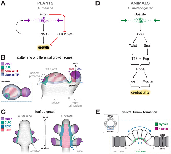
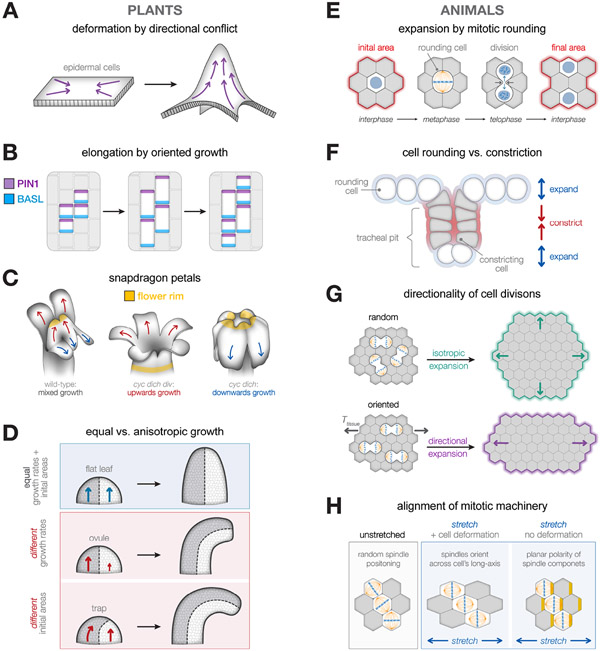
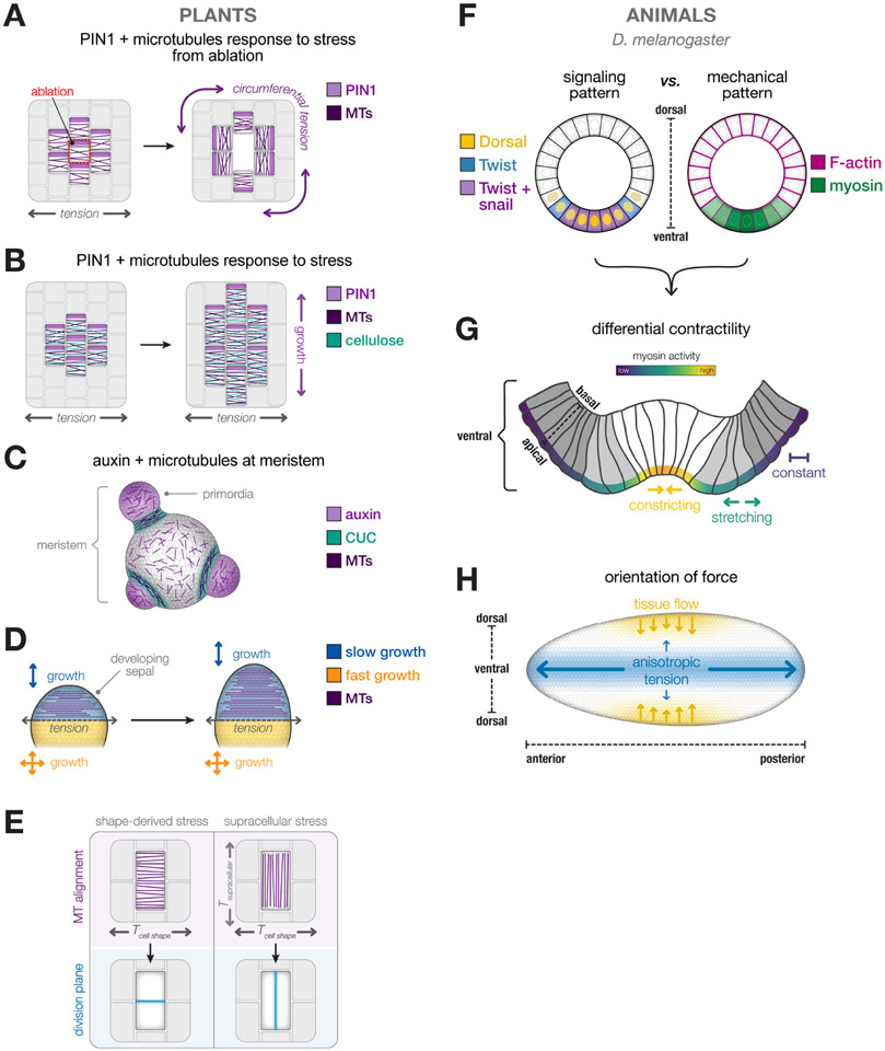
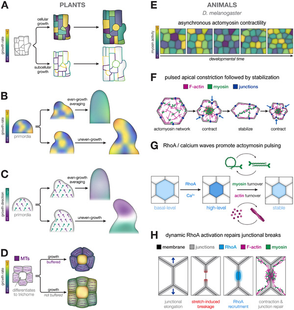
The emergence of tissue form in multicellular organisms results from the complex interplay between genetics and physics. In both plants and animals, cells must act in concert to pattern their behaviors. Our understanding of the factors sculpting multicellular form has increased dramatically in the past few decades. From this work, common themes have emerged that connect plant and animal morphogenesis-an exciting connection that solidifies our understanding of the developmental basis of multicellular life. In this review, we will discuss the themes and the underlying principles that connect plant and animal morphogenesis, including the coordination of gene expression, signaling, growth, contraction, and mechanical and geometric feedback.


Conflict of interest statement

Declaration of interests The authors declare no competing interests.

Figures

Figure 1:
Figure 1:. Transcriptional pre-patterning.
A) Positive feedback between auxin (plant hormone) and PIN1 (auxin transporter) create auxin signaling maxima and promote growth. CUC1,2,3 repress growth. B) Auxin maxima (purple spots) mark the location of organ primordia and cause outgrowth of the organ. CUC1,2,3 are expressed at the boundary of the organ to accentuate the different in growth rate between the organ and surrounding cells. The boundary between adaxial HD-ZIP class III transcription factors (TF, red stripes) and abaxial KANADI transcription factors (TF, navy stripes) determine the distance of the auxin maxima from the center of the meristem. C) In serrated Arabidopsis leaves (left), auxin maxima promote growth of the serration and CUC represses growth surrounding the serration. In Cardamine leaves (right) the growth difference is accentuated to form leaflets through the addition of STM which extends growing time and RCO which represses growth. D) Signaling pathway that leads to cytoskeleton activation and contractility within the Drosophila mesoderm. Expression and shuttling of the morphogen Spätzle (green spots) leads to high activation of Dorsal within cells along the ventral region of the embryo that will form the mesoderm. In response to high levels of Dorsal, Twist and Snail expression activates the RhoA pathway, which leads to actomyosin contractility. E) Schematic of the ventral side of a Drosophila embryo showing the invagination of the mesoderm during ventral furrow formation. Activation of F-actin (magenta) and myosin (green) promotes apical constriction of mesoderm cells (white), leading to the invagination of the tissue (blue arrows). Neighboring ectoderm cells (gray) are specified by low levels of Dorsal and therefore do not constrict.
Figure 2:
Figure 2:. Mechanisms for oriented growth:
A) A flat sheet of epidermis with cells growing in different directions causes deformation out of plane. B) Plant epidermal cells elongate, and then are partitioned by divisions. PIN1 (purple) localizes to the distal cell walls and BASL (blue) localizes to the proximal cell walls. C) Snapdragon petals of cyc dich div mutant grow upward (red arrows). Adding the transcription factor DIV promotes extra growth that turns the petals downward (blue arrows). WT has identity genes that cause the dorsal petals to grow upwards and the ventral petals to turn downwards. Snapdragon petals with growth in one orientation grow upward. Snapdragon petals with growth in two directions widens the petals, which causes petals to turn downwards. D) Equal growth of both halves of organ (ex. flat leaf) prevents curvature (top). Unequal growth between halves of an organ (ex. ovule) causes curvature (middle). Unequal initial areas of an organ that grow in the same direction (ex. carnivorous trap) causes curvature (bottom). E) Mitotic rounding of dividing cells results in expansion of cross-sectional cell area. During metaphase, the dividing cell (white) will round up and push against the surrounding non-dividing cells (gray). Once telophase is completed, two new daughter cells are formed, resulting in an increase in the total surface area of the tissue (outlined in red). F) Competition of expanding regions (blue) vs contracting regions (red) in developing Drosophila tracheal pit. Mitotic rounding of cells within and around the tracheal pit facilitate the invagination of the tissue by accelerating constriction of contracting cells within the pit. G) Orientation of cell divisions can influence the direction of tissue expansion. Random alignment of cell divisions leads to isotropic expansion of the tissue (green arrows). In contrast, when the orientation of division is aligned along a given axis, the tissue will expand anisotropically (purple arrows) in the same direction. H) Physical forces can influence the alignment of the mitotic machinery and cell division. In the absence of physical cues, cell shapes and mitotic spindles can lack alignment (left). When force is applied to the tissue and cells stretch, spindles will orient along the cell’s longest axis (Hertwig’s rule, middle), which are aligned to due to tissue forces. In some cases, stretch can induce alignment of the mitotic machinery via planar polarity of the spindle rotation machinery, in the absence or cell shape alignment (right, yellow stripes).
Figure 3:
Figure 3:. Mechanical feedback.
A) Ablation of an epidermal cell causes rearrangement of PIN1 (purple) expression and cortical microtubules in response to the new pattern of stress. B) Epidermal cells with medial lateral tension have PIN1 localization at the distal cell wall, cortical microtubule and cellulose (green) orientation parallel to the tension. This results in growth perpendicular to the tension. C) Mechanical feedback during organ initiation in which microtubules become anisotropic in the boundary region between the meristem and initiating organs. D) Fast growth in sepal morphogenesis recedes from distal to proximal and causes tension between the fast growing (yellow) and slow growing regions (blue). Cortical microtubules align parallel to the tension and inhibit widening of the sepal tip. E) Tension from cell shape promotes cortical microtubule orientation and division plane along the shortest axis. Tension from surrounding tissue promotes cortical microtubule orientation and division plane parallel to the supracellular tension. F) Differences between the patterning of morphogen signaling and cytoskeleton along the dorsal-ventral axis of a Drosophila embryo. G) Mechanics of mesoderm invagination, highlighting difference in levels of contractility. Mesoderm cells, which have the highest level of actomyosin activity, constrict (yellow arrows). Neighboring marginal mesoderm cells (gray) have lower levels of actomyosin activity and are able to stretch (green) in response to apically constricting cells. Ectoderm cells, which have little myosin activity but high F-actin levels, resist constricting forces and maintain their shape (purple). H). Orientation of anisotropic tension during gastrulation depends on embryo shape. Blue arrows denote direction of tension generated across the anterior-posterior axis on the ventral side of the embryo. Yellow arrows denote tissue flow, showing inward movement of the tissue towards the ventral surface.
Figure 4:
Figure 4:. Heterogeneity and collective cell behavior.
A) Epidermal cell growth rates (top) are heterogeneous within a tissue and epidermal cell wall growth rates are heterogenous within a cell and within a tissue. Growth rates are displayed as a heat map with fast growth in yellow and slow growth in purple. B) An organ with dynamic heterogenous growth rates that average spatially and temporally creates even, reproducible growth (top) vs an organ with static heterogenous growth rates that do not average spatially and temporally creates uneven, variable growth (bottom). C) An organ with dynamic heterogenous growth orientations that average spatially and temporally creates even, reproducible growth (top) vs an organ with static heterogenous growth orientations that do not average spatially and temporally creates uneven, variable growth (bottom). Growth direction is displayed as a heat map with left as purple and right as teal. D) Trichome cell in the center grows fast which creates mechanical stress. Cortical microtubule response to mechanical stress in surrounding cells slows their growth, creating heterogenous growth rates (top). If cortical microtubules do not respond to mechanical stress there is less heterogeneity in growth rates (bottom), but more influence on organ shape. E) Actomyosin contractility is heterogenous through the tissue. Apical constriction is staggered across cells, where some cells exhibit higher levels of myosin activity compared to their neighbors. F) Actomyosin contractility exhibits a pulsatile behavior. Myosin motors (green) pull on actin filaments (magenta) that are coupled to cellular junctions (dark blue) and constrict the apical surface of the cell. Constriction is followed by a period of stabilization, there the actomyosin network and cell shape is reinforced. G) Pulses of RhoA or Ca2+ can promote actomyosin pulsing and turnover. Cycling levels of RhoA or Ca2+ facilitates myosin turnover (activation/deactivation) or actin turnover (polymerization/depolymerization), respectively. In the Drosophila mesoderm, bursts of myosin activity are followed by periods of stabilization. H) Dynamic RhoA activation repairs breakages in cell junctions. When breaks occur in the junctional network, a burst of RhoA activity (blue) near the breakage site recruits F-actin (magenta) and myosin (green) to promote reinforcement and repair.

Similar articles

Cited by

References

    1. Somerville C, Bauer S, Brininstool G, Facette M, Hamann T, Milne J, Osborne E, Paredez A, Persson S, Raab T, et al. (2004). Toward a Systems Approach to Understanding Plant Cell Walls. Science 306, 2206–2211. 10.1126/science.1102765. - DOI - PubMed
    1. Cosgrove DJ (2014). Re-constructing our models of cellulose and primary cell wall assembly. Current Opinion in Plant Biology 22, 122–131. 10.1016/j.pbi.2014.11.001. - DOI - PMC - PubMed
    1. Cosgrove DJ (2018). Diffuse Growth of Plant Cell Walls. Plant Physiology 176, 16–27. 10.1104/pp.17.01541. - DOI - PMC - PubMed
    1. Coen E, and Cosgrove DJ (2023). The mechanics of plant morphogenesis. Science 379. 10.1126/science.ade8055. - DOI - PubMed
    1. Chugh P, and Paluch EK (2018). The actin cortex at a glance. Journal of Cell Science 131, jcs186254. 10.1242/jcs.186254. - DOI - PMC - PubMed

LinkOut - more resources