. 2023 Dec 26;120(52):e2314193120.
doi: 10.1073/pnas.2314193120. Epub 2023 Dec 18.

Defining a de novo non-RBM antibody as RBD-8 and its synergistic rescue of immune-evaded antibodies to neutralize Omicron SARS-CoV-2

Affiliations

Defining a de novo non-RBM antibody as RBD-8 and its synergistic rescue of immune-evaded antibodies to neutralize Omicron SARS-CoV-2

Xia Rao et al. Proc Natl Acad Sci U S A. .

Abstract

Currently, monoclonal antibodies (MAbs) targeting the SARS-CoV-2 receptor binding domain (RBD) of spike (S) protein are classified into seven classes based on their binding epitopes. However, most of these antibodies are seriously impaired by SARS-CoV-2 Omicron and its subvariants, especially the recent BQ.1.1, XBB and its derivatives. Identification of broadly neutralizing MAbs against currently circulating variants is imperative. In this study, we identified a "breathing" cryptic epitope in the S protein, named as RBD-8. Two human MAbs, BIOLS56 and IMCAS74, were isolated recognizing this epitope with broad neutralization abilities against tested sarbecoviruses, including SARS-CoV, pangolin-origin coronaviruses, and all the SARS-CoV-2 variants tested (Omicron BA.4/BA.5, BQ.1.1, and XBB subvariants). Searching through the literature, some more RBD-8 MAbs were defined. More importantly, BIOLS56 rescues the immune-evaded antibody, RBD-5 MAb IMCAS-L4.65, by making a bispecific MAb, to neutralize BQ.1 and BQ.1.1, thereby producing an MAb to cover all the currently circulating Omicron subvariants. Structural analysis reveals that the neutralization effect of RBD-8 antibodies depends on the extent of epitope exposure, which is affected by the angle of antibody binding and the number of up-RBDs induced by angiotensin-converting enzyme 2 binding. This cryptic epitope which recognizes non- receptor binding motif (non-RBM) provides guidance for the development of universal therapeutic antibodies and vaccines against COVID-19.

Keywords: Omicron BA.4/BA.5/BQ.1.1/XBB subvariants; RBD-8; SARS-CoV-2; cryptic epitope; neutralizing antibody.


Conflict of interest statement

Competing interests statement:The authors declare no competing interest.

Figures
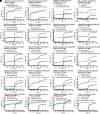
Fig. 1.
Fig. 1.
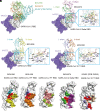
BIOLS56 and IMCAS74 are defined as RBD-8 class antibody. (A and B) No competitive binding between BIOLS56 antibody and hACE2 receptor. (C and D) No competitive binding of BIOLS56 with RBD-1 class antibody CB6 to SARS-CoV-2 PT RBD. (E and F) No competitive binding of BIOLS56 with RBD-2 class antibody REGN10933 to SARS-CoV-2 PT RBD. (G and H) No competitive binding of BIOLS56 with RBD-3 class antibody ADI-56046 to SARS-CoV-2 PT RBD. (I and J) No competitive binding of BIOLS56 with RBD-4 class antibody P2B-2F6 to SARS-CoV-2 PT RBD. (K and L) No competitive binding of BIOLS56 with RBD-5 class antibody REGN10987 to SARS-CoV-2 PT RBD. (M and N) No competitive binding of BIOLS56 with RBD-6 class antibody COVA1-16 to SARS-CoV-2 PT RBD. (O and P) No competitive binding of BIOLS56 with RBD-7 class antibody CR3022 to SARS-CoV-2 PT RBD. (Q and R) Competitive binding of BIOLS56 with IMCAS74 to SARS-CoV-2 PT RBD. (S and T) Competitive binding of BIOLS56 with S2H97 to SARS-CoV-2 PT RBD. Nonbiotinylated SARS-CoV-2 PT RBD was immobilized on NTA biosensor and then BIOLS56 antibody flowed through the tip in the presence of another antibody for real-time association and dissociation on Octet RED96.
Fig. 2.
Fig. 2.
Structural details between RBD-8 class antibodies and RBD. (A) Overall structures of hACE2, SARS-CoV-2 RBD, and RBD-8 class antibodies, BIOLS56, IMCAS74, and S2H97 (PDB code: 7M7W). (BD) Structural details between IMCAS74 Fab/SARS-CoV-2 Delta RBD (B) or BIOLS56 Fab/SARS-CoV RBD (C) or BIOLS56 Fab/SARS-CoV-2 PT RBD (D). Hydrogen bonds between amino acid residues are displayed by dashed lines. (E) Epitope of BIOLS56 in SARS-CoV RBD or SARS-CoV-2 PT RBD. SARS-CoV RBD epitopes recognized by the heavy chain, light chain, or both are colored in yellow, cyan, and red, respectively; for SARS-CoV-2 PT RBD, the epitopes recognized by the heavy chain, light chain, or both are colored in orange, light blue, and red, respectively. The conservative map of BIOLS56 epitope in SARS-CoV-2 PT RBD among RBD of SARS-CoV, SARS-CoV-2 variants and GX/P2V/2017 is displayed in the Right panel. The conserved and nonconserved amino acid residues of BIOLS56 epitope in RBD are colored in pink and light green, respectively. (F) Epitope of IMCAS74 in SARS-CoV-2 Delta RBD. The epitopes recognized by the heavy chain, light chain, or both are colored in dark purple, dark green, and red, respectively. (G) Epitope of S2H97 in SARS-CoV-2 PT RBD. The residues involved heavy chain, light chain or both are colored in light yellow, light purple, and red, respectively.
Fig. 3.
Fig. 3.
Binding dynamics of RBD-8 class representative antibodies BIOLS56, IMCAS74, and S2H97 to different RBDs. (A) SPR was used to detect the affinity between RBD-8 class representative antibodies and RBD in single-cycle mode on the BIAcore 8K system. Protein A biosensor chips were used to capture antibody and serial dilution RBD flowed through chip surface. The fitted curves of BIOLS56, IMCAS74, and S2H97 and the raw curves are represented by solid blue, red, green, and black lines, respectively. (B) Statistics of affinity between RBD-8 class representative antibodies and RBD. KD, ka, and kd are included. “#” represents KD value cannot be calculated due to no detectable dissociation between antibody and RBD. Data are presented as mean ± SD of three independent results.
Fig. 4.
Fig. 4.
Neutralization of RBD-8 class representative antibodies BIOLS56, IMCAS74, and S2H97. (A) Neutralization of RBD-8 class representative antibodies against pseudovirus was tested by the VSV-based pseudovirus system. Mixture of pseudovirus and serial dilution antibody was added into Vero (all tested variants except GX/P2V/2017) or Vero E6 cells (GX/P2V/2017). After 15 h incubation, cells were detected for green fluorescence value by CQ1 confocal image cytometer. (B) Statistics of IC50. Each neutralization experiment was performed twice with two replicates (n = 2). IC50 value is the representative data of two independent experiments.
Fig. 5.
Fig. 5.
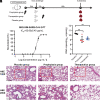
The prophylaxis and protection of BIOLS56 against SARS-CoV-2. (A) Experimental design for virus challenge in hACE2-transduced mice. hACE2-transduced mice were divided into three groups randomly. Mice in the prophylaxis group were administered one dose BIOLS56 at 24 h before the virus challenge while the mice in the therapeutics group were administered the same dose of BIOLS56 at 12 h after the virus challenge. Three days after the virus challenge, the lungs of three groups mice were collected to detect the RNA levels of the virus. (B) Neutralization of BIOLS56 antibody against SARS-CoV-2 PT authentic virus was tested in Vero E6 cells. IC50 was presented as mean ± SD of two independent results. (C) Virus titers of three groups (placebo, prophylaxis, therapeutics group) (n = 5) were measured by qRT-PCR using N gene primers, which was presented as RNA copies of per gram lung tissues. **P < 0.01. (DI) H&E staining of lung tissues in SARS-CoV-2 PT virus infected mice. Severe bronchopneumonia and interstitial pneumonia with bronchial epithelial cell desquamation (black arrow) and infiltration of interstitial inflammatory cells (red arrow) were observed in the placebo group (D and G). In the prophylaxis group (E and H), bronchopneumonia and interstitial pneumonia were significantly improved, with no bronchial epithelial cell desquamation and less infiltration of interstitial inflammatory cells (red arrow), while in the therapeutics group (F and I), no lesions were observed. The images and areas of interest (red boxes) are magnified 100× and 200×, respectively.
Fig. 6.
Fig. 6.
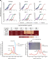
Neutralization effect rescue of RBD-5 MAb by scDb-Fc with an RBD-8 MAb. (A) Neutralization of representative antibodies against pseudovirus was tested by the VSV-based pseudotyping system. Mixture of pseudovirus and serial dilution antibody was added into Vero cells. After 15 h incubation, cells were detected for green fluorescence value by CQ1 confocal image cytometer. (B) Statistics of IC50. Each neutralization experiment was performed twice with two replicates (n = 2). IC50 value is the representative data of two independent experiments. (C and D) Gel filtration profiles of PT S (yellow), BIOLS56 Fab (cyan), and samples of PT S incubated with BIOLS56 Fab (red) are analyzed by size-exclusion chromatography as indicated. The SDS-PAGE analyses are shown with tubes 5 to 8, 13 to 18 of PT S/BIOLS56 Fab complex.
Fig. 7.
Fig. 7.
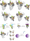
The proposed neutralization mechanism of RBD-8 class antibodies. (A) The epitope of BIOLS56 or IMCAS74 in “up” or “down” SARS-CoV-2 RBD. The color of the antibody chain is the same as Fig. 2. (BD) The structural superimposition of SARS-CoV-2 S trimer and RBD-8 class antibodies, BIOLS56 (B), IMCAS74 (C), and S2H97 (D) (PDB code: 7M7W). (E) Proposed neutralization mechanism models.

Similar articles

Cited by

References

    1. Tan W., et al. , A novel coronavirus genome identified in a cluster of pneumonia cases-Wuhan, China 2019–2020. China CDC Wkly. 2, 61–62 (2020). - PMC - PubMed
    1. Zhu N., et al. , A novel coronavirus from patients with pneumonia in China, 2019. N. Engl. J. Med. 382, 727–733 (2020). - PMC - PubMed
    1. Kirtipal N., et al. , From SARS to SARS-CoV-2, insights on structure, pathogenicity and immunity aspects of pandemic human coronaviruses. Infect. Genet. Evol. 85, 104502 (2020). - PMC - PubMed
    1. Lu R., et al. , Genomic characterisation and epidemiology of 2019 novel coronavirus: Implications for virus origins and receptor binding. Lancet 395, 565–574 (2020). - PMC - PubMed
    1. Wang P., et al. , Antibody resistance of SARS-CoV-2 variants B.1.351 and B.1.1.7. Nature 593, 130–135 (2021). - PubMed

Supplementary concepts