[Preprint]. 2023 Dec 7:2023.12.07.570613.
doi: 10.1101/2023.12.07.570613.

RAPIDASH: A tag-free enrichment of ribosome-associated proteins reveals compositional dynamics in embryonic tissues and stimulated macrophages

Affiliations

RAPIDASH: A tag-free enrichment of ribosome-associated proteins reveals compositional dynamics in embryonic tissues and stimulated macrophages

Teodorus Theo Susanto et al. bioRxiv. .

Update in

Abstract

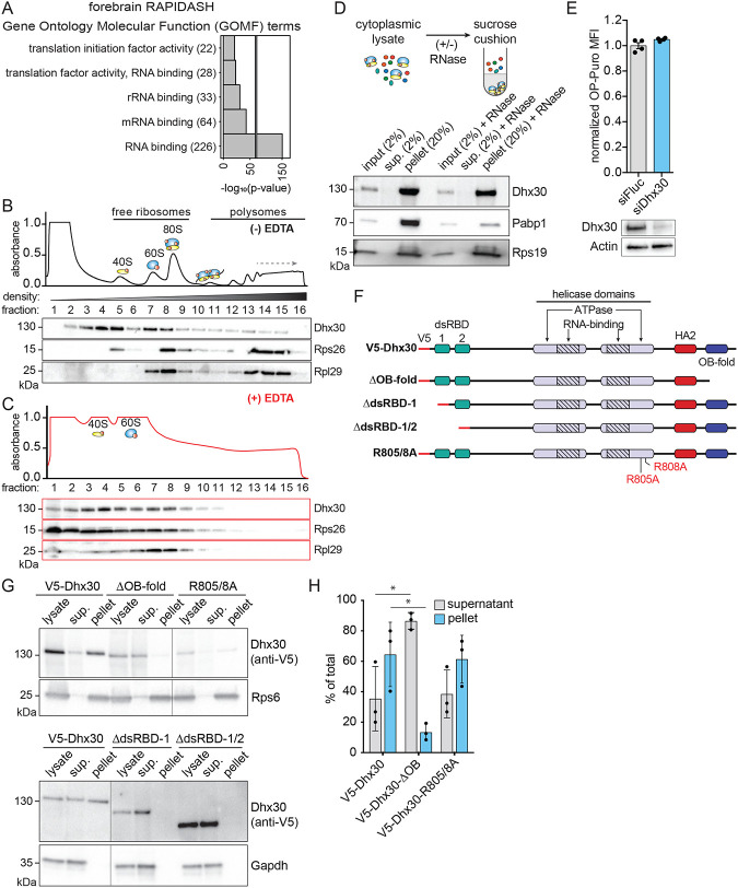
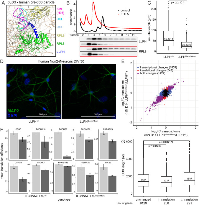
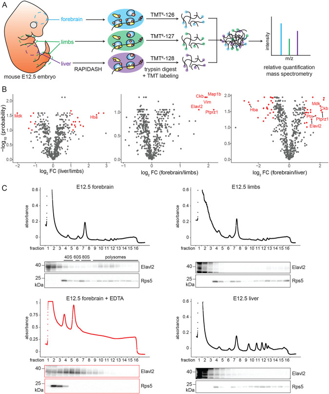
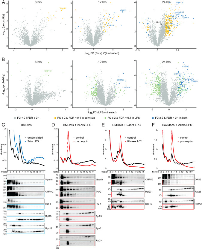
Ribosomes are emerging as direct regulators of gene expression, with ribosome-associated proteins (RAPs) allowing ribosomes to modulate translational control. However, a lack of technologies to enrich RAPs across many sample types has prevented systematic analysis of RAP number, dynamics, and functions. Here, we have developed a label-free methodology called RAPIDASH to enrich ribosomes and RAPs from any sample. We applied RAPIDASH to mouse embryonic tissues and identified hundreds of potential RAPs, including DHX30 and LLPH, two forebrain RAPs important for neurodevelopment. We identified a critical role of LLPH in neural development that is linked to the translation of genes with long coding sequences. Finally, we characterized ribosome composition remodeling during immune activation and observed extensive changes post-stimulation. RAPIDASH has therefore enabled the discovery of RAPs ranging from those with neuroregulatory functions to those activated by immune stimuli, thereby providing critical insights into how ribosomes are remodeled.

Keywords: embryonic development; macrophages; proteomics; ribosome; ribosome heterogeneity; ribosome-associated proteins; translational control.


Conflict of interest statement

Declaration of Interest The authors declare no competing interests.

Figures

Figure 1:
Figure 1:. Characterization of the ribosome-associated protein identification by affinity to sulfhydryl-charged resin (RAPIDASH) method in E14 mouse embryonic stem cells (mESCs)
(A) Schematic of the RAPIDASH protocol. This protocol can be applied to any biological sample, such as cells, patient biopsies, or organisms. Cytoplasmic lysates are subjected to sucrose cushion ultracentrifugation to enrich high density protein complexes. These then undergo enrichment for RNA-containing protein complexes by subjecting them to chromatography using sulfhydryl-charged resin. (B) Characterization of cysteine-charged sulfolink resin. Sucrose cushion pellet samples and poly(A) RNA isolated from mESCs were subjected to chromatography with cysteine-charged sulfolink resin. The percentage of RNA relative to input amount is plotted for the flowthrough, eluate, and bead-bound samples. N.D., not detected. Error bars are +/− standard error of the mean (SEM). (C) Characterization of RAPIDASH by sucrose gradient fractionation. Cytoplasmic lysate from E14 mouse embryonic stem cells (mESCs) was subjected to either enrichment with the sulfhydryl-charged resin or the entire RAPIDASH protocol. Each of these samples were fractionated on a sucrose density gradient to assess whether small ribonucleoproteins (gray) were depleted. (D) Boxplot of normalized log2 fold change (FC) of RAPIDASH eluate over sucrose cushion ultracentrifugation pellet tandem mass tag (TMT) mass spectrometry ratios from three biological replicates of mESCs. Ribosomal proteins (RPs; red) are significantly enriched over other proteins (gray) by RAPIDASH compared to sucrose cushion ultracentrifugation alone based on Welch’s t-test. p-values: rep1 = 1.24 × 10⁻¹⁸; rep2 = 4.39 × 10⁻²³; rep3 = 8.30 × 10⁻¹⁴. (E) Western blotting of mESC sucrose cushion pellet and RAPIDASH eluate samples for components of non-ribosomal complexes to assess the specificity of RAPIDASH. Approximately equal amounts of RPs for sucrose cushion pellet and RAPIDASH eluate samples, as shown by SYPRO Ruby (Supplementary Figure 1E), were analyzed by western blotting for Nup62, Atp5a1, and Tom20, components of the nuclear pore complex, ATP synthase, and the translocase of the outer membrane (TOM) complex, respectively. Cytoplasmic lysate was included as an input control. (F) Western blot detection of known RAPs enriched by RAPIDASH. A representative blot with 1% of the mESC cytoplasmic lysate volume and 35% of the RAPIDASH eluate volume was probed for the known RAPs Metap1, Ufl1, Upf1, Ddx1, and Nsun2,. (G) Bar graph showing the percentage of translational machinery identified by Ribo-FLAG immunoprecipitation (IP) or RAPIDASH. Ribo-FLAG IP proteins are those that were identified by FLAG IP liquid chromatography-tandem mass spectrometry (LC-MS/MS) of endogenously FLAG-tagged Rpl36 or Rps17 in E14 mESCs. Three biological replicates of RAPIDASH were performed for each of the same cell lines and analyzed by LC-MS/MS. Proteins identified with detectable peptide signal intensity in at least three out of the six RAPIDASH samples were compared against the Ribo-FLAG IP proteins. The percentage of 40S and 60S ribosomal proteins (RPs), translation elongation factors, translation initiation factors, and transfer RNA (tRNA) synthetases identified only in Ribo-FLAG IP (red), only in RAPIDASH (green), in both techniques (blue), or none (gray) are displayed. (H) Gene ontology (GO) term analysis of proteins identified by mESCs subjected to RAPIDASH. Proteins that were identified in RAPIDASH-enriched mESC samples were analyzed by Manteia. The ten most significant GO molecular function (GOMF) terms whose minimum level was 4 are shown.
Figure 2:
Figure 2:. Characterization of Dhx30 as a bona-fide mRNA-independent RAP
(A) Analysis of GO terms in forebrain MS data. RAPIDASH was performed on E12.5 mouse forebrain samples. These samples were analyzed by LC-MS/MS. The resulting proteins were analyzed by Manteia for GOMF terms level 4 or higher. The top 5 GOMF terms are shown. (B) Dhx30 co-fractionates with ribosomes, specifically the 40S fractions. Sucrose gradient fractionation was performed on E14 mESCs. The proteins from each fraction were precipitated and analyzed by western blotting. Dhx30 does not co-fractionate with the free fraction; instead, it largely co-fractionates with the 40S and 80S fractions. Rps26 and Rpl29 are shown as controls for small and large subunits, respectively. (C) E14 mESCs were treated with EDTA and subjected to sucrose gradient fractionation followed by western blotting as in (B). (D) Dhx30 still associates with the pellet fraction after RNase treatment. Top: illustration of sucrose cushion and RNase A treatment of E14 mESC cytoplasmic lysate. Bottom: western blot of control and RNase A-treated E14 mESC subjected to sucrose cushion ultracentrifugation. Pabp1 is a positive control for mRNA-dependent association with ribosomes. Rps19 is a positive control for ribosomes. Sup., supernatant. (E) Knockdown of Dhx30 by siRNA in mESC does not affect global protein synthesis. Protein synthesis was measured by O-propargyl-puromycin (OP-Puro) incorporation into the nascent proteome. Incorporated OP-Puro was fluorescently labeled using a click chemistry reaction. Translation activity was then measured using flow cytometry. Top: there is no change in OP-Puro median fluorescence intensity (MFI) between siFluc and siDhx30 (n=4). Bottom: western blotting of Dhx30 shows that siRNA knockdown is successful. (F) Illustration of Dhx30 constructs for transient transfection in E14 mESCs. Oligosaccharide binding (OB) fold domain; and double-stranded RNA-binding domains (dsRBDs). (G) Top: western blots of E14 mESCs transfected with V5-Dhx30, ΔOB-fold, and R805/8A constructs and subjected to sucrose cushion ultracentrifugation. Bottom: similar samples, but instead transfected with V5-Dhx30, ΔdsRBD-1, and ΔdsRBD-1/2 constructs. Loss of either OB-fold, dsRBD-1, or dsRBD-2 results in the loss of Dhx30 association with the ribosome. However, Dhx30 loss of function due to loss of helicase activity in R805/8A mutant does not affect its association with the ribosome. (H) Quantification of Dhx30 western blots shows a significant shift from pellet to supernatant in ΔOB-fold transiently transfected E14 mESCs but not R805/8A transfected E14 mESCs.
Figure 3:
Figure 3:. LLPH is a novel RAP with a role in neurodevelopment.
(A) LLPH binding location on the ribosome. Previously published cryo-EM data show LLPH binds near the sarcin-ricin loop of the human pre-60S particle, a highly conserved region in the ribosome critical for elongation (Protein Data Bank (PDB) ID: 6LSS). (B) LLPH cofractionates with ribosomes, specifically 60S fractions. Sucrose gradient fractionation was performed with (red) or without (black) EDTA in P493–6 cells where LLPH is highly expressed. The proteins from each fraction were precipitated and analyzed by western blotting. Rpl8 is a control for the large subunit. (C) Measurement of traced primary neurite lengths of individual LLPH+/+ and LLPHNterm/Nterm human Ngn2-induced neurons (hiNs) at days in vitro (DIV) 30. LLPHNterm/Nterm hiNs have shorter neurites, which may hint at neurodevelopmental defects. (D) Representative fluorescence images of fixed DIV 30 wild-type and LLPHNterm/Nterm hiNs. Wild-type and LLPHNterm/Nterm hiNs were fixed and stained with a primary antibody against MAP2 and 4’,6-diamidino-2-phenylindole) (DAPI). (E) Comparison of Ribo-seq and RNA-seq data for DIV 14 LLPH+/+ and LLPHNterm/Nterm hiNs (n = 3 each). Blue genes are those that significantly change (Benjamini-Hochberg procedure (FDR) < 0.1 and absolute fold change (FC) ≥ 2) in mRNA abundance only; red genes are those that change in ribosome occupancy only; purple genes are those that change in mRNA abundance and ribosome occupancy. (E) Representative genes with lower mean translation efficiency differences in LLPHNterm/Nterm vs. LLPH+/+h hiNs. Top: representative genes with downregulated translation efficiency in LLPHNterm/Nterm compared to LLPH+/+ that are involved in building the extracellular matrix. Bottom: representative genes known to be linked to neurodevelopmental defects related to growth cone defects or mRNA transport. (F) Genes downregulated for translation tend to have longer coding sequences (CDSs) than those that are translationally unchanged (Mann-Whitney U test, p = 0.0072). For clarity, only genes with CDS lengths shorter than 6000 are displayed. The median CDS length in each condition is displayed inside each boxplot. The full plot is shown in Supplementary Figure 2F.
Figure 4:
Figure 4:. Characterization of tissue-specific RAPs in the E12.5 mouse embryo.
(A) Schematic of the strategy to identify and quantify tissue-specific RAPs by tandem mass tag (TMT) mass spectrometry. Forebrain, limbs, and liver tissues were microdissected from E12.5 FVB/NJ mouse embryos and subjected to RAPIDASH. The enriched proteins were digested to peptides, which were labeled with TMT reagents to allow for relative quantification by LC-MS/MS. Four biological replicates were performed. (B) Volcano plots showing RAPs that are significantly enriched in one tissue compared to another. Putative RAPs identified in: liver to limbs (left), forebrain to limbs (center), forebrain to liver (right) are shown using volcano plots graphing −log10(p-value) against log2(FC). Proteins present in at least three out of four biological replicates with |log2FC| ≥ 1 and false discovery rate (FDR) < 0.10 are defined as significantly differentially enriched (red). (C) Elavl2 is a forebrain-enriched RAP. Forebrain, limb, and liver tissues from E12.5 mouse embryos were separated by sucrose gradient fractionation. An additional sample of forebrain tissue treated with EDTA as a control was also subjected to sucrose gradient fractonation. The protein from each fraction was precipitated and analyzed by western blotting for the presence of Elavl2 or Rps5, which served as a marker for the ribosome.
Figure 5.
Figure 5.. RAPIDASH identifies novel RAPs in macrophages following TLR stimulation.
(A-B) Murine bone marrow derived macrophages (BMDMs) were stimulated with lipopolysaccharide (LPS) (A) or polyinosinic-polycytidylic acid (poly(I:C)) (B) for 6, 12, or 24 hours prior to isolation of ribosome complexes for TMT-MS analysis. X-axes show the log2 FC of ribosome complex composition in activated versus unstimulated macrophages at 6 hours (left panels), 12 hours (middle panels), and 24 hours (right panels). (C-F) Lysates from unstimulated or LPS-stimulated BMDMs (C-E) or macrophages differentiated from HoxB8-immortalized progenitor cells (F) were subjected to polysome profiling analysis. Total protein was extracted from individual fractions and subjected to Western blot analysis for the indicated proteins. Puromycin and RNase treatments were performed as described in the Methods section.

Similar articles

References

    1. Genuth N.R., Shi Z., Kunimoto K., Hung V., Xu A.F., Kerr C.H., Tiu G.C., Oses-Prieto J.A., Salomon-Shulman R.E.A., Axelrod J.D., et al. (2022). A stem cell roadmap of ribosome heterogeneity reveals a function for RPL10A in mesoderm production. Nat Commun 13, 5491. 10.1038/s41467-022-33263-3. - DOI - PMC - PubMed
    1. Bartsch D., Kalamkar K., Ahuja G., Lackmann J.-W., Hescheler J., Weber T., Bazzi H., Clamer M., Mendjan S., Papantonis A., et al. (2023). mRNA translational specialization by RBPMS presets the competence for cardiac commitment in hESCs. Science Advances 9, eade1792. 10.1126/sciadv.ade1792. - DOI - PMC - PubMed
    1. Sampath P., Pritchard D.K., Pabon L., Reinecke H., Schwartz S.M., Morris D.R., and Murry C.E. (2008). A Hierarchical Network Controls Protein Translation during Murine Embryonic Stem Cell Self-Renewal and Differentiation. Cell Stem Cell 2, 448–460. 10.1016/j.stem.2008.03.013. - DOI - PubMed
    1. Ricciardi S., Manfrini N., Alfieri R., Calamita P., Crosti M.C., Gallo S., Müller R., Pagani M., Abrignani S., and Biffo S. (2018). The Translational Machinery of Human CD4+ T Cells Is Poised for Activation and Controls the Switch from Quiescence to Metabolic Remodeling. Cell Metabolism 28, 895–906.e5. 10.1016/j.cmet.2018.08.009. - DOI - PMC - PubMed
    1. Koppers M., Cagnetta R., Shigeoka T., Wunderlich L.C., Vallejo-Ramirez P., Qiaojin Lin J., Zhao S., Jakobs M.A., Dwivedy A., Minett M.S., et al. (2019). Receptor-specific interactome as a hub for rapid cue-induced selective translation in axons. eLife 8, e48718. 10.7554/eLife.48718. - DOI - PMC - PubMed

Publication types