[Preprint]. 2023 Dec 8:2023.12.07.570656.
doi: 10.1101/2023.12.07.570656.

Tail Length and E525K Dilated Cardiomyopathy Mutant Alter Human β-Cardiac Myosin Super-Relaxed State

Affiliations

Tail Length and E525K Dilated Cardiomyopathy Mutant Alter Human β-Cardiac Myosin Super-Relaxed State

Sebastian Duno-Miranda et al. bioRxiv. .

Update in

Abstract

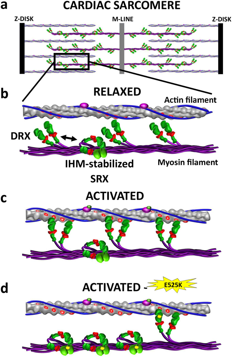
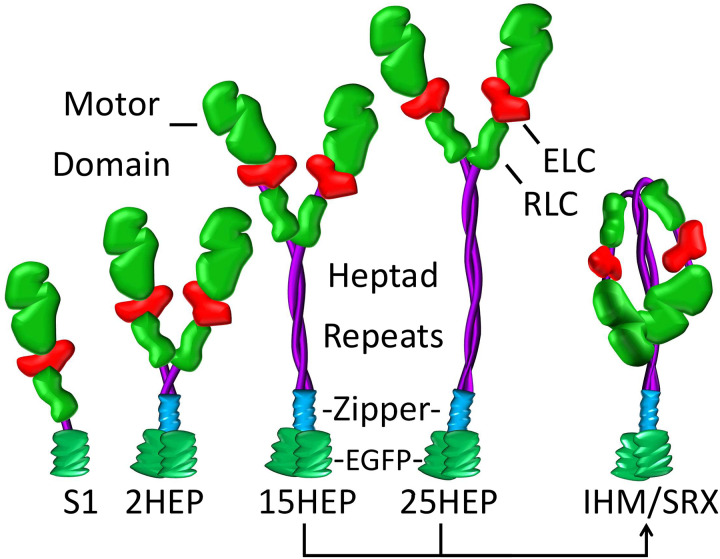
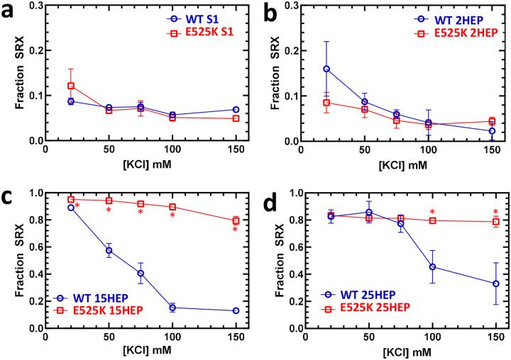
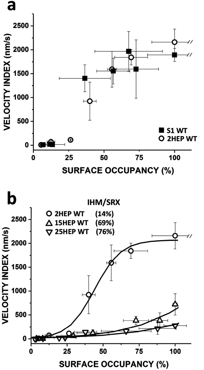
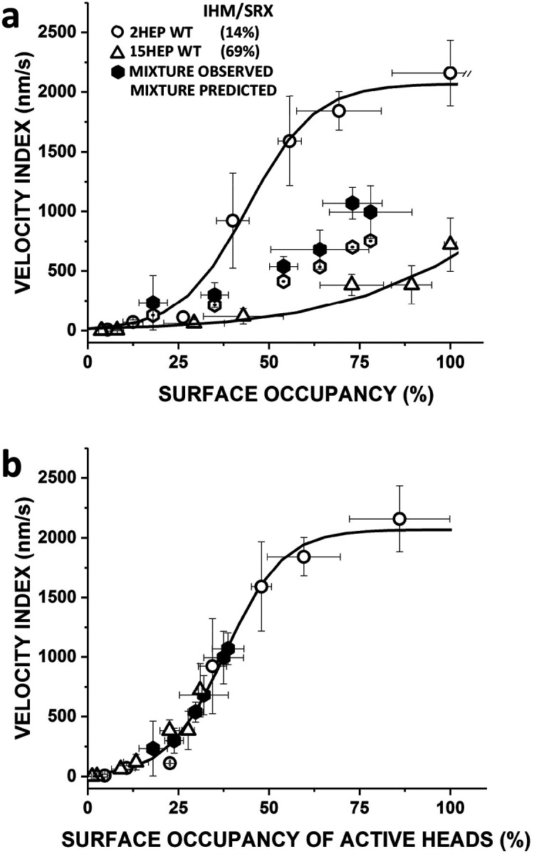
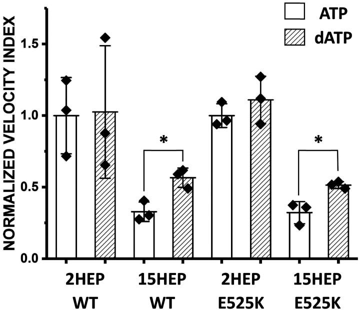
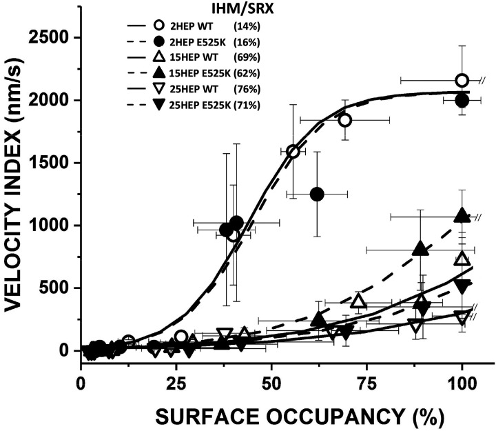
Dilated cardiomyopathy (DCM) is characterized by impaired cardiac function due to myocardial hypo-contractility and is associated with point mutations in β-cardiac myosin, the molecular motor that powers cardiac contraction. Myocardial function can be modulated through sequestration of myosin motors into an auto-inhibited "super relaxed" state (SRX), which is further stabilized by a structural state known as the "Interacting Heads Motif" (IHM). Therefore, hypo-contractility of DCM myocardium may result from: 1) reduced function of individual myosin, and/or; 2) decreased myosin availability due to increased IHM/SRX stabilization. To define the molecular impact of an established DCM myosin mutation, E525K, we characterized the biochemical and mechanical activity of wild-type (WT) and E525K human β-cardiac myosin constructs that differed in the length of their coiled-coil tail, which dictates their ability to form the IHM/SRX state. Single-headed (S1) and a short-tailed, double-headed (2HEP) myosin constructs exhibited low (~10%) IHM/SRX content, actin-activated ATPase activity of ~5s-1 and fast velocities in unloaded motility assays (~2000nm/s). Double-headed, longer-tailed (15HEP, 25HEP) constructs exhibited higher IHM/SRX content (~90%), and reduced actomyosin ATPase (<1s-1) and velocity (~800nm/s). A simple analytical model suggests that reduced velocities may be attributed to IHM/SRXdependent sequestration of myosin heads. Interestingly, the E525K 15HEP and 25HEP mutants showed no apparent impact on velocity or actomyosin ATPase at low ionic strength. However, at higher ionic strength, the E525K mutation stabilized the IHM/SRX state. Therefore, the E525K-associated DCM human cardiac hypo-contractility may be attributable to reduced myosin head availability caused by enhanced IHM/SRX stability.


Figures

Figure 1.
Figure 1.. Cardiac myosin conformations and interactions with actin filaments in relaxed and activated Muscle.
a) Representation of a cardiac sarcomere composed of interdigitated myosin filaments (purple) connected at the M-line and actin filaments anchored at the Z-disk. b) In relaxed muscle, myosin motors that emanate from the myosin filament backbone exist either in the enzymatically auto-inhibited, super-relaxed state (SRX), which can be stabilized by adopting the Interacting-Head-Motif (IHM) and precludes thin filament binding, or in the disordered-relaxed state (DRX) that is capable actin filament attachment and force generation upon cardiac activation. c) Once activated only DRX (i.e. active heads) myosin are recruited to generate force. d) Proposed mechanism by which the DCM-associated E525K M2β mutation (yellow starburst) stabilizes the IHM/SRX state, reducing the available pool of myosin motors for force generation, leading to cardiac hypo-contractility. Since most humans are heterozygote for cardiac myosin mutations, only one myosin head is depicted with the mutation.
Figure 2.
Figure 2.. Various human single- and double-headed M2β constructs.
Expressed human M2β constructs (single-headed: S1; double-headed with varying heptad (HEP) tail length: 2HEP, 15HEP, and 25HEP). All constructs have a regulatory light chain (RLC), essential light chain (ELC), and a C-terminal EGFP, with the double-headed constructs also containing a GCN4 leucine zipper to ensure dimerization. Only the 15HEP and 25HEP can adopt the IHM conformation, which presumably stabilizes the SRX state.
Figure 3.
Figure 3.. Steady-state ATPase activity of M2β constructs.
a–d. Steady-state ATPase kinetics of WT and E525K M2β constructs were determined using an NADH-coupled assay across varying actin concentrations. E525K mutations in S1 and 2HEP constructs significantly increased actin-activated ATPase activity (~3-fold) and decreased the actin concentration for half-maximal activity (KATPase) by ~18-fold. In contrast, 15HEP and 25HEP constructs showed reduced ATPase activities, with no significant difference between WT and E525K. Data are presented as mean ± SD of three experiments from separate protein preparations.
Figure 4.
Figure 4.. Assessment of IHM/SRX state fraction via single ATP turnover.
a–d. Single turnover of mantATP by M2β constructs was analyzed at different KCl concentrations to deduce the fraction of myosin heads in the IHM/SRX state. WT and E525K constructs at 0.25 μM were pre-incubated with mantATP (1 μM) for approximately 30 seconds before introducing saturating ATP (2 mM). The fluorescence transients were fitted to a two-exponential decay function to infer the SRX state’s fraction and kinetics. Notably, long-tailed (15 & 25HEP; c & d) constructs displayed a greater SRX fraction than S1 or 2HEP (a & b), which was sensitive to salt concentration only in the WT constructs with significant differences identified by asterisks (*) (15HEP, p<0.005; 25HEP, p<0.05). The mean ± SD of three experiments from separate protein preparations is reported.
Figure 5.
Figure 5.. M2β construct motility assay surface occupancy estimation.
a) M2β constructs were attached to the motility assay glass surface via their C-terminal EGFP to preadhered anti-GFP nanobodies. Increasing M2β binding showed increasing surface EGFP fluorescence intensity. b) Surface EGFP fluorescence intensity increased as a function of M2β construct concentration infused into motility assay flowcell. Fluorescence intensity followed saturable binding behavior with an apparent dissociation constant, Kapp(nM):KappS1WT=93±27, Kapp2HEPWT=116±42, Kapp15HEPWT=143±32, Kapp25HEPWT=181±36, Kapp2HEPE525K=152±38, Kapp15HEPE525K=191±45, Kapp25HEPE525K=153±34. No difference in the surface EGPF intensity across WT and E525K mutants (p = 0.98). c) Data in “b” normalized to fluorescence at 700 nM M2β concentration and converted to Surface Occupancy. Data are mean ± SD of at least three experiments from separate protein preparations.
Figure 6.
Figure 6.. Motility analysis of WT human M2β constructs.
a) The Velocity Index versus Surface Occupancy for WT S1 and 2HEP constructs showed a dose-response-like characteristic that saturated at ~2,000nm/s and were indistinguishable (p = 0.87). b) The Velocity Index versus Surface Occupancy for WT 2HEP, 15HEP, and 25HEP constructs. The longer-tailed WT 15HEP and 25HEP constructs had indistinguishable Velocity Indices (p = 0.87), but slower at each myosin surface occupancy compared to WT 2HEP. The WT 2HEP Velocity Index versus Surface Occupancy data were fitted to a Hill dose-response relationship (solid curve) and then used as the basis for an analytical model (see Results) to predict the IHM/SRX content for each construct. Predicted IHM/SRX percentages (legend in figure) and associated fitted curve (solid curve) are shown. All in vitro motility experiments were performed in low salt (25 mM KCl). Data points are presented as mean ± SD of at least three experiments from separate protein preparations.
Figure 7.
Figure 7.. Influence of IHM/SRX Content on Motility of WT 2HEP:15HEP Mixture.
a) The Velocity Index versus Surface Occupancy of WT 2HEP, 15HEP and a mixture of these two constructs. Addition of increasing amounts (0, 25, 50, 75, 100, 125 nM) of 15HEP WT (76% IHM/SRX) to a constant amount (25nM) of 2HEP WT (14% IHM/SRX) results in increasing Velocity Index values (filled octagons) that compare favorably to predicted values (open octagons; see Supplemental Table 1), supporting the model’s predictive nature, which assumes that a given construct has a defined IHM/SRX content and that the Velocity Index reflects actin filament motility generated by only active motors. b) Data in “a” were transformed to Velocity Index versus Surface Occupancy of Active Heads by replotting the Velocity Index data in “a” after calculating the surface occupancy of active (DRX) myosin heads from the product of observed total myosin surface occupancy and predicted percent DRX (i.e., 100%-(predicted %IHM/SRX content). The solid curve represents the 0% IHM/SRX curve (Supplemental Fig. 4). The 2HEP (open circles), 15HEP (open triangles), and observed mixture (closed hexagons) data, once transformed to represent only active motors, are well fit by the 0% IHM/SRX curve. Data points represent mean values ± SD of three experiments from separate protein preparations.
Figure 8.
Figure 8.. Deoxy-ATP (dATP) impact on IHM/SRX motors.
The SRX inhibitor, dATP, was exchanged for ATP in the motility assay with the WT and E525K 2HEP and 15HEP constructs at 100% myosin surface occupancy. Data are normalized to the respective (i.e., WT and E525K) 2HEP mean Velocity Index data in the presence of ATP. * identifies significantly different than control (p=0.01). Data points represent mean values ± SD of three experiments from separate protein preparations.
Figure 9.
Figure 9.. Impact of the E525K mutation on M2β motility.
Velocity Index versus Surface Occupancy for WT and E525K 2HEP, 15HEP, and 25HEP constructs. WT data and fitted curves from Figure 6b. All three E525K constructs were not significantly different than their WT controls (2HEP, p = 0.75; 15HEP, p = 0.75; 25HEP, p = 0.87). Dashed curves are the least squared error model fits to the Velocity Index versus Surface Occupancy relations in order to estimate the %IHM/SRX for the various E525K constructs (see legend in figure). The predicted %IHM/SRX for all of the E525K mutants are no different than WT. All in vitro motility experiments performed in low salt (25 mM KCl). Data points are presented as mean ± SD of at least three experiments from separate protein preparations.

Similar articles

References

    1. Rosenbaum AN, Agre KE, Pereira NL. Genetics of dilated cardiomyopathy: practical implications for heart failure management. Nat Rev Cardiol. 2020. May;17(5):286–297. doi: 10.1038/s41569-019-0284-0. Epub 2019 Oct 11. - DOI - PubMed
    1. McNally EM, Mestroni L. Dilated Cardiomyopathy: Genetic Determinants and Mechanisms. Circ Res. 2017. Sep 15;121(7):731–748. doi: 10.1161/CIRCRESAHA.116.309396. - DOI - PMC - PubMed
    1. Vincent JL. Understanding cardiac output. Crit Care. 2008;12(4):174. doi: 10.1186/cc6975. Epub 2008 Aug 22. - DOI - PMC - PubMed
    1. Brunello E, Fusi L. Regulating Striated Muscle Contraction: Through Thick and Thin. Annu Rev Physiol. 2023. Nov 6. doi: 10.1146/annurev-physiol-042222-022728. Epub ahead of print. - DOI - PubMed
    1. Palmiter KA, Tyska MJ, Dupuis DE, Alpert NR, Warshaw DM. Kinetic differences at the single molecule level account for the functional diversity of rabbit cardiac myosin isoforms. J Physiol. 1999. Sep 15;519 Pt 3(Pt 3):669–78. doi: 10.1111/j.1469-7793.1999.0669n.x. - DOI - PMC - PubMed

Publication types