. 2023 Nov 24;24(23):16720.
doi: 10.3390/ijms242316720.

Folate-Appended Hydroxypropyl-β-Cyclodextrin Induces Autophagic Cell Death in Acute Myeloid Leukemia Cells

Affiliations

Folate-Appended Hydroxypropyl-β-Cyclodextrin Induces Autophagic Cell Death in Acute Myeloid Leukemia Cells

Yasushi Kubota et al. Int J Mol Sci. .

Abstract

Acute myeloid leukemia (AML) is a heterogenous myeloid neoplasm that remains challenging to treat. Because intensive conventional chemotherapy reduces survival rates in elderly patients, drugs with lower toxicity and fewer side effects are needed urgently. 2-Hydroxypropyl-β-cyclodextrin (HP-β-CyD) is used clinically as a pharmaceutical excipient for poorly water-soluble drugs. Previously, we showed that HP-β-CyD exerts antitumor activity by disrupting cholesterol homeostasis. Recently, we developed folate-conjugated HP-β-CyD (FA-HP-β-CyD) and demonstrated its potential as a new antitumor agent that induces not only apoptosis, but also autophagic cell death; however, we do not know whether FA-HP-β-CyD exerts these effects against AML. Here, we investigated the effects of FA-HP-β-CyD on folate receptor (FR)-expressing AML cells. We found that the cytotoxic activity of FA-HP-β-CyD against AML cells was stronger than that of HP-β-CyD. Also, FA-HP-CyD induced the formation of autophagosomes in AML cell lines. FA-HP-β-CyD increased the inhibitory effects of cytarabine and a BCL-2-selective inhibitor, Venetoclax, which are commonly used treat elderly AML patients. Notably, FA-HP-β-CyD suppressed the proliferation of AML cells in BALB/c nude recombinase-activating gene-2 (Rag-2)/Janus kinase 3 (Jak3) double-deficient mice with AML. These results suggest that FA-HP-β-CyD acts as a potent anticancer agent for AML chemotherapy by regulating autophagy.

Keywords: 2-hydroxypropyl-β-cyclodextrin; Venetoclax; acute myeloid leukemia; autophagy; cholesterol; folate receptor; folic acid; metabolism; mitochondria; molecular targeting.


Conflict of interest statement

The authors declare no conflict of interest.

Figures

Figure 1
Figure 1
Cell surface expression of folate receptor (FR) α and β by AML cell lines. FRα expression by AML cells was measured by flow cytometry using an anti-FRα-PE antibody. Expression of FRβ was measured using an anti-FRβ-APC antibody. Blue: control; orange: sample stained with the anti-FRα or FRβ antibody. Median fluorescence intensity (MFI) for each cell lines is shown.
Figure 2
Figure 2
FA-HP-β-CyD induces apoptosis in HL-60, THP1, SKM1, and Kasumi1 cells. (A) HL-60, THP1, SKM1, and Kasumi1 cells were treated with 0 (medium only), 0.5, 1.0, and 1.5 mM of FA-HP-β-CyD for 72 h. After 72 h, cells were stained with Annexin V and PI. Representative FCM plots are shown (n = 3). (BE) Percentage of Annexin V-positive PI-negative cells after exposure to FA-HP-β-CyD for 72 h. (B) HL-60, (C) THP1, (D) Kasumi1, (E) SKM1 cells. Data represent the mean ± SEM of three independent experiments. * p < 0.05.
Figure 3
Figure 3
FA-HP-β-CyD does not affect cell cycle status in AML cells. HL-60, THP1 and Kasumi1 cells were treated for 24h with the indicated concentration of FA-HP-β-CyD and then subjected to flow cytometry analysis to detect PI-stained nuclei. (A) Representative flow cytometry histograms of PI-stained HL-60, THP1, and Kasumi1 cells. (BD) Percentage of viable HL-60 (B), THP1 (C), and Kasumi1 cells (D) cells in G0/G1, S, or G2/M phase. Data represent the mean ± SEM of three independent experiments.
Figure 4
Figure 4
FA-HP-β-CyD induces autophagy in AML cells. (A,B) HL-60 cells were incubated at 37 °C for 30 min with 0.1 μM DAPGreen working solution and then treated with FA-HP-β-CyD (1 or 10 mM) for 2h. Then, fluorescence was observed by flow cytometry (A) or fluorescence microscopy (B, original magnification ×400). The blue and orange histograms in (A) show conditions treated with 1 mM FA-HP-β-CyD and 10 mM FA-HP-β-CyD, respectively. Gray histogram is a negative control. (C,D) THP1 cells were incubated at 37 °C for 30 min with 0.1 μM DAPGreen working solution and then treated with FA-HP-β-CyD (1 or 10 mM) for 2h. Then, fluorescence was observed by flow cytometry (C) or fluorescence microscopy (D, original magnification ×400). The blue and orange histograms in (C) show conditions treated with 1 mM FA-HP-β-CyD and 10 mM FA-HP-β-CyD, respectively. Gray histogram is a negative control.
Figure 5
Figure 5
Autophagy inhibition in CyDs-treated AML cells. (A,B) Effects of chloroquine (20 μM), LY294002 (50 μM), and bafilomycin A1 (1 nM) on the antitumor activity of FA-HP-β-CyD (A) and HP-β-CyD (B) in HL-60 cells. (C,D) Effects of chloroquine (20 μM), LY294002 (50 μM), and bafilomycin A1 (1 nM) on the antitumor activity of FA-HP-β-CyD (A) and HP-β-CyD (B) in THP1 cells. Cells were treated with both 1 mM of FA-HP-β-CyD and 10 mM of HP-β-CyD at 37 °C for 2 h. Then, cells were incubated with the compounds for 24 h. Cell proliferation was assessed using Cell Counting Kit-8. Results are presented as the mean ± SEM (n = 3). * p < 0.05, compared with FA-HP-β-CyD without inhibitor.
Figure 6
Figure 6
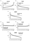
Combined effects of FA-HP-β-CyD and Ara-C or Venetoclax. (AC) FA-HP-β-CyD was combined with Ara-C. (DF) FA-HP-β-CyD was combined with Venetoclax. Cells were incubated for 72 h with five concentrations (0.25-, 0.5-, 0.75-, 1.0-, 1.5-fold the IC50) of each agent, or both in combination, using the constant ratio design, followed by modified MTT assay (Section 4.4). The IC50 values of Ara-C for HL-60, THP1 and SKM1 cells were 0.495 μM, 7.19 μM and 0.6 µM, respectively, and those of Venetoclax were 50.3 nM, 441.8 nM and 1.1 µM, respectively. The combination index (CI) was calculated using Calcusyn and plotted as a function of the fraction affected (Fa). For example, 50% growth inhibition would result in a Fa of 0.5. The synergistic (CI < 1), additive (CI = 1), or antagonistic (CI > 1) effects of combining multiple equal concentrations of drugs was assessed. Data are presented as the mean ± SEM of three independent experiments.
Figure 7
Figure 7
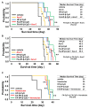
Effect of FA-HP-β-CyD on survival in leukemia mouse models and its effect in combination with Ara-C or Venetoclax. (A,B) Survival of mice transplanted with HL-60 cells (A) and THP12 cells (B). In these groups, the effect of Ara-C combination was also evaluated. HL-60 cells (5 × 10⁶) or THP1 cells (5 × 10⁶) were injected into BRJ mice. Three days later, mice received 200 μL of vehicle, 150 mM (2086.5 mg/kg) of HP-β-CyD, or 15 mM (249 mg/kg) of FA-HP-β-CyD via intraperitoneal injection (twice a day). Ara-C (100 mg/kg) was also injected intraperitoneally once a day. Administration continued for 20 days, and survival was checked daily. Black lines, yellow lines, blue lines, red lines, and green lines indicate survival of mice treated with vehicle, Ara-C, HP-β-CyD, FA-HP-β-CyD, and Ara-C plus FA-HP-β-CyD, respectively. (C) Survival curves of mice transplanted with HL-60 cells. In this group, the effect of Venetoclax combination was also evaluated. HL-60 cells (5×10⁶) were injected into BRJ mice. Three days after the injection, mice received 200 μL of vehicle, 150 mM (2086.5 mg/kg) of HP-β-CyD, or 15 mM (249 mg/kg) of FA-HP-β-CyD via intraperitoneal injection (twice a day). Venetoclax (25 mg/kg) was given daily by oral gavage. Administration was continued for 20 days, and survival was checked daily. Black lines, yellow lines, blue lines, red lines, and green lines indicate survival of mice treated with vehicle, Venetoclax, HP-β-CyD, FA-HP-β-CyD, and Venetoclax plus FA-HP-β-CyD, respectively. Survival data were analyzed using a log-rank nonparametric test and are shown as Kaplan–Meier survival curves (n = 6). * p < 0.05 and ** p <0.01 compared with vehicle group. Median survival time (day) for each group is shown.

Similar articles

References

    1. DiNardo C.D., Erba H.P., Freeman S.D., Wei A.H. Acute myeloid leukaemia. Lancet. 2023;401:2073–2086. doi: 10.1016/S0140-6736(23)00108-3. - DOI - PubMed
    1. Ohtake S., Miyawaki S., Fujita H., Kiyoi H., Shinagawa K., Usui N., Okumura H., Miyamura K., Nakaseko C., Miyazaki Y., et al. Randomized study of induction therapy comparing standard-dose idarubicin with high-dose daunorubicin in adult patients with previously untreated acute myeloid leukemia: The JALSG AML201 Study. Blood. 2011;117:2358–2365. doi: 10.1182/blood-2010-03-273243. - DOI - PubMed
    1. Choi J., Shukla M., Abdul-Hay M. Acute Myeloid Leukemia Treatment in the Elderly: A Comprehensive Review of the Present and Future. Acta Haematol. 2023 doi: 10.1159/000531628. - DOI - PubMed
    1. DiNardo C.D., Pratz K., Pullarkat V., Jonas B.A., Arellano M., Becker P.S., Frankfurt O., Konopleva M., Wei A.H., Kantarjian H.M., et al. Venetoclax combined with decitabine or azacitidine in treatment-naive, elderly patients with acute myeloid leukemia. Blood. 2019;133:7–17. doi: 10.1182/blood-2018-08-868752. - DOI - PMC - PubMed
    1. DiNardo C.D., Jonas B.A., Pullarkat V., Thirman M.J., Garcia J.S., Wei A.H., Konopleva M., Döhner H., Letai A., Fenaux P., et al. Azacitidine and Venetoclax in Previously Untreated Acute Myeloid Leukemia. N. Engl. J. Med. 2020;383:617–629. doi: 10.1056/NEJMoa2012971. - DOI - PubMed