Novel autosomal dominant TMC1 variants linked to hearing loss: insight into protein-lipid interactions
- PMID: 38066485
- PMCID: PMC10704677
- DOI: 10.1186/s12920-023-01766-7
Novel autosomal dominant TMC1 variants linked to hearing loss: insight into protein-lipid interactions
Abstract
Background: TMC1, which encodes transmembrane channel-like protein 1, forms the mechanoelectrical transduction (MET) channel in auditory hair cells, necessary for auditory function. TMC1 variants are known to cause autosomal dominant (DFNA36) and autosomal recessive (DFNB7/11) non-syndromic hearing loss, but only a handful of TMC1 variants underlying DFNA36 have been reported, hampering analysis of genotype-phenotype correlations.
Methods: In this study, we retrospectively reviewed 338 probands in an in-house database of genetic hearing loss, evaluating the clinical phenotypes and genotypes of novel TMC1 variants associated with DFNA36. To analyze the structural impact of these variants, we generated two structural models of human TMC1, utilizing the Cryo-EM structure of C. elegans TMC1 as a template and AlphaFold protein structure database. Specifically, the lipid bilayer-embedded protein database was used to construct membrane-embedded models of TMC1. We then examined the effect of TMC1 variants on intramolecular interactions and predicted their potential pathogenicity.
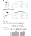
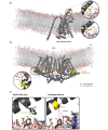
Results: We identified two novel TMC1 variants related to DFNA36 (c.1256T > C:p.Phe419Ser and c.1444T > C:p.Trp482Arg). The affected subjects had bilateral, moderate, late-onset, progressive sensorineural hearing loss with a down-sloping configuration. The Phe419 residue located in the transmembrane domain 4 of TMC1 faces outward towards the channel pore and is in close proximity to the hydrophobic tail of the lipid bilayer. The non-polar-to-polar variant (p.Phe419Ser) alters the hydrophobicity in the membrane, compromising protein-lipid interactions. On the other hand, the Trp482 residue located in the extracellular linker region between transmembrane domains 5 and 6 is anchored to the membrane interfaces via its aromatic rings, mediating several molecular interactions that stabilize the structure of TMC1. This type of aromatic ring-based anchoring is also observed in homologous transmembrane proteins such as OSCA1.2. Conversely, the substitution of Trp with Arg (Trp482Arg) disrupts the cation-π interaction with phospholipids located in the outer leaflet of the phospholipid bilayer, destabilizing protein-lipid interactions. Additionally, Trp482Arg collapses the CH-π interaction between Trp482 and Pro511, possibly reducing the overall stability of the protein. In parallel with the molecular modeling, the two mutants degraded significantly faster compared to the wild-type protein, compromising protein stability.
Conclusions: This results expand the genetic spectrum of disease-causing TMC1 variants related to DFNA36 and provide insight into TMC1 transmembrane protein-lipid interactions.
Keywords: DFNA36; Hearing loss; Protein-lipid interaction; Structural modeling; TMC1.
© 2023. The Author(s).
Conflict of interest statement
The authors declare no competing interests.
Figures


Similar articles
-
A novel DFNA36 mutation in TMC1 orthologous to the Beethoven (Bth) mouse associated with autosomal dominant hearing loss in a Chinese family.PLoS One. 2014 May 14;9(5):e97064. doi: 10.1371/journal.pone.0097064. eCollection 2014. PLoS One. 2014. PMID: 24827932 Free PMC article.
-
Mutations of TMC1 cause deafness by disrupting mechanoelectrical transduction.Auris Nasus Larynx. 2014 Oct;41(5):399-408. doi: 10.1016/j.anl.2014.04.001. Epub 2014 Jun 2. Auris Nasus Larynx. 2014. PMID: 24933710 Free PMC article.
-
Prevalence and clinical features of autosomal dominant and recessive TMC1-associated hearing loss.Hum Genet. 2022 Apr;141(3-4):929-937. doi: 10.1007/s00439-021-02364-2. Epub 2021 Sep 14. Hum Genet. 2022. PMID: 34523024 Free PMC article.
-
Mouse tales from Kresge: the deafness mouse.J Am Acad Audiol. 2003 Aug;14(6):296-301. J Am Acad Audiol. 2003. PMID: 14552423 Review.
-
Is TMC1 the Hair Cell Mechanotransducer Channel?Biophys J. 2016 Jul 12;111(1):3-9. doi: 10.1016/j.bpj.2016.05.032. Biophys J. 2016. PMID: 27410728 Free PMC article. Review.
Cited by
-
The Inheritance of Hearing Loss and Deafness: A Historical Perspective.Audiol Res. 2024 Jan 26;14(1):116-128. doi: 10.3390/audiolres14010010. Audiol Res. 2024. PMID: 38391767 Free PMC article. Review.
-
Genomic Landscape of Branchio-Oto-Renal Syndrome through Whole-Genome Sequencing: A Single Rare Disease Center Experience in South Korea.Int J Mol Sci. 2024 Jul 26;25(15):8149. doi: 10.3390/ijms25158149. Int J Mol Sci. 2024. PMID: 39125727 Free PMC article.
-
Functional pathogenicity of ESRRB variant of uncertain significance contributes to hearing loss (DFNB35).Sci Rep. 2024 Sep 11;14(1):21215. doi: 10.1038/s41598-024-70795-8. Sci Rep. 2024. PMID: 39261511 Free PMC article.
References
-
- Van Camp GSR. Hereditary Hearing Loss Homepage 2021 [Available from: https://hereditaryhearingloss.org.
-
- Marcovich I, Holt JR. Evolution and function of tmc genes in mammalian hearing. Curr Opin Physiol. 2020;18:11–9. doi: 10.1016/j.cophys.2020.06.011. - DOI
Publication types
MeSH terms
Substances
Supplementary concepts
Grants and funding
- FP-2022-00001-004/SNUH Kun-hee Lee Child Cancer & Rare Disease Project
- FP-2022-00001-004/SNUH Kun-hee Lee Child Cancer & Rare Disease Project
- FP-2022-00001-004/SNUH Kun-hee Lee Child Cancer & Rare Disease Project
- FP-2022-00001-004/SNUH Kun-hee Lee Child Cancer & Rare Disease Project
- FP-2022-00001-004/SNUH Kun-hee Lee Child Cancer & Rare Disease Project
- FP-2022-00001-004/SNUH Kun-hee Lee Child Cancer & Rare Disease Project
- FP-2022-00001-004/SNUH Kun-hee Lee Child Cancer & Rare Disease Project
- FP-2022-00001-004/SNUH Kun-hee Lee Child Cancer & Rare Disease Project
- FP-2022-00001-004/SNUH Kun-hee Lee Child Cancer & Rare Disease Project
- FP-2022-00001-004/SNUH Kun-hee Lee Child Cancer & Rare Disease Project
- 04-2022-4010/SNUH Research Fund
- 04-2022-4010/SNUH Research Fund
- 04-2022-4010/SNUH Research Fund
- 04-2022-4010/SNUH Research Fund
- 04-2022-4010/SNUH Research Fund
- 04-2022-4010/SNUH Research Fund
- 04-2022-4010/SNUH Research Fund
- 04-2022-4010/SNUH Research Fund
- 04-2022-4010/SNUH Research Fund
- 04-2022-4010/SNUH Research Fund
LinkOut - more resources
Full Text Sources
Molecular Biology Databases
Research Materials
Miscellaneous

