. 2023 Dec;104(12):001929.
doi: 10.1099/jgv.0.001929.

Inhibition of human cytomegalovirus replication by interferon alpha can involve multiple anti-viral factors

Affiliations

Inhibition of human cytomegalovirus replication by interferon alpha can involve multiple anti-viral factors

Shabab Chowdhury et al. J Gen Virol. 2023 Dec.

Abstract

The shortcomings of current direct-acting anti-viral therapy against human cytomegalovirus (HCMV) has led to interest in host-directed therapy. Here we re-examine the use of interferon proteins to inhibit HCMV replication utilizing both high and low passage strains of HCMV. Pre-treatment of cells with interferon alpha (IFNα) was required for robust and prolonged inhibition of both low and high passage HCMV strains, with no obvious toxicity, and was associated with an increased anti-viral state in HCMV-infected cells. Pre-treatment of cells with IFNα led to poor expression of HCMV immediate-early proteins from both high and low passage strains, which was associated with the presence of the anti-viral factor SUMO-PML. Inhibition of HCMV replication in the presence of IFNα involving ZAP proteins was HCMV strain-dependent, wherein a high passage HCMV strain was obviously restricted by ZAP and a low passage strain was not. This suggested that strain-specific combinations of anti-viral factors were involved in inhibition of HCMV replication in the presence of IFNα. Overall, this work further supports the development of strategies involving IFNα that may be useful to inhibit HCMV replication and highlights the complexity of the anti-viral response to HCMV in the presence of IFNα.

Keywords: human cytomegalovirus interferon alpha PML ZAP.


Conflict of interest statement

The authors declare no conflicts of interest.

Figures
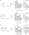
Fig. 1.
Fig. 1.
HCMV replication in HFF cells treated with IFNα or Ruxolitinib. (a) (i) and (iv) Diagram of experiments: HFF cells were treated with IFNα at the time of infection (or left untreated) or pre-treated for 24 h with IFNα (or left untreated) and then infected in the presence and absence of IFNα. Treatment of cells continued throughout infection with either AD169 or Merlin(R1111) for 96 h. (ii) and (v) Titre in p.f.u. ml−1 of each experiment. (iii) and (iv) Fold decrease in HCMV titre in the presence of IFNα compared to HCMV titre from infected untreated cells. (b) (i) Diagram of experiments: HFF cells were treated with Ruxolitinib (Ruxo) or the equivalent volume of DMSO at the time of infection or pre-treated with Ruxo or the equivalent volume of DMSO then infected in the presence of either Ruxo or DMSO. Treatment of cells continued throughout infection with either AD169 or Merlin(R1111). (ii) Titre in p.f.u. ml−1 of each experiment. (iii) Fold increase in HCMV titre in the presence of Ruxo compared to HCMV titre from infected cells treated with DMSO. In each figure data is representative of three independent experiments (black data points) and presented as the average (block) and standard deviation (error bars) of the data. In each experiment statistical relevance was examined using Student's t-test. ns=not significant (ns), P=<0.05 (*,**).
Fig. 2.
Fig. 2.
MxA expression in the presence and absence of IFNα. HFF cells were pre-treated for 24 h with IFNα (or left untreated) and then infected in the presence and absence of IFNα. Treatment of cells continued throughout infection with either AD169 or Merlin(R1111). HFF cell lysates were prepared for Western blotting at the time points indicated in the figure [hours post-infection (h p.i.)]. Uninfected HFF cell lysates were treated or untreated IFNα were also prepared for Western blotting at the time of infection (0 h p.i.). (a) Western blotting. Proteins recognized by the antibodies used in the experiment are indicated to the right of the figure. The positions of molecular weight markers (kDa) are indicated to the left of the figure. (b) Quantification of Western blotting. Relative band intensity [band intensity of MxA relative to β-actin signal in the same lane, arbitrary units (A.U.)] was analysed using ImageJ using data from two independent experiments. The mean of each data point from those experiments is shown. Hours post-infection (h p.i.).
Fig. 3.
Fig. 3.
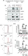
Western blotting and FACS analysis of HCMV infection in the presence and absence of IFNα. (a) Schematic of HCMV protein expression with relevant proteins grouped into kinetic classes. (b) HFF cells were pre-treated for 24 h with IFNα (or left untreated) and then infected in the presence and absence of IFNα. Treatment of cells continued throughout infection with either AD169 or Merlin(R1111). Cell lysates were prepared for Western blotting at 96 h post-infection (h p.i.). Uninfected cell lysates were treated or untreated IFNa were also prepared for Western blotting at the time of infection (0 h p.i.). Proteins recognized by the antibodies used in the experiment are indicated to the right of the figure and the positions of molecular weight markers (kDa) are indicated to the left of the figure. (c and d) HFF cells were pre-treated for 24 h with IFNα (or left untreated) and then infected in the presence and absence of IFNα. Treatment of cells continued throughout infection with AD169 or Merlin viruses expressing GFP. At 24 h post-infection cells were analysed using flow cytometry. (c) Flow-cytometry data from a representative experiment. (d) (i) number of cells expressing GFP (ii) mean fluorescent intensity of GFP expressing cells. Data is representative of three independent experiments (black data points) and presented as average (block) and standard deviation (error bars) of the data. Statistical relevance was examined using Student's t-test. ns=not significant (ns), P=<0.05 (*,**,***).
Fig. 4.
Fig. 4.
Western blotting of HCMV and PML proteins expressed in the presence and absence of IFNα. HFF cells were pre-treated for 24 h with IFNα (or left untreated) and then infected in the presence and absence of IFNα. Treatment of cells continued throughout infection with either AD169 or Merlin(R1111). Cell lysates were prepared for Western blotting at the time points indicated in the figure [hours post-infection (h p.i.)]. Uninfected cell lysates were treated or untreated IFNa were also prepared for Western blotting at the time of infection (0 h p.i.). Proteins recognized by the antibodies used in the experiment are indicated to the right of the figure and the positions of molecular weight markers (kDa) are indicated to the left of the figure.
Fig. 5.
Fig. 5.
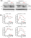
ZAP isoform expression in the presence and absence of IFNα. HFF cells were pre-treated for 24 h with IFNα (or left untreated) and then infected in the presence and absence of IFNα. Treatment of cells continued throughout infection with either AD169 or Merlin(R1111). HFF cell lysates were prepared for Western blotting at the time points indicated in the figure [hours post-infection (h p.i.)]. Uninfected HFF cell lysates were treated or untreated IFNα were also prepared for Western blotting at the time of infection (0 h p.i.). (a) Western blotting. Proteins recognized by the antibodies used in the experiment are indicated to the right of the figure (large and small ZAP isoforms, ZAP-L and ZAP-S, respectively). The positions of molecular weight markers (kDa) are indicated to the left of the figure. (b) Quantification of Western blotting. Relative band intensity [band intensity of each ZAP isoform relative to β-actin signal in the same lane, arbitrary units (A.U.)] was analysed using ImageJ using data from two independent experiments. Hours post-infection (h p.i.). The mean of each data point from those experiments is shown.
Fig. 6.
Fig. 6.
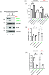
HCMV replication in CRISPR containing cells in the presence and absence of IFNα. (a) Uninfected HFF CRISPR-Luc (Luciferase) or CRISPR-ZAP HFF cells were treated with IFNα or left untreated.Cell lysates were prepared for Western blotting 24 h post-treatment. Proteins recognized by the antibodies used in the experiment are indicated to the right of the figure. The positions of molecular weight markers (kDa) are indicated to the left of the figure. (b) (i) HFF CRISPR-Luc or CRISPR-ZAP cells were pre-treated for 24 h with IFNα (or left untreated) and then infected in the presence and absence of IFNα. Treatment of cells continued throughout infection with either AD169 or Merlin(R1111) for 96 h. Titre in p.f.u. ml−1 of each experiment was calculated. Data is representative of three independent experiments (black data points) and presented as average (block) and standard deviation (error bars) of the data. Statistical relevance was examined using Student's t-test. ns=not significant (ns), P=<0.05 (*,**). (ii) Fold decrease in HCMV titre in the presence of IFNα compared to HCMV titre from infected untreated cells. (iii) Fold increase in HCMV titre in CRISPR-ZAP cells compared to CRISPR-Luc cells.
Fig. 7.
Fig. 7.
HCMV replication in HFF cells in the continuous and discontinuous presence of IFNα. (a) (i) HFF cells were pre-treated for 24 h with either IFNαor left untreated and then infected in the presence and absence of IFNα. Treatment of cells with IFNα continued throughout infection with either AD169 or Merlin(R1111) (+IFNα) or was discontinued at the time of infection (+/-IFNα). Titre in p.f.u. ml−1 of each experiment at 96 h post-infection was calculated. Data is representative of three independent experiments (black data points) and presented as the average (block) and standard deviation (error bars) of the data. Statistical relevance was examined using Student's t-test. ns=not significant (ns), P=<0.05 (*,**). (ii) Fold decrease in HCMV titre in the continuous presence of IFNα (+IFNα) compared to HCMV titre from infected untreated cells. (iii) Fold decrease in HCMV titre in the discontinuous presence of IFNα (+/-IFNα) compared to HCMV titre from infected untreated cells. (b) HFF cells were treated as in (a) and cell lysates were prepared for Western blotting at the time points indicated in the figure. Proteins recognized by the antibodies used in the experiment are indicated to the right of the figure. The positions of molecular weight markers (kDa) are indicated to the left of the figure.

Similar articles

Cited by

References

    1. Griffiths P, Reeves M. Pathogenesis of human cytomegalovirus in the immunocompromised host. Nat Rev Microbiol. 2021;19:759–773. doi: 10.1038/s41579-021-00582-z. - DOI - PMC - PubMed
    1. Coen DM, Schaffer PA. Antiherpesvirus drugs: a promising spectrum of new drugs and drug targets. Nat Rev Drug Discov. 2003;2:278–288. doi: 10.1038/nrd1065. - DOI - PubMed
    1. Strang BL. Toward inhibition of human cytomegalovirus replication with compounds targeting cellular proteins. J Gen Virol. 2022;103 doi: 10.1099/jgv.0.001795. - DOI - PubMed
    1. Rodriguez JE, Loepfe TR, Stinski MF. Human cytomegalovirus persists in cells treated with interferon. Brief report. Arch Virol. 1983;77:277–281. doi: 10.1007/BF01309276. - DOI - PubMed
    1. Mesev EV, LeDesma RA, Ploss A. Decoding type I and III interferon signalling during viral infection. Nat Microbiol. 2019;4:914–924. doi: 10.1038/s41564-019-0421-x. - DOI - PMC - PubMed

Publication types

MeSH terms

LinkOut - more resources