. 2023 Dec 8;33(3):e4855.
doi: 10.1002/pro.4855. Online ahead of print.

Direct evidence of lipid transport by the Drs2-Cdc50 flippase upon truncation of its terminal regions

Affiliations

Direct evidence of lipid transport by the Drs2-Cdc50 flippase upon truncation of its terminal regions

Sara Abad Herrera et al. Protein Sci. .

Abstract

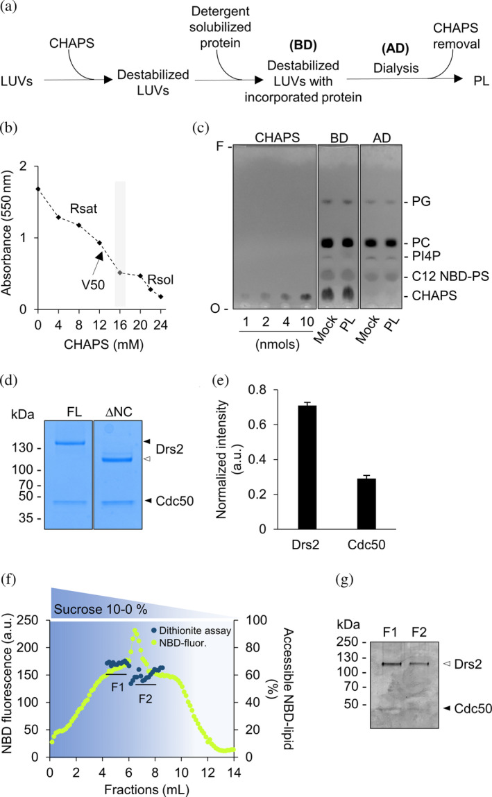
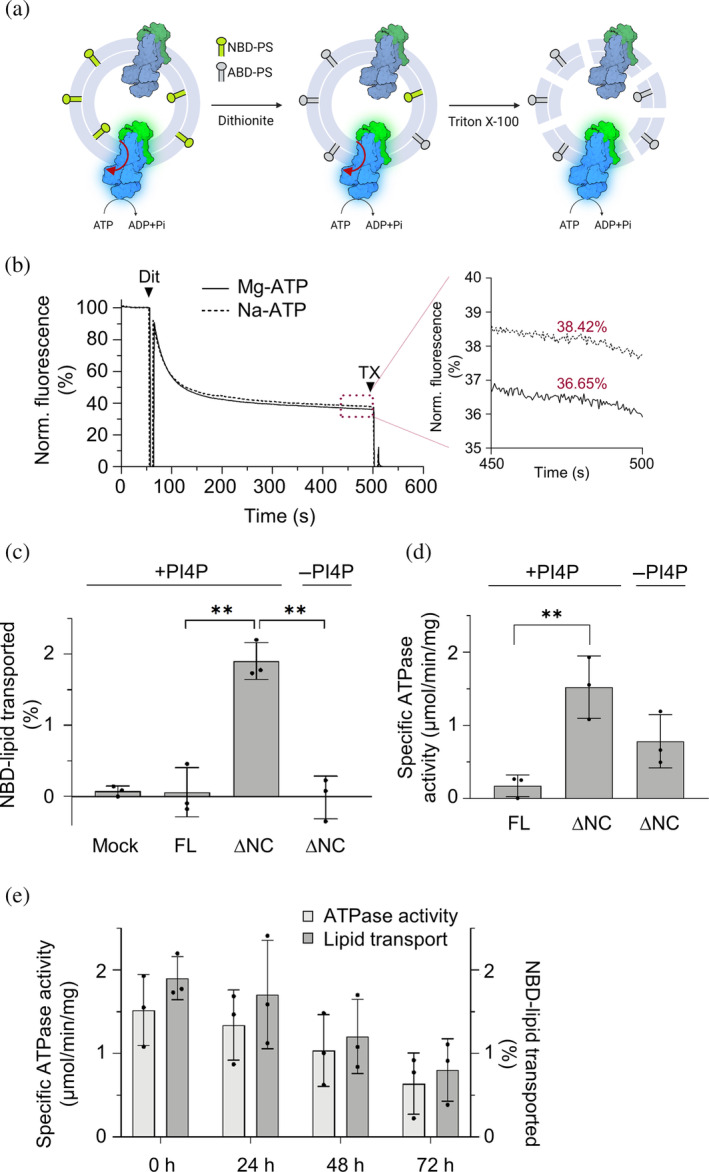
P4-ATPases in complex with Cdc50 subunits are lipid flippases that couple ATP hydrolysis with lipid transport to the cytoplasmic leaflet of membranes to create lipid asymmetry. Such vectorial transport has been shown to contribute to vesicle formation in the late secretory pathway. Some flippases are regulated by autoinhibitory regions that can be destabilized by protein kinase-mediated phosphorylation and possibly by binding of cytosolic proteins. In addition, the binding of lipids to flippases may also induce conformational changes required for the activity of these transporters. Here, we address the role of phosphatidylinositol-4-phosphate (PI4P) and the terminal autoinhibitory tails on the lipid flipping activity of the yeast lipid flippase Drs2-Cdc50. By functionally reconstituting the full-length and truncated forms of Drs2 in a 1:1 complex with the Cdc50 subunit, we provide compelling evidence that lipid flippase activity is exclusively detected for the truncated Drs2 variant and is dependent on the presence of the phosphoinositide PI4P. These findings highlight the critical role of phosphoinositides as lipid co-factors in the regulation of lipid transport by the Drs2-Cdc50 flippase.

Keywords: P4-ATPase; lipid flippase; membrane transporter; phosphoinositides; reconstitution.


Conflict of interest statement

The authors declare that they have no conflicts of interest with regard to the contents of this article.

Figures

FIGURE 1
FIGURE 1
Activation of purified Drs2–Cdc50 by phosphatidylserine fluorescent derivatives. (a) Topological illustration of Drs2 with 10 transmembrane segments (1–10) indicated in ochre and the cytosolic actuator (A), phosphorylation (P) and nucleotide binding (N) domains, indicated in yellow, blue, and red, respectively. The segments from the N‐terminus (N) and of the C‐terminus (C) that are cleaved off upon truncation of Drs2 are indicated as a dashed gray line with numbers corresponding to the residues missing in the truncated Drs2. The Cdc50 subunit is depicted in purple. (b) Coomassie blue‐stained SDS–PAGE of purified Drs2–Cdc50 complexes: full‐length (FL, closed arrow) and N‐ and C‐terminally truncated (∆NC, open arrow). Arrows indicate the positions of Drs2 and its subunit Cdc50. (c) Specific ATPase activity of the purified protein complexes in mixture with 0.98 mg mL−1 DDM and 0.25 mg mL−1 lipids (PC/PS, molar ratio 70:30; PC/PI4P/PS and PS‐derivatives, molar ratio 60:10:30) measured using the malachite green assay. For the full‐length (FL), PC/PI4P/PS were added. Data are from four independent experiments using protein from a single purification for the full‐length and truncated versions. Error bars indicate SD. The dotted line represents the activity measured for the ∆NC Drs2–Cdc50 in the presence of PC/PS and absence of additional PI4P. The asterisks denote a significant difference compared with the values for ∆NC Drs2–Cdc50 without PI4P. PC, phosphatidylcholine; PS, phosphatidylserine; C12‐NBD‐PS and ‐PC, 1‐palmitoyl‐2‐NBD‐dodecanoyl‐sn‐glycero‐3‐phosphoserine and ‐phosphocholine; C6‐NBD‐PS and ‐PC, 1‐palmitoyl‐2‐NBD‐hexanoyl‐sn‐glycero‐3‐phosphoserine and ‐phosphocholine; PI4P, brain l‐α‐phosphatidylinositol‐4‐phosphate.
FIGURE 2
FIGURE 2
Characterization of Drs2–Cdc50 reconstitution. (a) Schematic diagram illustrating the reconstitution of Drs2–Cdc50 using CHAPS. BD, before dialysis; AD, after dialysis. (b) Detergent destabilization of pre‐formed liposomes (10 mg mL−1; see Section 4) exposed to increasing concentrations of CHAPS. The concentration range used for reconstitution is highlighted in a light gray box; however, it was empirically tested for each LUV preparation before reconstitution. The dashed line serves as a guide to the eye. (c) Thin layer chromatography analysis before and after dialysis. CHAPS was compared to standards. O, origin; F, solvent front of the chromatogram. CHAPS, 3‐[(3‐cholamidopropyl)dimethylammonio]‐1‐propanesulfonate; PC, phosphatidylcholine; PG, phosphatidylglycerol; C12‐NBD‐PS, 1‐palmitoyl‐2‐NBD‐dodecanoyl‐sn‐glycero‐3‐phosphoserine; PI4P, brain l‐α‐phosphatidylinositol‐4‐phosphate. (d) Coomassie‐stained SDS‐PAGE of proteoliposomes containing the indicated Drs2–Cdc50 complexes: full‐length (FL) and N‐ and C‐terminally truncated wild type (∆NC). Closed arrowheads indicate the bands corresponding to both FL Drs2 and Cdc50; the empty arrowhead indicates ∆NC Drs2. (e) Normalized intensity of Drs2 and Cdc50 determined by densitometry analysis of Coomassie‐stained SDS‐PAGE of proteoliposomes reconstituted with truncated Drs2–Cdc50 (n = 3 independent reconstitutions). The normalized intensity was calculated based on the total intensity of the bands corresponding to Drs2 and Cdc50 set to 1. Coomassie dye binds mainly to basic residues; thus, considering the number for Drs2 (134 residues) and Cdc50 (55 residues), a band intensity of ~2.4‐times higher of Drs2:Cdc50 is expected for a 1:1 complex, in agreement with the results shown here. (f) Gradient flotation assay of proteoliposomes reconstituted with truncated Drs2–Cdc50. Fractions were collected from the bottom of the gradient and analyzed for NBD fluorescence (green dotted line) and NBD‐lipid accessibility by dithionite assay (blue dots). (g) SDS‐PAGE analysis of fraction pools (F1, F2) via silver staining. Arrows indicate the positions of purified truncated Drs2 and Cdc50.
FIGURE 3
FIGURE 3
Truncated but not full‐length Drs2–Cdc50 shows transport of C12‐NBD‐PS in the presence of PI4P. (a) Illustration of the flippase activity assay in proteoliposomes. The assay exploits the ability of dithionite to chemically reduce the NBD fluorophore, thereby irreversibly quenching the fluorescence of NBD‐lipids situated in the outer leaflet of the vesicles. Thus, proteoliposomes with lipid flippase activity show increased quenching after incubation with Mg‐ATP relative to the same liposomes after incubation with Na‐ATP. (b) Dithionite quenching of C12‐NBD‐PS fluorescence on proteoliposomes containing truncated Drs2–Cdc50 (∆NC) after 30 min of incubation with Mg‐ATP (solid trace) or Na‐ATP (dashed trace). Dithionite (Dit) was added at 60 s to quench the outer leaflet C12‐NBD‐PS, and Triton X‐100 (TX) was added at 500 s to quench the remaining inner leaflet NBD‐PS. (c) Flippase assay with protein‐free liposomes (mock) and liposomes containing the indicated Drs2–Cdc50 complexes: full‐length (FL) and N‐ and C‐terminally truncated (∆NC). Liposomes were prepared without (−) or with (+) PI4P. The increase in accessible C12‐NBD‐PS after a 30‐min incubation with Mg‐ATP relative to Na‐ATP was plotted. (d) Specific ATPase activity of the protein complexes reconstituted in liposomes. (e) ATPase (light gray) and lipid transport (dark gray) activities were measured every 24 h after reconstitution of the truncated Drs2–Cdc50 (∆NC). In panels c, d and, e, the data are from three independent reconstitution experiments using three independent LUV preparations. Error bars show SD, and statistical significance is indicated with asterisks.

Similar articles

References

    1. Azouaoui H, Montigny C, Ash MR, Fijalkowski F, Jacquot A, Grønberg C et al. A high‐yield co‐expression system for the purification of an intact Drs2p‐Cdc50p lipid flippase complex, critically dependent on and stabilized by phosphatidylinositol‐4‐phosphate. PLoS One. 2014;9(11):e112176. - PMC - PubMed
    1. Azouaoui H, Montigny C, Dieudonné T, Champeil P, Jacquot A, Vázquez‐Ibar JL, et al. High phosphatidylinositol 4‐phosphate (PI4P)‐dependent ATPase activity for the Drs2p‐Cdc50p flippase after removal of its N‐ and C‐terminal extensions. J Biol Chem. 2017;292(19):7954–7970. 10.1074/jbc.M116.751487 - DOI - PMC - PubMed
    1. Azouaoui H, Montigny C, Jacquot A, Barry R, Champeil P, Lenoir G. Coordinated overexpression in yeast of a P4‐ATPase and its associated Cdc50 subunit: the case of the Drs2p/Cdc50p lipid Flippase complex. Methods Mol Biol (Clifton, NJ). 2016;1377:37–55. 10.1007/978-1-4939-3179-8_6 - DOI - PubMed
    1. Bai L, Kovach A, You Q, Hsu H‐C, Zhao G, Li H. Autoinhibition and activation mechanisms of the eukaryotic lipid flippase Drs2p‐Cdc50p. Nat Commun. 2019;10(1):4142. 10.1038/s41467-019-12191-9 - DOI - PMC - PubMed
    1. Bryde S, Hennrich H, Verhulst PM, Devaux PF, Lenoir G, Holthuis JCM. CDC50 proteins are critical components of the human class‐1 P4‐ATPase transport machinery. J Biol Chem. 2010;285(52):40562–40572. 10.1074/jbc.M110.139543 - DOI - PMC - PubMed

LinkOut - more resources