. 2023 Dec 1;64(15):7.
doi: 10.1167/iovs.64.15.7.

Ontogenesis of the Mouse Ocular Surface Lymphatic Vascular Network

Affiliations

Ontogenesis of the Mouse Ocular Surface Lymphatic Vascular Network

Mariela Subileau et al. Invest Ophthalmol Vis Sci. .

Abstract

Purpose: Ocular lymphatic vessels play major physiological role in eye homeostasis and their dysfunction can contribute to the progression of several eye diseases. In this study, we characterized their spatiotemporal development and the cellular mechanisms occurring during their ontogenesis in the mouse eye.

Methods: Whole mount immunofluorescent staining and imaging by standard or lightsheet fluorescence microscopy were performed on late embryonic and early postnatal eye mouse samples.

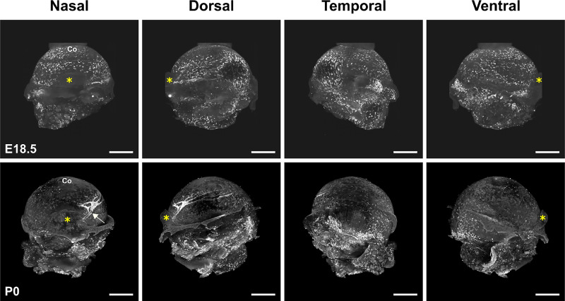
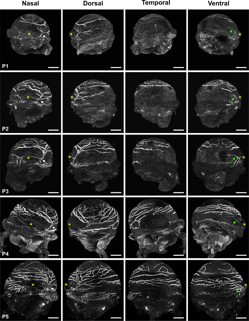
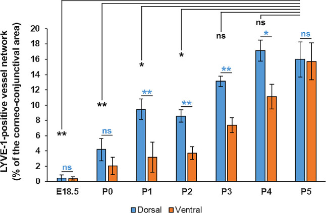
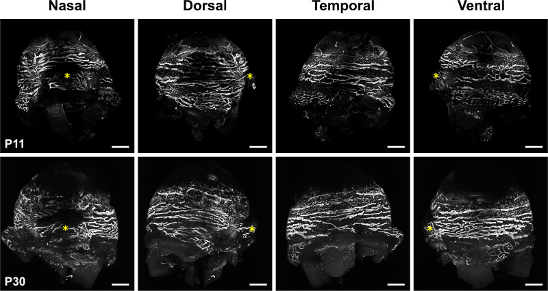
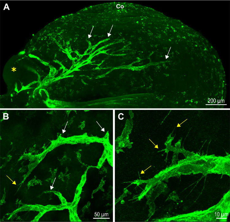
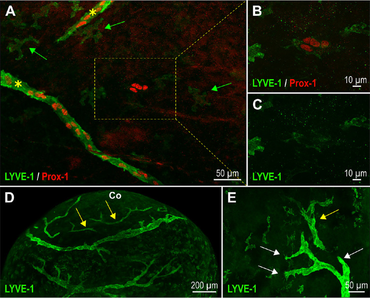
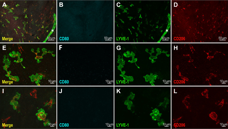
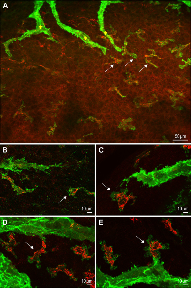
Results: We observed that the ocular surface lymphatic vascular network develops at the early postnatal stages (between P0 and P5) from two nascent trunks arising at the nasal side on both sides of the nictitating membrane. These nascent vessels further branch and encircle the whole eye surface by sprouting lymphangiogenesis. In addition, we got evidence for the existence of a transient lymphvasculogenesis process generating lymphatic vessel fragments that will mostly formed the corneolimbal lymphatic vasculature which further connect to the conjunctival lymphatic network. Our results also support that CD206-positive macrophages can transdifferentiate and then integrate into the lymphatic neovessels.

Conclusions: Several complementary cellular processes participate in the development of the lymphatic ocular surface vasculature. This knowledge paves the way for the design of new therapeutic strategies to interfere with ocular lymphatic vessel formation when needed.


Conflict of interest statement

Disclosure: M. Subileau, None; D. Vittet, None

Figures

Figure 1.
Figure 1.
Initiation of ocular lymphatic development at birth. LSFM representative images of the LYVE-1 whole mount immunofluorescent staining of dissected right eyes of an E18.5 mouse embryo and of a P0 neonate. The multiview images corresponding to the projections at the four mentioned cardinal axes are shown. The yellow asterisks mark the position of the nictitating membrane at the nasal side of the eye. The white arrow points to the first nascent emerging lymphatic trunk. Note the presence of remaining connective and muscular tissues in the lower part of the ocular globe. Co, cornea. Scale bars for all panels: 500 µm. For LSFM representative images of LYVE-1 antibody negative control staining, see Supplementary Figure S1.
Figure 2.
Figure 2.
Expansion of the ocular surface lymphatic vessel network during early post-natal steps. Representative LSFM images of the multiview projections at the four cardinal axes of the LYVE-1 immunofluorescent staining of right eyes from P1 to P5. Arrows pointed to the roots of the lymphatic trunks which develops at both sides of the nictitating membrane marked by yellow asterisks. White arrow: root of the trunk from which the corneolimbal and the dorsal conjunctival lymphatic networks form; Green arrow: root of the trunk from which the ventral conjunctival lymphatic network forms. Scale bars for all panels: 500 µm. For LSFM representative images of LYVE-1 antibody negative control staining, see Supplementary Figure S1.
Figure 3.
Figure 3.
Quantitative analysis of the early steps of the ocular surface lymphatic vessel network formation. The measurements were performed on the images corresponding to the ventral and the dorsal lymphatic vessel network, respectively, facing the left and the right sides of the nictitating membrane in right eyes. The lymphatic vessel density was measured using ImageJ as the percentage of the corneal and of the conjunctival area that was occupied by LYVE-1–positive vessels. Single LYVE-1–positive cells were excluded from the analysis by prior image treatment. Data are the mean ± SEM of three (E18.5, P3, P4), four (P1, P2, P5), or seven (P0) different right eye samples. Blue characters: ** P < 0.01, * P< 0.05, dorsal value significantly different from corresponding ventral value; ns, not significant; using an unpaired Student's t test. Black characters: ** P < 0.01, * P < 0.05, dorsal value significantly different from P5 dorsal value; ns, not significant; using an unpaired Student's t test.
Figure 4.
Figure 4.
Views of the final maturation and remodeling of the ocular surface lymphatic vessel network. Representative LSFM images of the LYVE-1 immunofluorescent staining of left eyes at P11 and P30. The yellow asterisks mark the position of the nictitating membrane. Scale bars for all panels: 500 µm. For LSFM representative images of LYVE-1 antibody negative control staining, see Supplementary Figure S1.
Figure 5.
Figure 5.
Sprouting lymphangiogenesis for ocular lymphatic vessel network expansion. (A) LSFM image of the sprouting LYVE-1-positive lymphatic vessel dorsal network of a right eye at P1. The yellow asterisk marks the nictitating membrane, and white arrows point to some sprout extremities. Co, cornea. (B, C) LYVE-1 immunofluorescent staining images of lymphatic sprouts observed at P1 after flat mountings of eye anterior segments. White arrows point to buds, and yellow arrows point to some filopodia.
Figure 6.
Figure 6.
Lymphvasculogenesis constitute a cellular process for ocular lymphatic vessel development. (A) Visualization of the presence of a LYVE-1+ Prox-1+ cell cluster (box) at the P0 ocular surface after whole mount immunofluorescent staining of the eye anterior segment and flat-mounting. The yellow asterisks mark the location of two lymphatic sprouts. The green arrows point to some isolated LYVE-1–positive cells that correspond to macrophages. (B, C) Imaging of the boxed LYVE-1+ Prox-1+ lymphatic cell cluster at a higher magnification. (D) LSFM imaging of some LYVE-1-positive lymphatic vessel fragments (yellow arrows), which have formed at the corneal and corneolimbal surfaces of a P1 neonate. Co, cornea. (E) LYVE-1 immunofluorescence staining image obtained at P1, allowing the view of both lymphangiogenic sprouting (sprout extremities are pointed by white arrows), and a lymphatic vessel fragment, which could have been formed by lymphvasculogenesis (yellow arrow).
Figure 7.
Figure 7.
Ocular surface–scattered LYVE-1–positive cells display CD206 M2-macrophage antigenic marker expression. Illustrations of M1 (CD80) and M2 (CD206) macrophage subtype markers expressions in LYVE-1–positive cells after whole mount immunofluorescence staining and flat mounting of the anterior segments of the eyes at P1. The asterisk marks an LYVE-1–positive lymphatic vessel sprout.
Figure 8.
Figure 8.
Morphological evidence for potential M2-macrophage contribution to ocular lymphatic vessel formation. Illustrations of immunofluorescent staining for LYVE-1 (green) and CD206 (red) at P1. (A) View of a corneolimbal surface part showing both lymphatic sprouts and single M2-macrophages. Note the high LYVE-1– and CD206-positive macrophage (white arrows) density and their close vicinity to the sprouts extremities marked by yellow asterisks. (B–E) Images evoking the association (B, C), the further attachment (D) and integration (E) into a developing lymphatic sprout, of an LYVE-1– and CD206-positive macrophage (white arrow) initially present in close proximity to a lymphatic sprout extremity.
Figure 9.
Figure 9.
Evidence for the existence of mixed Prox-1– and CD206-positive cells in lymphatic neovessels. Whole mount double immunofluorescence staining with LYVE-1 and CD206 antibodies (A–C) or with Prox-1 and CD206 antibodies (D–F), of the developing ocular surface lymphatic vasculature at P1. The yellow asterisk marks a CD206-positive macrophage displaying a low LYVE-1 expression when compared to the LYVE-1 expression level of cells constituting the lymphatic neovessel. The white arrows point to the CD206 immunoreactivity in some LYVE-1– (upper panels) or Prox-1–positive cells (lower panels) of the lymphatic neovessel.

Similar articles

References

    1. Oliver G, Kipnis J, Randolph GJ, Harvey NL.. The lymphatic vasculature in the 21(st) century: novel functional roles in homeostasis and disease. Cell . 2020; 182: 270–296. - PMC - PubMed
    1. Schwager S, Detmar M.. Inflammation and lymphatic function. Front Immunol . 2019; 10: 308. - PMC - PubMed
    1. Kraft JD, Blomgran R, Lundgaard I, et al. .. Specialized pro-resolving mediators and the lymphatic system. Int J Mol Sci . 2021; 22: 2750. - PMC - PubMed
    1. Liu X, De la Cruz E, Gu X, et al. .. Lymphoangiocrine signals promote cardiac growth and repair. Nature . 2020; 588(7839): 705–711. - PMC - PubMed
    1. Niec RE, Chu T, Schernthanner M, et al. .. Lymphatics act as a signaling hub to regulate intestinal stem cell activity. Cell Stem Cell . 2022; 29: 1067–1082.e1018. - PMC - PubMed