Paranasal sinus angiosarcoma with facial paralysis as a novel manifestation: a case report and literature review
- PMID: 38042771
- PMCID: PMC10693057
- DOI: 10.1186/s12883-023-03482-2
Paranasal sinus angiosarcoma with facial paralysis as a novel manifestation: a case report and literature review
Abstract
Background: Paranasal sinus angiosarcoma is an uncommon malignancy, with only a few reported cases worldwide. Although it exhibits multiple symptoms, facial paralysis has not been previously documented as a noticeable presentation.
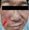
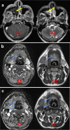
Case presentation: In this case, we report a 40-year-old male who presented with facial numbness and pain for one month, weakness of his facial muscles for 15 days, and recurrent right epistaxis for 1 year. He had a history of nasal inflammatory polyps with chronic sinusitis. Computed tomography and magnetic resonance imaging showed space-occupying lesions in the right nasal cavity and maxillary sinus, with bone destruction occurring in the sinus wall and turbinate. This patient then underwent endoscopic surgery. According to the histopathological and immunohistochemical results, he was eventually diagnosed with paranasal sinus angiosarcoma in April 2021. To date, this patient has not initiated any radiotherapy or chemotherapy and has survived with lymphatic metastasis for at least 3 years.
Conclusions: This manuscript suggests that paranasal sinus angiosarcoma can present with facial paralysis. Moreover, pathological and immunohistochemical tests are still vital for diagnosing paranasal sinus angiosarcoma and differential diagnosis. Additionally, regular follow-up is crucial for patients with paranasal sinus angiosarcoma, enabling monitoring of recurrence, metastasis, and recovery while contributing valuable clinical data to understanding this rare disease and associated research endeavours.
Keywords: Case report; Facial paralysis; Immunohistochemistry; Paranasal sinus angiosarcoma; Pathology.
© 2023. The Author(s).
Conflict of interest statement
The authors declare no competing interests.
Figures


Similar articles
-
A Rare Case of Giant Cavernous Hemangioma of the Maxillary Sinus.Am J Case Rep. 2022 Oct 9;23:e937191. doi: 10.12659/AJCR.937191. Am J Case Rep. 2022. PMID: 36209361 Free PMC article.
-
Destructive Fibrosarcoma of the Maxillary Sinus.J Craniofac Surg. 2018 May;29(3):e226-e228. doi: 10.1097/SCS.0000000000004173. J Craniofac Surg. 2018. PMID: 29283942
-
Angiosarcoma of the maxillary sinus: literature review and case report.Head Neck Surg. 1979 Jan-Feb;1(3):274-80. doi: 10.1002/hed.2890010311. Head Neck Surg. 1979. PMID: 574132
-
Multiple metastases of clear-cell renal cell carcinoma to different region of the nasal cavity and paranasal sinus 3 times successively: A case report and literature review.Medicine (Baltimore). 2018 Apr;97(14):e0286. doi: 10.1097/MD.0000000000010286. Medicine (Baltimore). 2018. PMID: 29620646 Free PMC article. Review.
-
Angiosarcoma of the maxillary sinus.J Laryngol Otol. 2000 May;114(5):381-4. doi: 10.1258/0022215001905634. J Laryngol Otol. 2000. PMID: 10912272 Review.
Cited by
-
Imaging features of pediatric angiosarcomas: clinical, pathologic, and radiological review.Pediatr Radiol. 2024 Oct;54(11):1873-1883. doi: 10.1007/s00247-024-06041-0. Epub 2024 Sep 3. Pediatr Radiol. 2024. PMID: 39223383
-
Angiosarcoma of the Maxillary Sinus: A Case Report.Cureus. 2024 Jun 25;16(6):e63131. doi: 10.7759/cureus.63131. eCollection 2024 Jun. Cureus. 2024. PMID: 39055444 Free PMC article.
References
-
- Rufus J, Mark MD, et al. Angiosarcoma A Repott of 67 patients and a review of the literature. Cancer. 1996. - PubMed
Publication types
MeSH terms
Grants and funding
LinkOut - more resources
Full Text Sources
Miscellaneous

