. 2023 Dec 13;480(23):1909-1928.
doi: 10.1042/BCJ20230277.

A guide to ERK dynamics, part 2: downstream decoding

Affiliations

A guide to ERK dynamics, part 2: downstream decoding

Abhineet Ram et al. Biochem J. .

Abstract

Signaling by the extracellular signal-regulated kinase (ERK) pathway controls many cellular processes, including cell division, death, and differentiation. In this second installment of a two-part review, we address the question of how the ERK pathway exerts distinct and context-specific effects on multiple processes. We discuss how the dynamics of ERK activity induce selective changes in gene expression programs, with insights from both experiments and computational models. With a focus on single-cell biosensor-based studies, we summarize four major functional modes for ERK signaling in tissues: adjusting the size of cell populations, gradient-based patterning, wave propagation of morphological changes, and diversification of cellular gene expression states. These modes of operation are disrupted in cancer and other related diseases and represent potential targets for therapeutic intervention. By understanding the dynamic mechanisms involved in ERK signaling, there is potential for pharmacological strategies that not only simply inhibit ERK, but also restore functional activity patterns and improve disease outcomes.

Keywords: cell proliferation; eukaryotic gene expression; extracellular signal-regulated kinases; gene regulatory networks; receptor tyrosine kinases.


Conflict of interest statement

The authors declare that there are no competing interests associated with the manuscript.

Figures

Figure 1.
Figure 1.. Differential gene expression responses to ERK signaling.
(A) Differential regulation of EGR1, FOSL1, and FOS expression. In each case, ERK phosphorylates a transcription factor (TF) that binds to the gene promoter. Left: EGR1 protein binds to its own gene promoter, inhibiting further transcription. Middle: ERK initiates FOSL1 transcription and also stabilizes FRA-1 protein. Right: ERK initiates FOS expression, and stabilizes FOS protein through phosphorylation; sustained ERK activation also apparently induces a FOS transcriptional repressor that has not been definitively identified [36]. (B) Sustained signaling generates high concentrations of FRA-1 and FOS protein. Subsequently, the FOS repressor inhibits further expression of FOS. After the initial peak of EGR1 concentration, EGR1 represses its own transcription (auto-inhibition), thus returning to an equilibrium with low baseline expression. (C) Pulsatile signaling generates short bursts of EGR1 accumulation. Since EGR1 transcription is brief and the protein is unstable, auto-inhibition does not persist. This allows EGR1 levels to track closely with ERK activity pulses. Conversely, pulsatile signaling weakly induces FRA-1 and FOS expression because sustained signaling is required for protein stabilization. Figure adapted in part from [24].
Figure 2.
Figure 2.. Functions of ERK dynamics during development and tissue homeostasis.
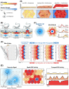
(A) ERK activity increases the probability of cell division. When exposed to different EGF concentrations (bottom panel, blue), single-cell ERK activity within a population varies from cell-to-cell (cell colors), increasing the average rate of proliferation [59,107]. (B) Spatially restricted ERK activity indicates cellular position during development. Within the C. elegans gonad, the anchor cell (AC, blue) is the point source for EGF ligand secretion, creating a gradient beneath the basement membrane (blue triangles below cells). Cells closest to the anchor cell receive the most stimulation and thus have the highest ERK activity (individual cell colors, red to yellow), inducing differential cell differentiation programs to create the mature vulva [80,81]. (C) Secretion of EGFR ligands from apoptotic cells creates a radial gradient around the dying cell. Cells closest to the center (red) have high ERK activity, which conveys a survival advantage, while cells farther from the dying cell receive a lower concentration of ligands (blue halo in left panel) resulting in lower and delayed ERK activity. Individual ERK activity traces are shown to the right. Figure adapted from Gagliardi et al. [87]. (D) During tissue injury, ERK activity provides both directional and migratory signals. Initially, cells adjacent to the injury have high ERK activity (first row, red cells) and secrete a short-range ERK stimulus gradient that activates its neighbors (abutting gradient, blue). Once the neighbors are activated, the ERK activity in the previous cell layer decays, ultimately creating a wave of ERK activity (second through fourth panel). ERK activation waves repeatedly propagate from the site of injury. This directs cells to migrate toward the site of injury (red arrows below) resulting in gap closure and wound healing [55,93,94]. (E) Multiple sources of ERK activation generate sporadic spatiotemporal ERK activation. Cells that actively shed EGFR ligands create transient, stimulatory microenvironments (blue gradients), in contrast with (A) which depicts uniform stimuli. These dynamic and overlapping microenvironments create varying temporal patterns of ERK activity, generating diverse gene expression profiles within the population [24].
Figure 3.
Figure 3.. Alterations in ERK dynamics as a result of oncogenic mutations and inhibitors.
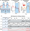
(A) In cells with wild type (WT) pathway genes, ERK undergoes fast activation and deactivation in response to optogenetic stimulation of SOS. BRAF G491A mutation leads to slower deactivation time (top). When stimuli are frequent, ERK responses are elongated into sustained activity (bottom), leading to aberrant transcriptional regulation of target genes [23]. (B) Mutations in KRAS drive increased baseline ERK activity. Maximum activity levels upon stimulation remain similar [68,110]. (C) Malignant cells release paracrine signals that stimulate sporadic and pulsatile ERK activity in neighboring wild-type cells [24,68]. (D) Modulation of ERK dynamics by pharmacological inhibitors. In wild-type cells, the pattern of normal pulsatile signaling (typically resulting from paracrine stimuli [55]) can be altered by the addition of various pharmacological inhibitors (dashed vertical line). EGFR inhibition (EGFRi) quickly abrogates ERK activation, but pulsatile signaling re-emerges within several hours [37]. RAF inhibitors (RAFi) can generate low-frequency ERK pulses [126], potentially as a result of paradoxical activation of RAF. Non-EGFR receptor tyrosine kinase inhibitors(RTKi) result in high-frequency pulses of ERK [126]. In cells carrying oncogenic RAS or RAF mutations, baseline ERK signaling is elevated and inhibition of RAF or MEK (RAFi/MEKi) suppresses ERK signaling initially. A rebound of ERK activity occurs in a subpopulation of cells (solid red line) within several hours, while other cells remain sensitive (dashed red line) [109,128]. All curves shown are approximated depictions of experimental data.

Similar articles

Cited by

References

    1. Lavoie, H., Gagnon, J. and Therrien, M. (2020) ERK signalling: a master regulator of cell behaviour, life and fate. Nat. Rev. Mol. Cell Biol. 21, 607–632 10.1038/s41580-020-0255-7 - DOI - PubMed
    1. Vasudevan, H.N. and Soriano, P. (2016) Chapter twenty-three - a thousand and one receptor tyrosine kinases: wherein the specificity? Curr. Top. Dev. Biol. 117, 393–404 10.1016/bs.ctdb.2015.10.016 - DOI - PMC - PubMed
    1. Vasudevan, H.N., Mazot, P., He, F. and Soriano, P. (2015) Receptor tyrosine kinases modulate distinct transcriptional programs by differential usage of intracellular pathways. eLife 4, e07186 10.7554/eLife.07186 - DOI - PMC - PubMed
    1. Chen, J.-Y., Lin, J.-R., Cimprich, K.A. and Meyer, T. (2012) A two-dimensional ERK-AKT signaling code for an NGF-triggered cell-fate decision. Mol. Cell 45, 196–209 10.1016/j.molcel.2011.11.023 - DOI - PMC - PubMed
    1. Kholodenko, B.N., Hancock, J.F. and Kolch, W. (2010) Signalling ballet in space and time. Nat. Rev. Mol. Cell Biol. 11, 414–426 10.1038/nrm2901 - DOI - PMC - PubMed

Publication types

MeSH terms

Substances