. 2023 Nov 2:16:1241432.
doi: 10.3389/fnmol.2023.1241432. eCollection 2023.

The role of primed and non-primed MSC-derived conditioned media in neuroregeneration

Affiliations

The role of primed and non-primed MSC-derived conditioned media in neuroregeneration

Nikola Hudakova et al. Front Mol Neurosci. .

Abstract

Introduction: With growing significance in nervous system repair, mesenchymal stem cell-derived conditioned media (MSCCM) have been used in cell-free therapies in regenerative medicine. However, the immunomodulatory and neuroregenerative effects of MSCCM and the influence of priming on these effects are still poorly understood.

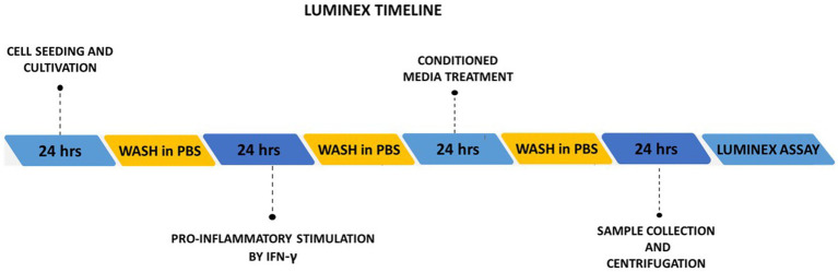
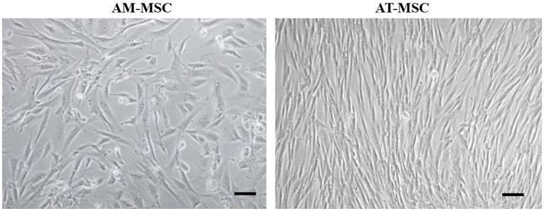
Methods: In this study, by various methods focused on cell viability, proliferation, neuron-like differentiation, neurite outgrowth, cell migration and regrowth, we demonstrated that MSCCM derived from adipose tissue (AT-MSCCM) and amniotic membrane (AM-MSCCM) had different effects on SH-SY5Y cells.

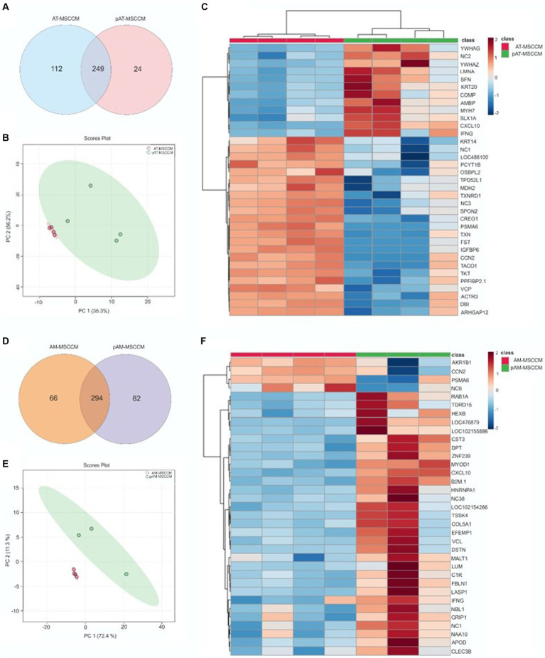
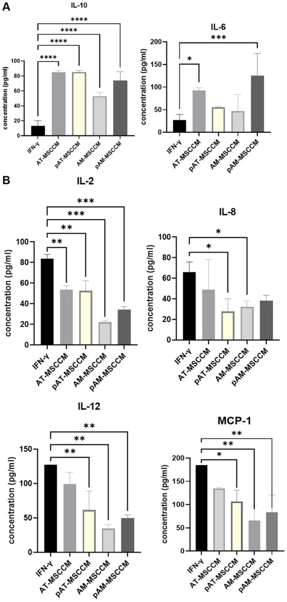
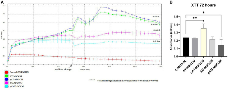
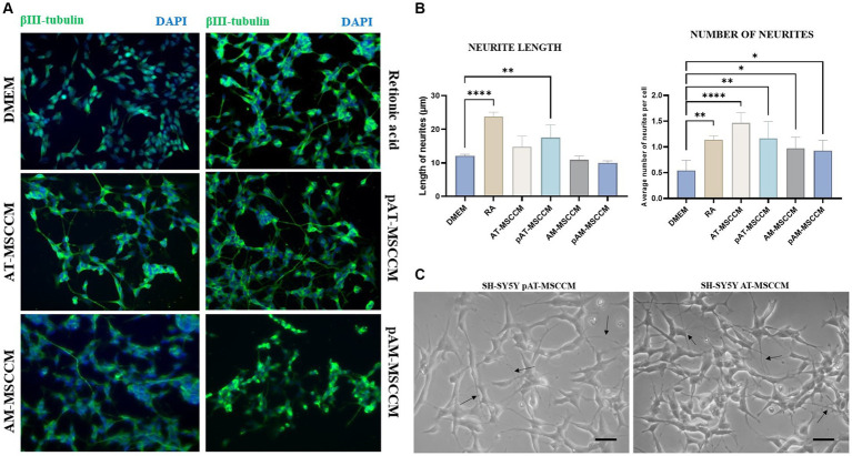
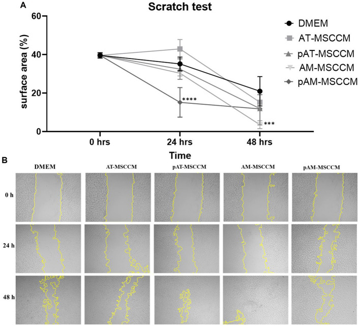
Results and discussion: AT-MSCCM was found to have a higher proliferative capacity and the ability to impact neurite outgrowth during differentiation, while AM-MSCCM showed more pronounced immunomodulatory activity, migration, and re-growth of SH-SY5Y cells in the scratch model. Furthermore, priming of MSC with pro-inflammatory cytokine (IFN-γ) resulted in different proteomic profiles of conditioned media from both sources, which had the highest effect on SH-SY5Y proliferation and neurite outgrowth in terms of the length of neurites (pAT-MSCCM) compared to the control group (DMEM). Altogether, our results highlight the potential of primed and non-primed MSCCM as a therapeutic tool for neurodegenerative diseases, although some differences must be considered.

Keywords: IFN-γ; MSC; conditioned medium; neurotrophic effect; priming.


Conflict of interest statement

The authors declare that the research was conducted in the absence of any commercial or financial relationships that could be construed as a potential conflict of interest.

Figures

Figure 1
Figure 1
Schematic illustration of the experimental timelines of Luminex assay.
Figure 2
Figure 2
Morphology of canine AM-MSC and AT-MSC. AM-MSC exhibited fibroblast-like morphology, while AT-MSC were spindle-shaped, both typical for mesenchymal stromal perivascular cells. Scale bar 50 μm.
Figure 3
Figure 3
FACS analysis of the expression of the AT-MSC and AM-MSC surface markers at passage 3. (A) AT-MSC and AM-MSC expressed typical mesenchymal markers CD29, CD44, and CD90 but not CD34 and CD45. (B) The negative control is represented by unlabeled cells to exclude autofluorescence.
Figure 4
Figure 4
Multilineage differentiation of cells in passage 3. Canine AT-MSC and AM-MSC successfully underwent osteogenic (A,D), chondrogenic (B,E), and weaker adipogenic differentiation (C,F). Respective negative controls (C1,C2,C3) are shown as AM-MSC cells where no differentiation media was used.
Figure 5
Figure 5
LC/MS–MS analysis of MSCCM and pMSCCM. (A,D) Venn diagrams representing the total amount of identified proteins in MSCCM and pMSCCM. (B,E) Principal component analysis scores plot showing clustering of MSCCM and pMSCCM. (C,F) Heat maps of differentially expressed proteins Red and blue colors represent up-regulated and down-regulated proteins, respectively.
Figure 6
Figure 6
(A) Graphs showing changes in amounts of anti-inflammatory cytokines IL-10 and IL-6 (both pro and anti-inflammatory properties) detected by Luminex analyses. (B) Graphs showing changes in amounts of pro-inflammatory cytokines IL-2, IL-8, IL-12, and MCP-1 detected by Luminex analyses. Analytes released by SH-SY5Y cells after 24-h induction with IFN-γ and SH-SY5Y cultured in IFN-γ and with subsequent treatment with MSCCM for 24 h were measured. ANOVA Dunett’s multiple comparisons tests *p < 0.05, **p < 0.01, ***p < 0.001, ****p < 0.0001.
Figure 7
Figure 7
The effect of conditioned media on proliferation and metabolic activity. (A) Time-dependent proliferation profile of SH-SY5Y cultured in MSCCM. Proliferation curves of SH-SY5Y cells as generated by xCELLigence RTCA during 120 h. (B) Cell proliferation and metabolic activity were determined by the XTT assay after 72 h of incubation in DMEM medium supplemented with ATB/ATM and for experimental groups in conditioned media (AT-MSCCM, pAT-MSCCM, AM-MSCCM, pAM-MSCCM). *p < 0.05, **p < 0.01, ***p < 0.001, ****p < 0.0001. In both cases GraphPad Prism software was used for statistical analysis, utilizing the ANOVA Dunett’s multiple comparisons tests (*p < 0.05, **p < 0.01, ***p < 0.001, ****p < 0.0001).
Figure 8
Figure 8
Effect of conditioned media on SH-SY5Y differentiation and neurite outgrowth. (A) Representative immunofluorescence images of β- III- tubulin positive cells (green) in differentiation media (Retinoic acid) and conditioned media (AT-MSCCM, pAT-MSCCM, AM-MSCCM, pAM-MSCCM) treatment groups. (B) Quantitative analyses of the average length of neurites and the average number of neurites per cell in different groups (ANOVA Dunett’s multiple comparisons tests * indicates p < 0.05). (C) Representative images of SH-SY5Y after culture in MSCCM for 12 days, note the long outgrowths in the group cultured in pAT-MSCCM (indicated by arrows) and the high number of neurites per cell in SH-SY5Y treated with AT-MSCCM (indicated by arrows). Scale bar: 20 μm.
Figure 9
Figure 9
Cell migration detected by the scratch test (magnification, × 5). (A) Shows the statistical analysis of the separation distance of cells in all groups (percentage of empty area per field in all groups) by ANOVA (Dunett’s multiple comparisons tests, ***p < 0.001, ****p < 0.0001). (B) Representative photos of wells where we performed the Scratch test followed by 48-h treatment in Control medium (DMEM with ATB/ATM) and conditioned media (AT-MSCCM, pAT-MSCCM, AM-MSCCM, pAM-MSCCM).

Similar articles

References

    1. Ahmadbeigi N., Shafiee A., Seyedjafari E., Gheisari Y., Vassei M., Amanpour S., et al. . (2011). Early spontaneous immortalization and loss of plasticity of rabbit bone marrow mesenchymal stem cells. Cell Prolif. 44, 67–74. doi: 10.1111/j.1365-2184.2010.00731.x, PMID: - DOI - PMC - PubMed
    1. Al Delfi I. R., Sheard J. J., Wood C. R., Vernallis A., Innes J. F., Myint P., et al. . (2016). Canine mesenchymal stem cells are neurotrophic and angiogenic: An in vitro assessment of their paracrine activity. Vet. J. 217, 10–17. doi: 10.1016/j.tvjl.2016.09.003 - DOI - PubMed
    1. Andreeva E. R., Udartseva O. O., Zhidkova O. V., Buravkov S. V., Ezdakova M. I., Buravkova L. B. (2018). IFN-gamma priming of adipose-derived stromal cells at “physiological” hypoxia. J. Cell. Physiol. 233, 1535–1547. doi: 10.1002/jcp.26046, PMID: - DOI - PubMed
    1. Beletskiy A., Chesnokova E., Bal N. (2021). Insulin-like growth factor 2 as a possible neuroprotective agent and memory enhancer—its comparative expression, processing and signaling in mammalian CNS. Int. J. Mol. Sci. 22:1849. doi: 10.3390/ijms22041849, PMID: - DOI - PMC - PubMed
    1. Berebichez-Fridman R., Montero-Olvera P. R. (2018). Sources and clinical applications of mesenchymal stem cells. Sultan Qaboos Univ. Med. J. 18, e264–e277. doi: 10.18295/squmj.2018.18.03.002, PMID: - DOI - PMC - PubMed

Grants and funding

This study was supported by APVV-19-0193 (DC), VEGA 1/0376/20 (DC), VEGA 2/0129/21 (PM). This work was supported by National Laboratory for Pesticides of the University of Veterinary Medicine and Pharmacy in Kosice, Slovakia.

LinkOut - more resources