. 2023 Nov 9:16:1258615.
doi: 10.3389/fnmol.2023.1258615. eCollection 2023.

Impaired synaptic incorporation of AMPA receptors in a mouse model of fragile X syndrome

Affiliations

Impaired synaptic incorporation of AMPA receptors in a mouse model of fragile X syndrome

Magdalena Chojnacka et al. Front Mol Neurosci. .

Abstract

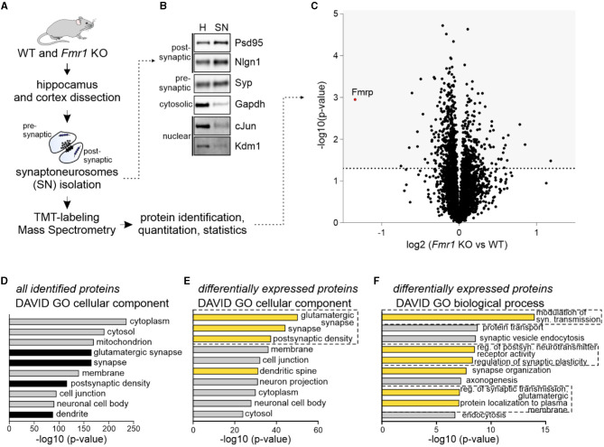
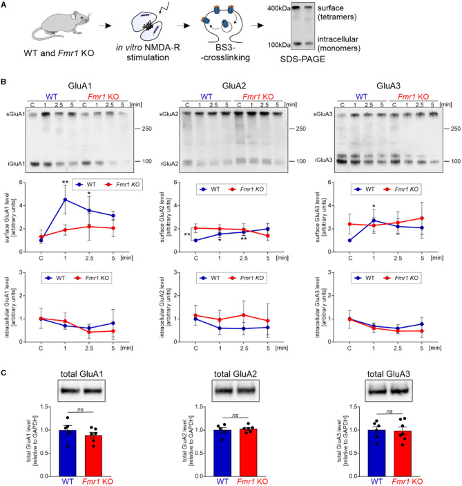
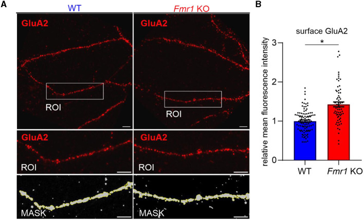
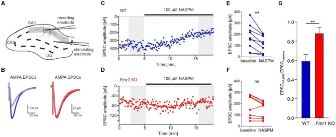
Fragile X syndrome (FXS) is the most common monogenetic cause of inherited intellectual disability and autism in humans. One of the well-characterized molecular phenotypes of Fmr1 KO mice, a model of FXS, is increased translation of synaptic proteins. Although this upregulation stabilizes in adulthood, abnormalities during the critical period of plasticity have long-term effects on circuit formation and synaptic properties. Using high-resolution quantitative proteomics of synaptoneurosomes isolated from the adult, developed brains of Fmr1 KO mice, we show a differential abundance of proteins regulating the postsynaptic receptor activity of glutamatergic synapses. We investigated the AMPA receptor composition and shuttling in adult Fmr1 KO and WT mice using a variety of complementary experimental strategies such as surface protein crosslinking, immunostaining of surface receptors, and electrophysiology. We discovered that the activity-dependent synaptic delivery of AMPARs is impaired in adult Fmr1 KO mice. Furthermore, we show that Fmr1 KO synaptic AMPARs contain more GluA2 subunits that can be interpreted as a switch in the synaptic AMPAR subtype toward an increased number of Ca2+-impermeable receptors in adult Fmr1 KO synapses.

Keywords: AMPA receptors; FXS; Fmr1 KO; GluA2; brain; synaptic plasticity.


Conflict of interest statement

The authors declare that the research was conducted in the absence of any commercial or financial relationships that could be construed as a potential conflict of interest.

Figures

Figure 1
Figure 1
Quantitative mass spectrometry analysis of synaptoneurosomes from adult Fmr1 KO mice. (A) Schematic illustration of the experimental workflow depicting synaptoneurosomes (SNs) isolation and proteomics. (B) Western blot on fractions obtained during SN preparation reveals the enrichment of both pre- and postsynaptic markers in the SN fraction. Cytosolic and nuclear markers were depleted in the SN. (C–F) Results of high-resolution quantitative mass spectrometry. (C) Volcano plot showing an abundance of identified proteins in Fmr1 KO SNs as compared to WT SNs. The vertical line defines the p-value statistical significance cutoff (-log10 p-value > 1.3; Student t-test, n = 4 per group). (D, E) DAVID gene ontology analysis of “cellular component” annotation of proteins. (D) In synaptoneurosomal samples, the top categories were cytoplasmic, mitochondrial, synaptic, and membrane proteins. (E, F) DAVID analysis of differentially expressed proteins in Fmr1 KO SNs showed the top three “cellular component” categories as glutamatergic synapse, synapse, and postsynaptic density. Among the top “biological process” categories proteins involved in the modulation of glutamatergic synaptic transmission, the regulation of postsynaptic neurotransmitter receptor activity, synapse organization, and protein localization to the plasma membrane were identified.
Figure 2
Figure 2
Fmr1 KO synaptoneurosomes display increased surface levels of GluA2 at the basal state and do not increase/accumulate surface AMPAR in response to stimulation. (A) Schematic illustration of the experimental workflow depicting synaptoneurosomes (SNs) isolation and NMDAR in vitro stimulation, followed by surface protein crosslinking, SDS-PAGE, and Western blotting. (B) WT and Fmr1 KO synaptoneurosomes at the basal state (control, C, basal state) and stimulated for 1, 2.5, and 5 min were subjected to surface protein crosslinking. Representative immunoblots show surface AMPARs (tetramers) at ~400 kDa and intracellular AMPARs (monomers) at ~100 kDa. Band intensities were calculated according to WT C, which was set as “1”. Data are presented as mean ± SEM. In the WT synaptoneurosomes after the stimulation, a rapid increase in the surface levels of AMPAR was observed (GluA1: n = 3; 1′ vs. C **p = 0.0066; 2.5′ vs. C *p = 0.0278; GluA2: n = 3; 1′ vs. C *p = 0.0423; 2.5′ vs. C **p = 0.0097; GluA3: n = 4; 1′ vs. C *p = 0.0358; repeated measures two-way ANOVA, post-hoc Tukey's multiple comparisons test). In contrast, in Fmr1 KO samples, SN stimulation did not influence AMPAR surface levels at any analyzed timepoints (RM two-way ANOVA, post-hoc Tukey's multiple comparisons test, p > 0.05). Interestingly, we observed increased levels of GluA2-containing AMPARs at the basal state in the Fmr1 KO SNs (GluA2: n = 3; WT C vs. KO C **p = 0.0074; RM two-way ANOVA, post-hoc Sidak's multiple comparisons test) (see Supplementary Figure S1). (C) Analysis of total GluA1, GluA2, and GluA3 protein levels in WT and Fmr1 KO SNs did not reveal any significant changes in AMPAR subunits among the two genotypes (WT, n = 5; Fmr1 KO, n = 6; unpaired t-test; p > 0.05).
Figure 3
Figure 3
Increased levels of surface GluA2 in hippocampal neurons of Fmr1 KO mice. (A) Upper: Representative immunofluorescence images of secondary dendrites from WT and Fmr1 KO hippocampal neurons at 19 DIV stained for surface GluA2. Scale bar: 5 μm. Lower: Magnified images of boxed areas. A thresholding mask was created to segment the target dendrite, and the mean fluorescent intensity was measured within the mask. (B) The graph shows the results of the quantification of the mean surface GluA2 fluorescent intensity. Values were relativized to the average GluA2 intensity in WT, and data are presented as mean ± SEM (*p = 0.015; nested t-test; n = 66–102 ROIs analyzed/genotype, N = 4 independent neuronal cultures/genotype).
Figure 4
Figure 4
Increased levels of Ca2+-impermeable AMPAR (GluA2-containing AMPAR) at the synapses of Fmr1 KO mice. (A) Diagram showing the positions of the stimulating and recording electrodes in the CA1 field of the hippocampus. (B) Example AMPAR-mediated EPSCs averaged from the first 60 traces (navy blue, WT, or dark red, Fmr1 KO) and the last 60 traces (light blue or light red) of the recordings shown in (C, D). (C, D) Representative recordings of AMPA receptor-mediated EPSCs of CA1 neurons from WT (blue) and Fmr1 KO (red) illustrate the effect of NASPM after 10 min of bath application. (E, F) Averaged amplitudes of each recorded cell before and after the application of NASPM (grayed areas from C, D panels) showing a decrease of EPSCs amplitude following bath application of 100 μM NASPM in WT (**p = 0.0034, paired t-test) but not Fmr1 KO (ns, p = 0.067, paired t-test). (G) Bar graph summarizing NASPM-induced decrease of AMPARs EPSCs amplitudes, suggesting higher abundance of GluA2 subunits in the CA1 of Fmr1 KO mice (unpaired t-test, **p = 0.0069).

Similar articles

References

    1. Achuta V. S., Moykkynen T., Peteri U. K., Turconi G., Rivera C., Keinanen K., et al. . (2018). Functional changes of AMPA responses in human induced pluripotent stem cell-derived neural progenitors in fragile X syndrome. Sci Signal. 11, 8784. 10.1126/scisignal.aan8784 - DOI - PubMed
    1. Antoine M. W., Langberg T., Schnepel P., Feldman D. E. (2019). Increased excitation-inhibition ratio stabilizes synapse and circuit excitability in four autism mouse models. Neuron. 101, 648–661.e4. 10.1016/j.neuron.2018.12.026 - DOI - PMC - PubMed
    1. Ascano M., Jr., Mukherjee N., Bandaru P., Miller J. B., Nusbaum J. D., Corcoran D. L., et al. . (2012). FMRP targets distinct mRNA sequence elements to regulate protein expression. Nature. 492, 382–386. 10.1038/nature11737 - DOI - PMC - PubMed
    1. Banke T. G., Barria A. (2020). Transient enhanced GluA2 expression in young hippocampal neurons of a fragile X mouse model. Front. Synaptic Neurosci. 12, 588295. 10.3389/fnsyn.2020.588295 - DOI - PMC - PubMed
    1. Bassell G. J., Warren S. T. (2008). Fragile X syndrome: loss of local mRNA regulation alters synaptic development and function. Neuron. 60, 201–214. 10.1016/j.neuron.2008.10.004 - DOI - PMC - PubMed

Grants and funding

The author(s) declare financial support was received for the research, authorship, and/or publication of this article. This study was mainly supported by NCN Grant 2019/35/B/NZ4/04355 for MD.